Biology Reference
In-Depth Information
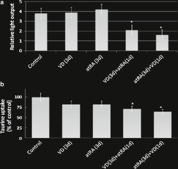
Fig. 27.3
Regulation of
TauT
by VD and RA in MCF-7 cells. MCF-7 cells were cultured in
medium containing VD, RA, or VD plus RA for stated times, and then expression of TauT was
determined. (
a
) Reporter gene assay. (
b
) Taurine uptake. (
c
) Western blot analysis. (
d
) Relative
density of C. (
e
) Effect of 9-cis RA on taurine uptake
27.4
Discussion
The impact of 1,25(OH)
2
D
3
and all-
trans
retinoic acid on the
TauT
gene in MCF-7 cells
is the opposite of that in renal cells:
TauT
is downregulated. The MCF-7 human breast
carcinoma cell line has an interesting biologic feature in that it demonstrates epithelial
cell polarity (van Deurs et al.
1987
). In contrast to most breast carcinoma cell lines,
MFC-7 cells form apical tight junctions that do not permit entry of a ricin-horseradish
peroxidase conjugate, the binding site for which is found on the apical surface of the
cells. These MFC-7 cells align as human mammary cells do in vivo. Moreover, these
MFC-7 cells express the human milk fat globule membrane antigen, which is found on
the apical surface of nonmalignant human breast cells. Hence, MFC-7 cell lines are
likely to be informative about polarized taurine transport in vitro.
MCF-7 cells demonstrate estrogen-dependent growth and are responsive to anties-
trogenic therapy (Demirpence et al.
1994
) . All-
trans
retinoic acid (RA) and 1,25(OH)
2
D
3
Search WWH ::

Custom Search