Biomedical Engineering Reference
In-Depth Information
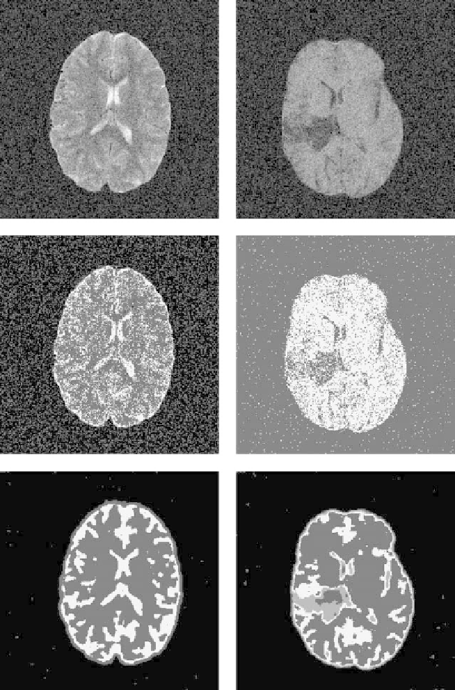
Figure 9.20:
Brain tumor MRI examples. Upper row: Original MR images cor-
rupted with salt and pepper noise. Middle row: the segmented images using FCM
without any neighborhood consideration. Bottom row: The segmented images
using BCFCM (
α
=
0
.
85).