Biomedical Engineering Reference
In-Depth Information
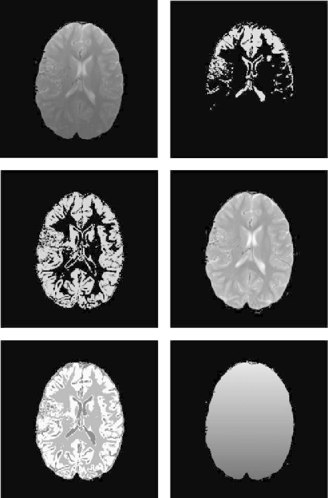
Figure 9.19:
Brain MRI example: (upper left) the original MR image corrupted
with intensity inhomogeneities. (Upper right) crisp gray matter membership
using traditional FCM. (Middle left) crisp gray matter membership using the
proposed BCFCM algorithm. (Middle right) the bias-field corrected image using
BCFCM. The segmented image and bias field estimate using BCFCM are shown
in bottom left and bottom right, respectively.