Biomedical Engineering Reference
In-Depth Information
Node 10
Node 8
Node 7
1
1
1
Monopolar
Bipolar
Tripolar
Monopolar
Bipolar
Tripolar
Monopolar
Bipolar
Tripolar
0.8
0.8
0.9
0.6
0.6
0.8
0.4
0.4
0.7
0.2
0.2
0.6
0
0
0.5
−0.2
−0.2
0.4
−0.4
−0.4
0.3
−0.6
−0.6
−0.8
0.2
0
5
10
15
20
25
30
35
0
5
10
15
20
25
30
35
0
5
10
15
20
25
30
35
[mm]
[mm]
[mm]
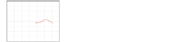
Fig. 9
Extracellular voltage at nodes 10, 8 and 7 with a modiolar electrode insertion for monopolar,
bipolar and tripolar stimulation mode. For better presentation, the extracellular voltage has been
normalized for each stimulation mode
Ta b l e 4
Spread of excitation width in (mm) at different nodes and for different stimulation modes
Node 10 (mm)
Node 8 (mm)
Node 7 (mm)
Monopolar
1.78
1.3
2.79
Bipolar
1.25
0.98
3.5
Tripolar
0.78
0.9
2.1
excitation width for the different stimulation modes and for different degrees of
dendrite degeneration.
FromTable
4
and Fig.
9
it can be observed that for no nerve degeneration (node 10),
monopolar stimulation produces wider spread of excitation than bipolar or tripolar.
However, as the dendrites are more retracted (node 7) the differences in spread of
excitation width for the different stimulation modes are less marked.
3.3 Experiment 3: Simulation of the Voltage Distribution
for Different Cochlear Anatomies and Stimulation Modes
This experiment investigates the effect of cochlear size on extracellular voltage along
the spiral ganglion. Two cochlear geometries were used: “C1” and “C2” using a mid-
scalar electrode placement for both cochleae. The extracellular voltagewas computed
for monopolar, bipolar and tripolar stimulation modes using the same configuration
as in Experiment 2. The results obtained are presented in Fig.
10
. The results show
differences in the shape of the excitation pattern for the two cochlear geometries. For
example, for the cochlea “C1” the peaks of the extracellular voltage for monopolar
and bipolar stimulation are shifted with respect to cochlea “C2”. Furthermore, for
the cochlea “C2” monopolar stimulation produces a narrower spread of excitation
than for cochlea “C1”. From these experiments, it seems that the size and shape
of the cochlea can also contribute to explain the inter subject variability in speech
intelligibility typically observed in CIs users.