Biomedical Engineering Reference
In-Depth Information
(a)
(b)
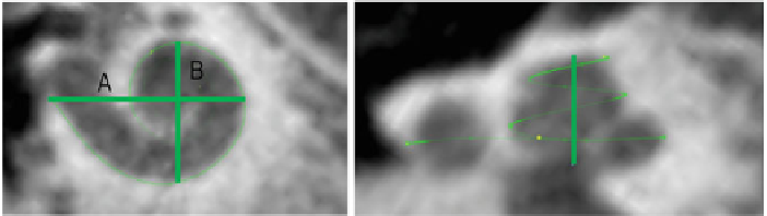
Fig. 1 a
Cochlear dimensions estimated from clinical computer tomography.
A-
and
B-
value in
CBCT data as clinical metric parameters.
b
Height of the cochlea starting from the lowest basal
point to the apex
Ta b l e 1
Cochlear dimensions for the cochleas “C1” and “C2”
0.5 TL
1TL
1.5 TL
2TL
2.5 TL
A-value
B-value
Height
(mm)
(mm)
(mm)
C1
11.61
22.39
32.44
39.02
43.95
7.74
5.21
6.74
C2
14.05
25.27
35.44
43.08
48.03
8.43
5.93
5.37
Additionally the cochlear length at different places (0.5, 1.5, 2, 2.5 turns of the
cochlea) and the height of the cochlea can be adapted from clinical cone beam
computed tomography (CBCT) data (Fig.
1
a, b). As an example, the dimensions for
two cochleas (“C1” and “C2”) are given in Table
1
.
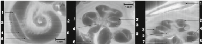
Next, a CI electrode array was modelled using ball/sphere electrodes positioned
inside the scala tympani. This simple approachwas taken because it allows for an easy
parameterization of experiments by simply shifting the positions of the electrodes.
Additionally, the positions of the electrodes can be adapted to the individual position
of each CI user by analyzing post-operative CT scans (Fig.
2
). Each electrode was
placed at a distance of 1.5mmwith respect to the next electrode. In total 16 electrodes
were placed inside the scala tympani.
Fig. 2
Left
Cochlea in sagital cross-section.
1
Basilar membrane,
2
Modiolus,
3
Spiral ligament,
4
Scala vestibuli,
5
Scala tympani,
6
Limbus laminae spiralis
Center
Cochlea in axial cross-section.
1
Spiral ligament,
2
Scala vestibuli,
3
Scala tympani,
4
Modiolus,
5
Osseous spiral lamina in contact
to the basilar membrane,
6
Cochlear nerve in internal auditorymeatus
Right
Cochlea in sagital cross-
section
1
Facial nerve in facial nerve canal,
2
Scala vestibuli,
3
Spiral ligament,
4
Osseous spiral
lamina in contact to the basilar membrane,
5
Modiolus,
6
Scala tympani,
7
Cochlear nerve in
internal auditory meatus