Biomedical Engineering Reference
In-Depth Information
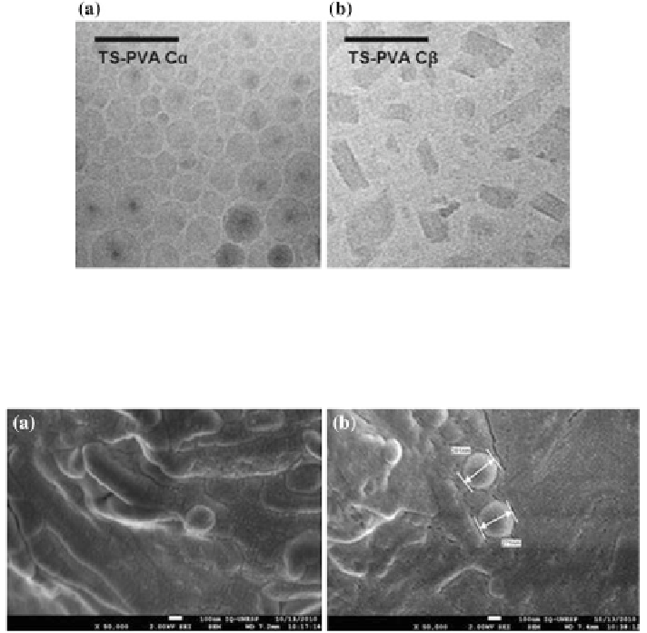
Fig. 4.5
Cryo-TEM images of tristearin nanoparticles stabilized with poly(vinyl) alcohol (
PVA
).
a
Tristearin with
α
α-modification; C
α
and
b
tristearin with
β
β-modification, C
β
. C
α
particles had
a
circular shape
and C
β
particles had
angularly-shaped
platelets. Reprinted from Eur J Pharm
Biopharm, Petersen et al. (
2011
), with permission from Elsevier
Fig. 4.6
SEM images of unloaded (
a
) and praziquantel-loaded (
b
) solid lipid nanoparticles.
(Composition: 5 % stearic acid, 1 % Poloxamer 188). Reprinted from J Therm Anal Calorim, de
Souza et al. (
2012
), with permission from Springer
in their natural state (dispersed in water) by cryo-FESEM (Saupe et al.
2006
).
Figure
4.7
displays an FESEM image of lipid nanoparticles.
4.2.3 Atomic Force Microscopy
Atomic force microscopy (AFM) has been commonly used to investigate the
morphology of lipid nanoparticles (Aji Alex et al.
2011
; de Mendoza et al.
2008
;
Dubes et al.
2003
; Olbrich et al.
2001
; Shahgaldian et al.
2003
; Sitterberg
et al.
2010
; Tabatt et al.
2004a
). AFM provides a high-resolution image of the par-
ticle surface and is an important characterization tool for particulate or biological
samples as it allows imaging under hydrated conditions. However, probe-sample
Search WWH ::

Custom Search