Biology Reference
In-Depth Information
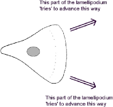
FIGURE 11.2
The 'tug of war' between rival parts of a lamellipodium.
and against which the actin/myosin filaments of the rest of the cell pull (see Chapter 8).
Strong adhesions will support vigorous protrusion and drawing forward of the cell, whereas
weaker ones that slip under mechanical load will be less efficient. In the tug of war between
rival parts of the leading edge, those parts that have the strongest grip on the substrate would
be expected to win (
Figure 11.3
). This type of guidance is and has been demonstrated in
a variety of culture systems.
One classical method for demonstrating haptotaxis is to produce patterned substrates
in
vitro
, on which cells will be confronted by boundaries of different adhesiveness as in
Figure 11.3
. The relative adhesiveness of different substrates can be assessed semi-
quantitatively by plating cells on to them and then by trying to blow them off by directing
a jet of fluid at them from a fine pipette (
Figure 11.4
a): the closer the pipette has to be brought,
the more adhesive the substrate. Such simple assays demonstrate that the growth cones of
chick embryo dorsal root ganglion cells stick very well to poly-ornithine, a little less well to
collagen, less well still to palladium and tissue culture plastic, and very poorly to bacteriolog-
ical plastic
2
. Substrates can be coated with palladium using equipment originally designed to
produce thin conductive films of metal on samples for scanning electron microscopy; if a fine
metal grid is interposed between the coating gun and the substrate it will cast palladium-free
'shadows' in a grid pattern on an otherwise palladium-coated surface. The growth cones of
ganglion cells plated on such a substrate will therefore be presented with many boundaries
between palladium and an alternative. If the alternative is tissue culture plastic, which is about
equally adhesive as palladium, the growth cones migrate randomly across boundaries. If the
alternative is a more adhesive substrate such as poly-ornthine, they will instead remain on or
cross on to the polyornithine and will then follow it, even performing right-angled turns in
preference to straying on to the less adhesive substrate
2,3
(
Figure 11.4
b). At a boundary, filopo-
dia explore the less adhesive as well as the more adhesive substrate, but then slip back. Similar
behaviour can be observed in other cells such as fibroblasts,
4
although their courses of migra-
tion are less obvious than those of growth cones because they leave no axon behind them.