Biomedical Engineering Reference
In-Depth Information
Figure 13. (a) Photograph of a BEC-enclosing Ca-Alg/gelatin fiber. Bar: 500 μm. (b) Diameters of Ca-
Alg/gelatin hydrogel fibers as a function of the flow rate of the Na-Alg/gelatin mixture and the diameter
of the needles, (■) 270, (●) 480 and (▲) 940 μm in diameter) from which the mixture was extruded.
The flow rate of the 100 mM CaCl
2
solution was fixed at 33.9 cm/s (Reproduced with permission, from
Sakai S et al. Biomacromolecules [75] @ 2008 American Chemical Society).
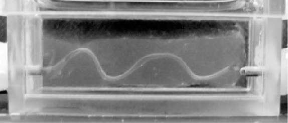
Figure 14. Photograph of the wavy-shaped channel developed in collagen gel by degrading the cell-
enclosing alginate fiber (Reproduced with permission, from Sakai S et al. Biomacromolecules [75] @
2008 American Chemical Society).
Cell Encapsulation in Ca-alginate Gel Fibers
It was also of interest to determine whether it is possible to prepare cell-enclosing
microcapsules using the aqueous solution containing dissolved multivalent cations such as
calcium ions as an ambient co-flowing liquid. It was not possible because the extruded
sodium-alginate solution instantaneously gelates after contacting the multivalent cations in
aqueous solution. We obtained cell-enclosing alginate fibers when we injected cell-
suspending aqueous alginate solution into a co-flowing 100 mM CaCl
2
solution (Figure 13a).
Similar to the droplet production in liquid paraffin, the diameter of the fiber is controllable by
changing the flow rate of ambient liquid and the diameter of the needle from which the cell-
suspending alginate solution is extruded (Figure 13b). One possible application of this cell-
enclosing alginate fiber is use it as a template for a vascular-like structure previously
developed using biocompatible hydrogels
in vitro
[75]. Due to the flexibility of alginate gel
fibers and their ability to degrade using alginate lyase, it is possible to develop a tubular