ABSTRACT
In this paper, we describe a theoretical foundation for and experimental implementation of a novel 360-degree stereo digital image correlation (DIC) method for quantifying shape and deformation of quasi-cylindrical specimens along their full length and around their circumference. The proposed approach has been implemented for in-vitro experiments on arteries immersed within a physiologic solution (PS) that maintains unaltered native biomechanical properties. To this end, we also address the ubiquitous issue of refraction in non-contacting optical measurements on biological samples immersed in PS by developing and contrasting three different approaches for the correction of the refraction error.
Introduction
Quantifying the regionally varying properties of arteries is of primary importance to investigate the mechanical response of arteries in order to better understand disease progression and response to clinical intervention [1]. Towards this end, in vitro experiments represent an irreplaceable tool to collect the needed data over a full range of multiaxial loads, but they are typically performed on arterial specimens that are nearly straight and cylindrical [2]. Stereo-systems tracking a given set of markers affixed to the surface of complex shaped specimens [3, 4] demonstrated to be able to give information on potential regional differences in the tissue mechanical properties when used within sub-domain inverse finite elements frameworks [5, 6]. However, the recent move toward the use of mouse model to study changes in arterial properties due to genetic mutations or disease progression [7-9] makes it cumbersome to apply a large number of markers on the arterial surface to sample the displacement field with a sufficient resolution and without affecting the estimated wall properties.
To address the above mentioned issues, a panoramic Digital Image Correlation (DIC)-based system for quantifying full surface strain fields along the full length and around the entire circumference of small arteries having complex geometries has been developed and tested [10, 11]. The strength point of this method relies in the capability to map the whole 3D deformation of arterial specimens in their native (e.g. curved) or altered (e.g. with aneurism) geometries with the high spatial resolution typical of the DIC measurement.
In this paper we present an improved version of this method that includes a more efficient calibration procedure. Moreover, since living arteries need to be tested while immersed in physiological solution, three methods for correcting the measurement error due to refraction at the air/water interface of the specimen bath have been developed and compared.
Materials and Methods
A detailed description of the theoretical rational behind the panoramic DIC method together with the results of the experimental campaign carried out to assess accuracy and resolution of the measurement can be found in [10, 11]. Briefly, measurement of full surface strain fields of quasi-cylindrical objects can be achieved if the specimen is placed nearly coaxial within a 45° concave conical mirror. When viewed from above, the mirror reflects the surface of the specimen over the full 360 degrees; moreover, when at least two different points of view are used, 3-D information can be obtained using the principles of Stereophotogrammetry [12]. In particular, using a DIC-based procedure [13] to match paired stereo-images allows full reconstruction of the surface in reference and deformed configurations, which in turn permits full analysis of the surface strain field with high spatial resolution.
In the original set-up presented in [10, 11], a four-view stereo-system was realized by placing a 45° gimbal-mounted flat mirror between the camera and the conical mirror and sequentially tilting the mirror to enable a single camera to view four nearly polar-symmetrical images: right (R), left (L), up (U), and down (D). This peculiar scheme of the image formation, in fact, disallows the use of a standard lateral view stereo-system with two fixed cameras since the stereo-angle required to get satisfactory images needs to be less than 1°. An immediate consequence of the adoption of a very small stereo-angle is a large reconstruction error for the object points close to the plane containing the axes of the two cameras (called hereafter the ‘stereo-plane’). An evenly accurate reconstruction of the sample can be obtained only by adopting a four-view set-up composed by two stereo-systems (namely RL and UD) with their stereo-planes fairly perpendicular to each others. Merging the two sets of data points sufficiently far for the stereo-planes in a complementary way, in fact, allows us to reconstruct the full 360-deg shape of quasi-cylindrical specimens with an accuracy of 10-2 mm [10, 11].
In this work we address two issues critical for the implementation of the methodology presented in [10, 11]: calibration and correction of the refraction error. In particular, we test an alternative procedure to calibrate the cameras more accurately and we implement and contrast three different methods to correct the error due to the refraction at the air/water interface of the bath containing the specimen.
Experimental system set-up and calibration
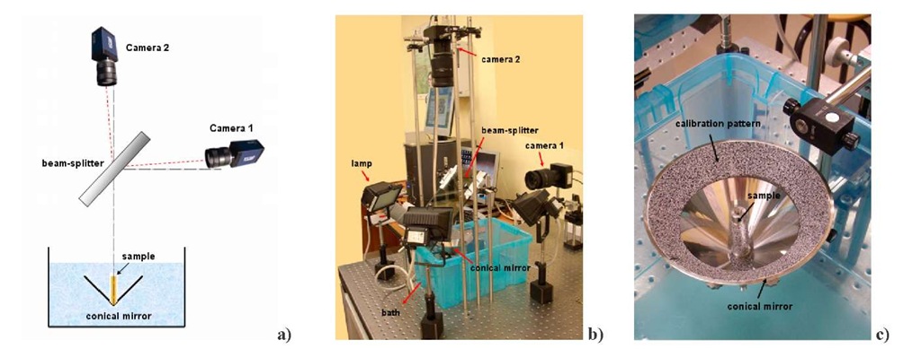
Figure 1a shows a scheme of the experimental set-up adopted in this work. We chose a two-view stereo-system with two fixed cameras in order to not include in the analysis the eventual error due to repositioning of the cameras of the original four-view arrangement. A beam splitter has been initially positioned at 45° with respect to the axis of the cone and the axes of two fixed cameras as depicted in Fig. 1a. Then, the cameras were slightly tilted in order to realize the desired stereo-angle. A steel post with a printed random pattern on it was placed coaxially with the conical mirror. On the uppermost portion of the mirror, another speckle pattern was glued to serve as a calibration target. A preliminary calibration procedure was needed prior to performing the fine alignment of the system. Conversely to the procedure adopted in [10, 11], in fact, calibration of the cameras and the position of the conical mirror surface into the global reference system was achieved by using a speckle pattern instead of a dot calibration pattern. This allowed us to obtain better efficiency in aligning and calibrating the system as will be discussed later. The developed calibration/alignment procedure can be summarized as follows:
- The two cameras where aligned in order to form a stereo-angle of about 16°.
- A steel bracket with a regular dot pattern on it was placed inside the cone and served as a target to retrieve the position of the cameras in a global reference system conveniently joint to the bracket (named GRS_1) via calibration with the Direct Linear Transformation Method (DLT) [14].
- The bracket was removed and the position of 6000 points (SET_1) of the speckle pattern glued on the uppermost portion of the mirror was retrieved with great accuracy (due to the large stereo-angle adopted) in the GRS_1.
- An optimization procedure allowed to find the components of the rotation matrix and the translational vector needed to transform the coordinates of the points belonging to the cone from the GRS_1 to another system (GRS_2) in which the cone equation can be written as![]() The angle y of aperture of the cone has been included among the design variables of the optimization procedure.
The angle y of aperture of the cone has been included among the design variables of the optimization procedure.
- The so obtained new coordinates of the 6000 points (SET_2) belonging to the calibration pattern on the cone can be used to find intrinsic and extrinsic parameters of the two cameras in the new global reference system GRS_2.
- The two cameras were progressively moved by means of multiaxial translational and rotational stages in order to finely align the system to the final configuration. In particular, the stereo-angle was reduced to 1° as required from the panoramic measurement and the two cameras were aligned in order to be symmetrical with respect to the axis of the cone. The exact position of the cameras was retrieved at each step of the alignment procedure by calibration via the SET_2 of points and calculating from the DLT parameters the position of sensor centers and pin-holes and visualizing them into a CAD environment. The stages were thus progressively tuned in order to move the axes of the cameras in the desired final position.
This procedure possesses several advantages with respect to the calibration reported in [10, 11]. In the previous work in fact, a dot pattern was glued on the uppermost portion of the cone and the ‘actual’ position of the dots was deduced on the basis of geometrical considerations starting from the position of the dots of the unrolled printed pattern. As can be easily understood, however accurate the positioning of the pattern on the cone can be, the exact theoretical position of the dots cannot be practically obtained. This obviously leads to an inaccurate calibration of the stereo-system and to an erroneous evaluation of the position of the mirror surface in the global reference system. This has effects mainly on the reconstruction of the points close to the stereo-planes for which the ‘virtual’ stereo-angle coincide with the actual stereo-angle (see [10, 11] for details). With the present calibration procedure, instead of using a centroid-seeking algorithm, the more efficient DIC algorithm is used to match the two views of the stereo-system to retrieve the position of 6 times more points than in the previous calibration pattern. The actual position of these points (SET_1) in the global reference system is evaluated by using a stereo-system with a large stereo-angle and a more reliable ‘bracket-like’ calibration target. The expected augmented efficiency of the calibration procedure used in this work was confirmed by a strongly reduced error on the stereo-plane observed in the reconstruction of the steel post (less than 5% on radius).
Fig. 1 a) Scheme of the optical set-up adopted in this work; b) picture of the experimental set-up; c) detailed view of the conical mirror with the steel post used in this work as the calibration sample.
Correction of the refraction error
Once the system has been calibrated, a series of experiments were run to evaluate the relative merits of three different methodologies to correct the refraction error at the air/water interface of the specimen bath. This methodology, in fact, has been conceived to be used for measuring the full surface strain field on living arteries which necessarily must be tested while immersed within the physiological solution. To evaluate the effect of the refraction on the panoramic measurement, two couples of images have been captured with the cone and sample immersed into the water (IW1 and IW2 from camera 1 and 2, respectively) and in air (IA1 and IA2). The three approaches are here briefly described and discussed.
- DLT-based approach.
The two cameras are calibrated via standard DLT by using the images captured into the water IWj and IW2, and the object is reconstructed by using the set of the 11 DLT parameters so obtained. The straightforward use of this approach implies to make a theoretical mistake since the DLT method relies on the pin-hole projection scheme and thus on the collinearity condition that considers the image point, the pin-hole and the object point belonging on the same line. This condition is not satisfied in the presence of refraction. Indeed, it is well known that when light crosses an air/water interface, its direction of travel deviates according to the Snell’s law na sin 6a = nw sin 0w where 0a is the incident angle, 0w is the refracted angle, and na and nw are indices of refraction for air and water respectively (na = 1.0 and nw = 133).
Hence, when using the DLT approach to calibrate the cameras position by using the images captured in air, the set of the DLT parameters obtained represent a solution not physically feasible. In other words, if the DLT parameters are used to calculate the sensor center or the pin-hole position, they yield to locate the camera in an unfeasible position in the reference system. However, since the DLT parameters represent the solution in a least square sense of a largely redundant system (6000 points for 11 unknowns) it is possible that, under certain conditions, they can be effectively used to reconstruct the points falling within the calibration area. The very good results obtained here by using this method (see Fig. 2b and 3b) can be explained by considering that: i) the calibration points lie on the same surface that contains the object points; ii) the calibration pattern surrounds the area of measurement and occupies a small central region of the camera sensor; iii) the angle of stereo vision is very small, i.e. the rays are almost perpendicular to the air/water interface with a consequent small difference between incident and refracted angles.
Fig. 2 Sample reconstruction. a) The sample reconstructed in air (black dots) vs the sample under water reconstructed without correcting refraction (red dots); b) correction with the DLT approach (purple dots); c) correction with the RP approach (green dots); d) correction with the ES approach (blue dots).
- Refraction plane (RP) approach.
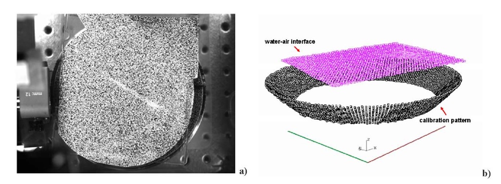
This approach aims to find the actual position of the air/water interface in the global reference system in order to allow the reconstruction of the object via a ray-tracing procedure. A flat plastic sheet with a random pattern is let to float on the experimental bath and reconstructed in the global reference system (Fig. 4). The coefficients of this plane are used as starting values in an optimization routine aimed to find the new set of coefficients of the actual air/water interface. The objective function to be minimized is the sum of the distances between the points of the calibration target as reconstructed in air and their counterparts obtained from the images of the pattern under water. In particular, the travel of the light from each image point is traced from the sensor, through the pin-hole and then deviated at the current air/water interface according to Snell’s law. The refraction index of the water has been included among the optimization variables (here, a value of nw = 1 327 has been obtained). Once the optimal air/water interface has been found, reconstruction of the sample is performed via ray-tracing by using the DLT parameters from calibration in air (Fig. 2c and Fig.3c).This approach yields to the larger error in reconstruction among the three methods considered for the analysis in spite of being the most ‘physically’ meaningful. This may be due to the inherent sensitivity of any optimization procedure to the starting point and to the adopted optimization algorithm.
Fig. 3 Plots of the reconstruction error (calculated as the distance between reconstructed and target points). a) reconstruction without correction of the refraction error; b) correction with the DLT approach; c) correction with the RP approach; d) correction with the ES approach.
- Error surfaces (ES) approach.
Generally speaking, it is not possible to express the refraction error as a function of the image coordinates alone since two separate object points P1 and P2 that map to the same image point IW have two different refraction errors A/1 and AI2 with respect to their image points in air /A1 and /A2 that depend on their 3D position in the space. However, in our case, all object-rays hitting the camera sensors come from a surface (the conical mirror surface) whose position is fixed and known with respect to the stereo system. This allows us to implement a more straightforward approach for reducing the error due to refraction as described in detail in [10, 11]. Briefly, the two couples of images IWr IAi and IW2-IA2 are correlated via DIC. This allows us to quantify the effect of the refraction by mapping the displacement u^ and un [pixel] along the x and y directions for a set of points belonging to the calibration pattern applied on the uppermost part of the cone. If a distortion function is determined for all points belonging to the conical surface and for each camera sensor, it could be used to convert images of the specimen immersed in water to corresponding distortion-free images as they would have been captured in air.
To this aim, the values of u( and un for points on the inner portion of the mirror are extrapolated from those on the upper annular portion by fitting the displacement maps with a Non-Uniform Rationale B-Spline (NURBS) in a CAD environment. The so obtained ‘distortion functions’ are used to correct the position of the image points of the sample that is finally reconstructed by using the DLT set of parameters of calibration in air (Fig. 2d and Fig.3d).
Several trial tests showed the results of this procedure to be strongly dependent from the setting parameters of the NURBS (stiffness and u-v spans) since displacement values of the order of 10-1 pixel demonstrated to yield to an error of several percents on the radius of the sample. This limit could be overcome by utilizing a speckle pattern on the whole surface of the cone and thus evaluating the distortion functions directly from the images and avoiding extrapolation from the outer annular pattern [10, 11]. However, this would imply a longer and more tedious procedure not free from additional sources of errors.
Fig. 4 a) The plastic sheet floating on the specimen bath used for calculating the initial values of the design variables in the optimization procedure of the RP method; b) the air/water interface as reconstructed in the GRS_2.
Conclusion
In this work, we presented an improved panoramic DIC system for quantifying shape and deformation of quasi-cylindrical specimens along their full length and around their circumference. In particular we introduced a more efficient calibration procedure that demonstrated to improve reconstruction accuracy and investigated the different merits of three approaches for correcting the refraction error. Future work will focus on investigating the possibilities of the DLT localized method for correcting the refraction error and on improving the efficiency of the optimization procedure at the basis of the RP method.


