Connections of the Neostriatum With the Substantia Nigra
The substantia nigra has two principal components: a region of tightly compacted cells, called the pars compacta, and a region just ventral and extending lateral to the pars compacta, called the pars reticulata (Fig. 20-2B). Fibers arising from the neostriatum project to the pars reticulata. Transmitters identified in this pathway are GABA and substance P. The pathway from the substantia nigra to the neostriatum arises from the pars compacta and uses dopamine as its neurotransmitter (Fig. 20-5). The pars reticulata also gives rise to efferent fibers (which are likely inhibitory) that project to the thalamus, superior colliculus, and locally to the pars compacta. In this manner, the pars reticulata and internal pallidal segment appear to be functionally analogous because both regions provide outputs to the cerebral cortex via the thalamus. Additional functions of these pathways are discussed later in this topic.
Connections Between the Globus Pallidus and Subthalamic Nucleus
As noted earlier, the globus pallidus shares reciprocal connections with the subthalamic nucleus. The lateral segment of the globus pallidus (which receives GABAergic and enkephalinergic inputs from the neostriatum) projects to the subthalamic nucleus. GABA also mediates this pathway. In turn, the subthalamic nucleus projects back to the medial segment of globus pallidus. This pathway, however, is mediated by glutamate.
Output of The Basal Ganglia
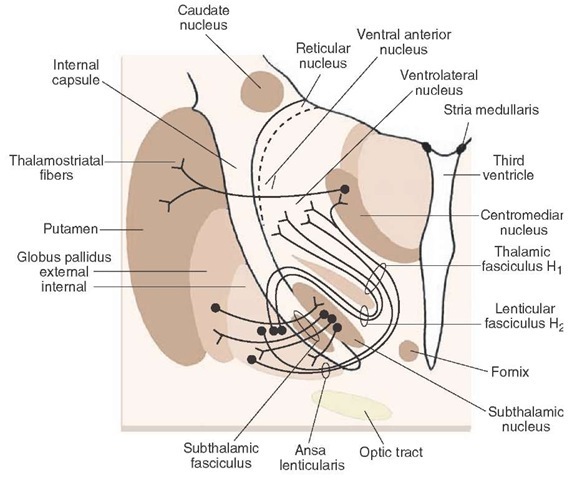
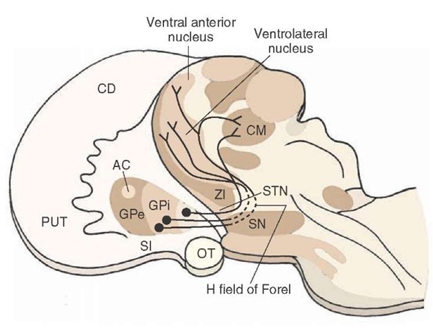
As indicated earlier, the basal ganglia influence motor functions primarily by acting on motor neurons of the cerebral cortex via relay nuclei of the thalamus. The output pathways of the basal ganglia achieve this.The first pathway, the ansa lenticularis, arises from the ventral aspect of the medial pallidal segment. It passes caudally towards the red nucleus and then turns rostrally to enter the thalamus. The second pathway, the lenticular fasciculus, which arises from the medial pallidal segment, exits the pallidum dorsally. As the fibers run caudally in the direction of the midbrain (adjacent to the red nucleus), they pass between the subthalamic nucleus and zona incerta. As the fibers approach the red nucleus, they also abruptly turn rostrally and enter the thalamus (Fig. 20-7).
In addition to the internal pallidal segment, the basal ganglia can also influence motor functions through the output pathways of the substantia nigra. We have already noted the important dopaminergic pathway from the pars compacta to the neostriatum. The pars reticulata contributes at least two pathways. One pathway projects to the ventral anterior and ventrolateral nuclei of the thalamus, and its effects upon thalamic neurons are mediated by GABA. Thus, the overall influence of the pars reticulata on motor functions of the cerebral cortex is made possible by the synaptic connections between nigral fibers and these thalamic relay nuclei. A second pathway projects to the superior colliculus. Because the superior colliculus is involved in the integration of saccadic eye movements and tracking, this pathway can influence motor functions related to reflex and voluntary control of eye movements. The efferent pathways of the substantia nigra are summarized in Figure 20-5.
FIGURE 20-6 Efferent projections of the pallidum. Note that the fibers of medial pallidal segment use two pathways to supply the ventrolateral, ventral anterior, and centromedian nuclei (the ansa lenticularis and the lenticular fasciculus or H2 field of Forel). The region where fibers of the ansa lenticularis, lenticular fasciculus, and cerebelloth-alamic merge is the H, field of Forel.The subthalamic fasciculus represents reciprocal connections between the globus pallidus and subthalamic nucleus and serves as the anatomical substrate for the indirect pathway.
FIGURE 20-7 Outputs of the basal ganglia and their trajectories. The origins and trajectories of the major outputs of the globus pallidus (GP) to the thalamus are diagrammed. Note that the pathways initially pass caudally toward the red nucleus of the midbrain before reversing their course (at the H field of Forel) to pass rostrally into the ventrolateral, ventral anterior, and centromedian (CM) thalamic nuclei. AC = anterior commissure; CD = caudate nucleus; GPi = globus pallidus internal; GPe = globus pallidus external; OT = optic tract; PUT = putamen; SN = substantia nigra; STN = subthalamic nucleus; ZI = zona incerta.
Functional Mechanisms of the Basal Ganglia
Possible Role of Intrinsic Circuits
The information presented thus far has enabled us to identify the major circuitry of the basal ganglia. This includes the primary input pathways and the internal circuitry of the basal ganglia and their output pathways. In terms of the functional properties of this system, they may be best understood by following the flow-through relationships beginning with the inputs from the cerebral cortex.
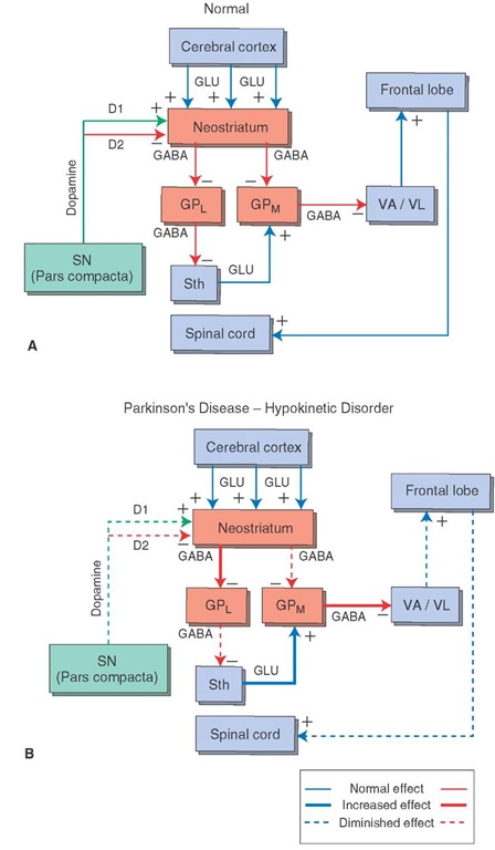
As noted earlier, there are both direct and indirect pathways from the striatum to the thalamus, and the functional effects of each are different. Concerning the direct pathway, cortical inputs have excitatory effects on the neostriatum, and the neurotransmitter is glutamate. Excitation of the neostriatum by the cortex results in dis-inhibition of thalamic nuclei. This can be understood in terms of the following relationships. The projection from the neostriatum to the medial pallidal segment is inhibitory, and, likewise, the projection from the medial pallidal segment to the thalamus is inhibitory. GABA mediates both inhibitory pathways. In this manner, activation of the cortex causes the neurons in the medial pallidal segment to be inhibited by the GABAergic projection from the neostriatum. When the neurons of the medial pallidal segment are inhibited, the thalamic neurons that project to motor regions of the cortex are then released from the inhibition normally imposed on them by the medial palli-dal segment. Because the projections from the thalamus to cortex are direct and excitatory, movement is then facilitated by the actions of the thalamocortical projection, which excites motor regions of cortex and their descending pathways to the brainstem and spinal cord. Thus, this feedback circuit can be interpreted to mean that movement follows activation of the neostriatum by the cortex. The functional relationships of the overall circuit for the direct pathway could now be summarized as follows, with ( + ) indicating excitation and (-) indicating inhibition (Fig. 20-8A): cerebral cortex (glutamate) — ( + ) neostriatum (GABA) — (-) internal pallidal segment (GABA) — (-) thalamus — ( + ) the motor regions of cortex. The net effect is excitation of the motor regions of cortex.
FIGURE 20-8 Schematic diagram depicting the possible mechanisms for hypokinetic and hyperkinetic disorders. (A) Normal input-output relationships of the basal ganglia. GLU = glutamate; GABA = gamma aminobutyric acid; GPL = lateral pallidal segment; GPM = medial pallidal segment; SN = substantia nigra; Sth = subthalamic nucleus; VA = ventral anterior thalamic nucleus; VL = ventrolateral thalamic nucleus. (B) Hypokinetic disorder (Parkinson’s disease). This model proposes that the hypokinetic effect is manifested by the reduced quantities of dopamine released, which act on dopamine D, and D2 receptors in the neostriatum. Dopamine acting through D, receptors in the neostriatum is excitatory to GABAergic neurons, which project to the GPM (direct pathway). Moreover, dopamine acting through D2 receptors in the neostriatum inhibits GABAergic neurons, which project to the GPL segment (indirect pathway). When there is diminished dopamine in the neostriatum released onto D, receptors (dotted line), the resulting inhibitory output of the neostriatum on the GPM is diminished. This allows for an enhancement of the inhibitory output of the GPM to the thalamus to be increased (thicker arrow). Because the thalamus normally excites the cortex, greater inhibition on the thalamic nuclei by the GPM will cause a weakened input to motor regions of the cortex. Likewise, the projection from the neostriatum, acting through D2 receptors, is inhibitory to the GPL. In turn, the GPL is normally inhibitory to the Sth. Thus, the diminished inhibitory input into the neostriatum from SN allows the neostriatum to exert greater inhibition on GPL, which, in turn, results in a weakened inhibition upon the Sth. Because the Sth normally excites the GPM via a glutamatergic mechanism, the reduced amount of inhibition on the Sth would allow it to exert a greater excitatory effect on the GPM. Therefore, the combined effects of reduced dopaminergic inputs into the neostriatum affect both the direct and indirect pathways in such a way as to allow for enhancement of the inhibitory outputs of the GPM on the thalamic projection nuclei and their target regions in motor cortex, causing the hypokinetic movement disorder.
FIGURE 20-8 (C) Hyperkinetic disorder (Huntington’s disease). Inhibitory influences mediated from the caudate and putamen to the external segment of the glo-bus pallidus are diminished. Because of a reduction in GABA in the neostriatum, tonic inhibition mediated from the GPl to the Sth is enhanced, thus reducing the excitation from the Sth to the GPM. The diminished excitatory input to the GPm results in increased excitation to the VA and VL thalamic nuclei as well as the cortical receiving areas of these thalamic nuclei. The overall effect mediated on the motor regions of the cerebral cortex is an increase in excitation and greater (inappropriate) motor activity..
In contrast, activation of the indirect pathway has a different effect on cortical neurons. The indirect pathway involves the following elements: (1) a GABAergic projection from the neostriatum to the external pallidal segment, (2) a GABAergic projection from the external pallidal segment to the subthalamic nucleus, and (3) a glutamatergic projection from the subthalamic nucleus to the internal pallidal segment. In this circuit, cortical activation initially results in activation of a GABAergic pathway from the neostriatum to the external pallidal segment, which inhibits neurons in this region. Because the pathway from the external pallidal segment to the subthalamic nucleus is also GABAergic and inhibitory, activation of striatal neurons results in inhibition of the (inhibitory) output pathway from the external pallidal segment to the subthalamic nucleus. As a consequence of activation of this circuit, the subthalamic nucleus is released from inhibition and can then provide a heightened glutamate-mediated excitatory input to the internal pallidal segment. Activation of the internal pallidal segment results in inhibition (via a GABAergic pathway) of the thalamic nuclei to which the neurons of the internal pallidal segment project. Thus, when this circuit is stimulated, the overall effect is to cause thalamic relay neurons to be inhibited, resulting in the reduction of excitation to the motor regions of cortex. Therefore, activation of the indirect pathway results in just the opposite effect (i.e., inhibition) on cortical motor neurons compared with the direct pathway. The functional relationships of the indirect pathway can now be summarized as follows, with ( + ) indicating excitation and (-) indicating inhibition: cerebral cortex (glutamate) — ( + ) neostriatum (GABA) — (-) external pallidal segment (GABA) — (-) subthalamic nucleus (glutamate) — ( + ) internal pallidal segment (GABA) — (-) thalamus — (+) motor regions of cortex. The net effect is inhibition of the motor regions of the cortex.
Modulatory Role of Dopamine
In addition to the direct and indirect pathways that significantly influence motor functions of the cortex, other modulatory systems are present as well. The most prominent of these systems is the dopamine pathway from the substantia nigra to the neostriatum. Current research suggests that the dopamine pathway has opposing effects on the direct and indirect pathways (i.e., activation of the dopaminergic pathway excites the direct pathway but inhibits the indirect pathway). As indicated earlier, the direct pathway causes excitation of the motor regions of the cortex and thus facilitates movement, but the indirect pathway inhibits cortical-induced movement. Therefore, the action of dopamine is to facilitate movement because it excites the facilitatory (direct) pathway via its excitatory effects on dopamine D1 receptors in regions of the neostriatum that project to the medial pallidal segment, while inhibiting the inhibitory (indirect) pathway that projects initially to the lateral pallidal segment because of the inhibitory effects on dopamine D2 receptors in the neostriatum. The result is diminution of excitation from the glutamate-mediated pathway arising from the subthalamic nucleus to the medial pallidal segment. In this manner, the overall output of the medial pallidal segment to the thalamus is diminished, resulting in greater excitation of the motor regions of cortex, the consequence of which is an increase in movement. From an overall perspective, we may speculate that purposeful movement requires a sequence of directed responses balanced by other responses, which are appropriately inhibitory. It may be that the direct pathway potentiates normally directed movements, whereas the indirect pathway enhances inhibitory sequences.


