One of the unique features of nerve cells is their capability to communicate with each other with great precision even when they are separated by long distances. The mechanism by which neurons communicate with each other is called synaptic transmission, which is defined as the transfer of signals from one cell to another. A synapse is described as a special zone of contact at which one neuron communicates with another. In this topic, the different types of synapses and various aspects of synaptic transmission are described.
Types of Synaptic Transmission
Two types of synaptic transmission—electrical and chemical—are recognized in the nervous system. It should be noted that the electrical synapses are relatively less common than the chemical synapses in the mammalian nervous system.
Electrical Transmission
In electrical transmission between nerve cells, the current generated by an impulse in one neuron spreads to another neuron through a pathway of low electrical resistance. Electrical synapses occur at gap junctions (described later in this section). In an electrical synapse, ion channels connect the cytoplasm of the presynaptic and postsynaptic cells. In the adult mammalian central nervous system (CNS), electrical synapses are present where the activity of neighboring neurons needs to be highly synchronized. For example, hormone-secreting neurons in the mammalian hypothalamus are connected with electrical synapses so that they fire almost simultaneously and secrete a burst of hormone into the circulation.
At an electrical synapse, the current generated by voltage-gated channels at the presynaptic neuron flows directly into the postsynaptic neuron. Therefore, transmission at such a synapse is very rapid (<0.1 msec). At some synapses (e.g., in the giant motor synapse of crayfish), the current can pass in one direction (from presynaptic to postsynaptic neuron) but not in the reverse direction. Such synapses are called rectifying or unidirectional synapses. At other synapses, the current can pass equally well in both directions. Such synapses are called nonrectifying or bidirectional synapses. Most electrical synapses in the mammalian nervous system are believed to be the nonrec-tifying type.
At this point, it is important to understand the morphology of a gap junction. The area where the two neurons are apposed to each other, at an electrical synapse, is called a gap junction (Fig. 7-1A). The channels that connect the neurons at the gap junction are called gap junction channels (Fig. 7-1B). The extracellular space between presynaptic and postsynaptic neurons at an electrical synapse is 3 to 3.5 nm, which is much smaller than the usual extracellular space (about 20-50 nm) between neurons. The narrow space between these neurons is bridged by gap junction channels. These channels are formed by two hemichannels, one in the presynaptic neuron and the other in the postsynaptic neuron. Each hemichannel is called a connexon (Fig. 7-1C), which is composed of six subunits of identical proteins called connexins (Fig. 7-1D). In the connexon, the connexins are arranged in a hexagonal pattern (Fig. 7-1C). Each connexin consists of four membrane-spanning regions (Fig. 7-1E). The hemichan-nels located in presynaptic and postsynaptic neurons meet each other at the gap between the membranes of the two neurons and form a conducting channel. Rotation of the connexins, in a manner similar to the opening of a shutter in a camera, results in the opening of the pore of the gap junction channel.
FIGURE 7-1 Morphology of a gap junction. (A) Gap junction. (B) Gap junction channel. (C) Hemichannel (connexon). (D) Connexin. (E) Membrane-spanning regions of connexin.
It should be noted that gap junctions are relatively rare in the adult mammalian nervous system. However, they are more common in nonneural cells, such as the epithelial cells, smooth and cardiac muscle cells, liver cells, some glandular cells, and glia.
Chemical Transmission
At chemical synapses (Fig. 7-2A), there is no continuity between the cytoplasm of the presynaptic terminal and postsynaptic neuron. Instead, the cells are separated by synaptic clefts, which are fluid-filled gaps (about 20-50 nm). The presynaptic and postsynaptic membranes adhere to each other due to the presence of a matrix of extracellular fibrous protein in the synaptic cleft. The presynaptic terminal contains synaptic vesicles that are filled with several thousand molecules of a specific chemical substance, the neurotransmitter. Pyramid-like structures consisting of proteins arise from the intracellular side of the presynaptic terminal membrane and project into the cytoplasm of the presynaptic terminal. These pyramids and the membranes associated with them are called active zones and are the specialized release sites in the presynap-tic terminal. The vesicles containing the neurotransmitter are aggregated near the active zones.
FIGURE 7-2 Morphology of a chemical synapse. (A) The presynaptic terminal and postsynaptic neuron are separated by a fluid-filled synaptic cleft. Note that the presynaptic terminal contains synaptic vesicles, which contain neurotransmitter and active zones. Receptors for the transmitter are located on the postsynaptic membrane. Different types of central nervous system synapses include (B) axodendritic synapse, (C) axosomatic synapse, and (D) axoaxonic synapse. (E) In a symmetrical synapse, the presynaptic and postsynaptic membranes are similar in thickness. (F) In an asymmetrical synapse, the postsynaptic membrane of a synapse is thicker than the presynaptic membrane.
Types of Central Nervous System Synapses
Axodendritic synapse: A synapse in which the post-synaptic membrane is on a dendrite of another neuron (Fig. 7-2B).
Axosomatic synapse: A synapse in which the postsynaptic membrane is on the cell body (soma) of another neuron (Fig. 7-2C).
Axoaxonic synapse: A synapse in which the postsynaptic membrane is on the axon of another neuron (Fig. 7-2D). Dendrodentritic synapse: A synapse in which dendrites of specialized neurons form synapses with each other. Symmetric synapse: A synapse in which the postsynaptic and presynaptic membranes are similar in thickness. This type of synapse is usually inhibitory (Fig. 7-2E). Asymmetrical synapse: A synapse in which the postsyn-aptic membrane of a synapse is thicker than the presynap-tic membrane. This type of synapse is usually excitatory (Fig. 7-2F).
Receptors
Receptors consist of membrane-spanning proteins. The recognition sites for the binding of the chemical transmitter are located on the extracellular components of the receptor. As indicated earlier, when a neurotransmitter binds to its receptor, the result is opening or closing of ion channels on the postsynaptic membrane. The ion channels are gated either directly or indirectly (by activating a second-messenger system within the postsynaptic cell).
Directly Gated Synaptic Transmission at a Peripheral Synapse (Neuromuscular Junction)
The cell bodies of motor neurons are located in the ventral horn of the spinal cord (Fig. 7-3A). At the neuromuscular junction, the axons of motor neurons innervate skeletal muscle fibers (Fig. 7-3B). As the motor axon reaches a specialized region on the muscle membrane, called the motor end-plate, it loses its myelin sheath and gives off several fine branches. Many varicosities (swellings), called synaptic boutons, are present at the terminals of these branches (Fig. 7-3C). These boutons lie over depressions in the surface of the muscle fiber membrane. At these depressions, the muscle fiber membrane forms several folds called post-synaptic junctional folds (Fig. 7-3D) that are lined by a basement membrane (connective tissue consisting of collagen and glycoproteins).
The presynaptic boutons enclose the synaptic vesicles that contain acetylcholine (Ach). When the motor axon is stimulated, an action potential reaches the axon terminal and depolarizes the membrane of the presynaptic bou-ton; the result is that the voltage-gated Ca2+ channels open. Influx of Ca2+ (calcium) into the terminal promotes fusion of the vesicle with the terminal membrane and subsequent release of Ach by exocytosis. Ach acts on the nicotinic cholinergic receptors located at the crest of the junctional folds to produce an excitatory postsynaptic potential in the muscle fiber, which is generally referred to as an end-plate potential (EPP).
During the EPP, Na+ (sodium) flows into the postsynaptic cell, while K+ (potassium) flows out of the cell because the transmitter-gated ionic channel at the motor end-plate is permeable to both Na+ and K+. The current for the EPP is determined by: (1) the total number of end-plate channels, (2) the probability of the opening of the channel, (3) the conductance of each open channel, and (4) the driving forces acting on the ions. In the junctional folds, the muscle cell membrane has a high density of voltage-gated Na+ channels. The amplitude of the EPP is large enough (about 70 mV) to activate the voltage-gated Na+ channels in the junctional folds and generate an action potential that then propagates along the muscle fiber and brings about muscle contraction. Released Ach is then inactivated by the enzyme acetylcholinesterase that is present in the basement membrane at the end-plate. Ace-tylcholinesterase is synthesized in the endoplasmic reticu-lum of the presynaptic neuronal cell body. The enzyme is transported to its active site (e.g., presynaptic axonal terminal) by the axonal microtubules.
The following features of the transmission at the nerve-muscle synapse (peripheral synapse) contribute to its relative simplicity compared to the transmission at a central synapse: (1) only one motor neuron innervates a muscle fiber, (2) only excitatory input (no inhibitory input) is received by each muscle fiber, (3) only one neurotransmitter (Ach) activates the muscle fibers, and (4) only one kind of receptor channel (nicotinic acetylcholine receptor [nAChR] channel) mediates the actions of Ach.
Directly Gated Transmission at a Central Synapse
The directly gated synaptic transmission at a central synapse is more complex than that at the nerve-muscle synapse. The transmission at a synapse in the CNS involves many inhibitory as well as excitatory inputs to a central neuron. These inputs release different transmitters that are targeted for different receptor channels in the neuronal membrane.
FIGURE 7-3 Mechanism of directly gated synaptic transmission at a neuromuscular junction. (A) Cell bodies of motor neurons. (B and C) Myelinated axons of motor neurons innervate skeletal muscle fibers. As the motor axon reaches a specialized region on the muscle membrane (motor end-plate), it loses its myelin sheath and gives off several fine branches. Presynaptic boutons (swellings) are present at the terminals of these branches. (D) The presynaptic boutons have synaptic vesicles containing acetylcholine.
When a neuron is stimulated (e.g., by a neurotransmitter), graded potentials are produced, which are brief local changes in membrane potential that occur in neuronal dendrites and cell bodies but not in axons. They are called graded potentials because their amplitude is directly proportional to the intensity of the stimulus; the larger the stimulus, the greater the change in membrane potential. Graded potentials travel through the neuron until they reach the trigger zone. In the efferent neurons, the trigger
zone is at the axon hillock. The purpose of the graded potentials is to drive the axon hillock to threshold membrane potential so that an action potential is generated. Threshold is a membrane potential at the trigger zone at which action potentials become self-propagating (self-generating), which means that an action potential automatically triggers the adjacent membrane regions into producing an action potential.An action potential is a brief all-or-nothing reversal in membrane potential that is brought about by rapid changes in membrane permeability of Na+ and K+. When multiple signals arrive at the trigger zone, they are superimposed (summed). In spatial summation, the multiple signals arrive simultaneously, whereas in temporal summation, the signals arrive at different times. Comparison of the graded and action potentials is shown in Table 7-1.
TABLE 7-1 Comparison of Graded and Action Potentials
|
Graded Potentials |
Action Potentials |
|
Amplitude varies with the intensity of stimulus, i.e., the response is graded. |
Once the threshold is reached, the amplitude of an action potential is not dependent on the initial stimulus, i.e., it is an all-or-none phenomenon. |
|
There is no threshold. |
There is a threshold. |
|
There is no refractory period. |
There is a refractory period. |
|
Duration is dependent on the initial stimulus. |
Duration is constant. |
|
Conduction decreases with distance (decremental conduction). |
Conduction is not decremental. |
|
Can be depolarizing or hyperpolarizing. |
Are always initiated by depolarization. |
|
Summation can occur. |
No summation occurs. |
|
Are mediated by a receptor. |
Are mediated by voltage-gated ion channels. |
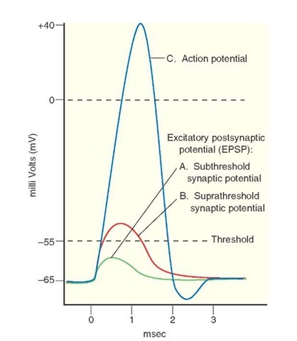
FIGURE 7-4 Generation of an action potential by an excitatory postsynaptic potential (EPSP). (A) Subthreshold EPSPs do not elicit an action potential. (B) When the EPSP is large enough, the membrane potential of the axon hillock of the spinal motor neuron is raised beyond the threshold (suprathreshold synaptic potential), and (C) an action potential is generated.
Neurotransmitters produce depolarizing graded potentials when Na+ channels open. A depolarizing graded potential that drives the membrane potential towards the threshold and excites the neuron is called an excitatory postsynaptic potential (EPSP). For example, an EPSP is elicited in a spinal motor neuron following the stimulation of afferent fibers arising from one of the thigh muscles (e.g., quadriceps). This EPSP is generated by the opening of directly gated ion channels, which permit influx of Na+ and efflux of K+. Subthreshold depolarizations (synaptic potentials) do not elicit an action potential (Fig. 7-4). When the depolarization produced by the EPSP is large enough, the membrane potential of the axon-hillock of the spinal motor neuron is raised beyond threshold (suprathreshold synaptic potential), and an action potential is generated. In the CNS, glutamate is one of the major excitatory neurotransmitters.
Neurotransmitters can also produce graded potentials that may be hyperpolarizing (i.e., K+ or Cl- channels open). A hyperpolarizing graded potential that drives the membrane potential away from the threshold and inhibits the neuron is called an inhibitory postsynaptic potential (IPSP). In the CNS, gamma aminobutyric acid and glycine are major inhibitory neurotransmitters.


