The Brain
Myelencephalon (Medulla)
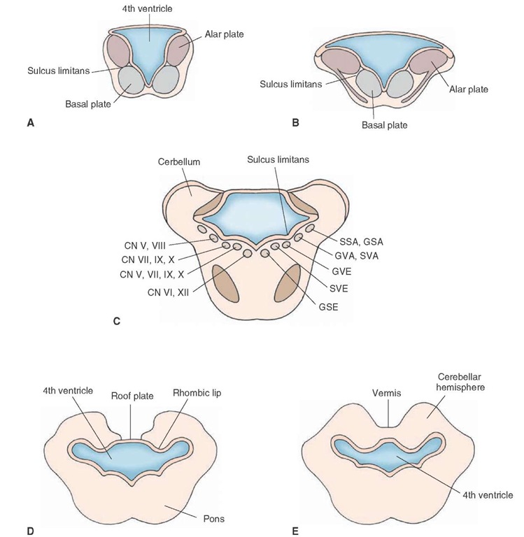
Recall that the alar and basal plates of the mantle layer, which are separated by a shallow groove, the sulcus limi-tans, form the walls of the neural canal of the developing nervous system. Although the size of the neural canal remains relatively small in the developing spinal cord, this is not the case for the brainstem. In the part of the developing brainstem that contains the fourth ventricle, the roof plate expands greatly so that the alar plate becomes located lateral to the basal plate (Fig. 2-4, A and B).
Based on the previous discussion, we can say that structures associated with motor functions tend to lie medial to structures associated with sensory functions. More precisely, the motor and sensory neurons are arranged in columns that are oriented in a medial-to-lateral fashion. The following descriptions illustrate this organization. Cranial nerve motor nuclei, which supply neurons to skeletal muscles of somite origin such as the hypoglossal nucleus, are classified as GSE fibers and lie near the midline (Fig. 2-4C). Cranial nerve motor nuclei, such as CN IX (glossopharyn-geal) and CN X (vagus), also supply axons that innervate skeletal muscle derived from the pharyngeal arches. They lie in a column situated relatively more laterally but, nevertheless, medial to sensory neurons. Such neurons are classified as special visceral efferent (SVE) neurons.
Like the developing spinal cord, structures that mediate autonomic functions develop from neurocytes situated close to the sulcus limitans. Here, neurons that mediate autonomic functions are located in the general position between sensory and somatic motor structures. This column contains neurons that innervate visceral organs and glands and include components of CN VII (metencephalon; see next section), IX, and X (myelencephalon). These neurons are classified as GVE neurons.
FIGURE 2-4 Development of the brainstem. Panels A-C depict the development of the lower brainstem. The nuclear organization is transformed from the dorsal-to-ventral orientation in the spinal cord to a medial-to-lateral orientation shown in panels B and C. Panel C depicts how cranial nerve nuclei are organized in the brainstem in terms of their medial-to-lateral position. Here, it can be seen that motor nuclei (GSE, SVE) are situated medial to the sulcus limitans, sensory nuclei (SSA, GSA) are located lateral to the sulcus limitans, and autonomic nuclei (GVA, SVA, GVE) are found in the region adjoining the sulcus limitans. Panels D and E depict development of cerebellum. Note the development and formation of the cerebellum from the rhombic lips that become fused at the midline.
Immediately lateral to the sulcus limitans lies the next column, which includes a class of sensory neurons classified as GVA and special visceral afferent (SVA) neurons. GVA neurons are associated with autonomic functions (such as baroreceptors, which sense changes in blood pressure and heart rate) and receive visceral afferent information from CN IX and X associated with such processes as changes in blood pressure and functions of the body viscera. SVA neurons are concerned with components of CN VII, IX, and X that receive information from peripheral chemoreceptors (i.e., receptors that respond to changes in the chemical milieu of their environment).
Sensory functions that involve mainly sensory components of the trigeminal nerve lie in a column positioned further laterally within the medulla and are classified as GSA neurons. The sensory column located most laterally concerns neurons that are associated with special senses. Within the brainstem, these include auditory and vestibular nuclei of CN VIII and are classified as special sensory afferent (SSA) neurons.
The size of the neural canal has now expanded greatly and will become the fourth ventricle (Fig. 2-4D). The roof plate also shows great expansion, as if it were stretched in a lateral plane to a position where it becomes connected with the lateral aspect of the alar plate. Only the floor plate remains relatively fixed in its original position.
The walls of the cavity that will become the fourth ventricle include mesenchymal tissue that will become highly vascular. This tissue will become attached to the ependymal wall of the ventricle and will generate pia mater as well. This vascular tissue will also become relatively pronounced along the central portion of the roof of the ventricle where it invaginates into the developing ventricle and will become the choroid plexus. During later periods of development (i.e., months 4 and 5), a pair of foramina develops along the roof of the ventricle. The foramina come to be situated laterally and are called the lateral apertures or foramina of Luschka. Another foramen becomes situated medially and is called the median aperture or foramen of Magendie.
The marginal layer of the developing myelencephalon, like that of the spinal cord, contains considerable amounts of white matter. In particular, on the ventral aspect of the medulla, one can identify large groups of axons that arise from prominent cells of the cerebral cortex, called pyramidal cells, which are distributed to regions throughout the brainstem and spinal cord. At the level of the medulla, these nerve fibers are referred to as the pyramids. The lateral aspect of the medulla also contains fibers that are part of the marginal layer and, to some extent, represent an extension of the fibers contained in the marginal layer of the spinal cord. Thus, this aspect of the marginal layer contains such ascending systems from the spinal cord as the spinocerebellar and spinothalamic pathways.
Metencephalon
The metencephalon consists of two principal components: the pons and cerebellum. The pons contains two basic divisions: a dorsal region called the tegmentum, which is an extension of the myelencephalon, and a ventral region called the basilar pons.
Pons. With respect to the tegmentum, the same principle applies to this region that was described earlier in attempting to understand the organization of the medulla. In general, developing neurons that lie within the basal plate tend to form the medial half of the tegmentum, whereas neurons that are part of the alar plate are more or less distributed throughout more lateral aspects of the tegmen-tum. In this manner, motor neurons, such as the nuclei of CN VI (GSE) and CN VII and V (SVE), are situated along a diagonal plane relatively medial to the planes in which sensory neurons, such as the lateral and superior vestibular nuclei or the spinal, main sensory, or mesen-cephalic nuclei of CN V (GSA), are located. Additionally, autonomic nuclei of the pons (i.e., GVE neurons of CN VII) tend to lie in a diagonal plane somewhat interposed between the planes containing sensory and somatic motor neurons (Fig. 2-4C).
The basilar portion of the pons (Fig. 2-4D) is derived mainly from neurons migrating from the alar plate. The neurocytes that are found in this region give rise to axons that grow in a transverse direction, ultimately extending beyond the body of the pons to form a major peduncle of the cerebellum called the middle cerebellar peduncle. This pathway then becomes a major route by which information from the pons can enter the cerebellum. Other fiber bundles contained within the basilar portion of the pons include descending axons that originate from the cerebral cortex and that are destined to supply nuclei of the lower brainstem and spinal cord. Therefore, these neurons evolve as part of the development of the cerebral cortex, which is described later in this topic.
Cerebellum. The cerebellum is derived from the dorsal aspect of the alar plate. Cells from the alar plate migrate further laterally and dorsally until they become situated dorsal and lateral to the lateral walls and lateral aspect of the roof of the developing fourth ventricle, respectively (Fig. 2-4D). As the medial aspect of the developing roof bends further medially, it thins out to form a narrow roof plate of the ventricle. This transitional region is referred to as the rhombic lips (Fig. 2-4D). As the rhombic lips proliferate, they extend over the roof plate, and cells from each side of the developing brain begin to approach each other. After 3 months of development, these groups of cells ultimately merge and fuse (Fig. 2-4E). The cells formed in the central region are referred to as the vermis, and those in the lateral region constitute the cerebellar hemispheres. The vermal region will display little additional growth; whereas, in contrast, the hemispheres will continue to expand considerably. At approximately the fourth month of development, fissures begin to develop with respect to the anterior lobe of cerebellum, and, by the seventh month, other aspects of the cerebellar hemispheres are apparent.
Cellular development of the cerebellum occurs in a variety of ways. Some cell types, such as those found near the surface of the developing cerebellar cortex, migrate inward to form a granule cell layer. Purkinje cells, which also appear quite early, contribute to the development of the cerebellar cortex by forming a distinctive layer called the Purkinje cell layer just superficial to the granule cell layer. The outer layer contains mainly the axons of granule cells and the apical dendrites of Purkinje cells which extend vertically toward the surface of the cortex. These axons are called parallel fibers because they run parallel to the cortical surface. They remain in the superficial surface region of the cortex, in contrast to their cell bodies, which have migrated inward. Because of the paucity of cell bodies and the extensive presence of fibers near the cortical surface, this layer is called the molecular layer. The neurons of the cerebellar cortex do not project out of the cerebellum. However, several of these cells can contact other cells, which show little migration from their original positions and remain close to the fourth ventricle.
Deep cerebellar nuclei give rise to axons which project out of the cerebellum by growing into portions of the brainstem and forebrain. For example, neurons of the developing dentate and interposed nuclei grow within a fiber bundle that later in development is called the superior cerebellar peduncle. This growth is directed toward the midbrain where fibers of the interposed nuclei reach the red nucleus (involved in motor functions); other fibers originating from the dentate nucleus extend beyond the midbrain into the lateral thalamus.Other fibers originating from the fastigial nucleus display a different trajectory in their growth patterns. Axons of these cells emerge from the cerebellum in bundles that pass close to the inferior cerebellar peduncle and reach the lower brainstem where they make synaptic connections with neurons of the reticular formation and ves-tibular nuclei of the pons and medulla.
Mesencephalon (Midbrain)
The midbrain can be divided into three general regions: a tectal region, located dorsally; a tegmentum, which is a continuation of the tegmentum of the pons and medulla and is located in an intermediate position; and a peduncular region, which is located in a ventral position.
The cavity of the midbrain vesicle, in contrast to the large size of the fourth ventricle found at the level of the upper medulla and pons, will continue to remain narrow and constitute a channel by which cerebrospinal fluid (CSF) can flow from the forebrain into the fourth ventricle. It is referred to as the Aqueduct of Sylvius (cerebral aqueduct).
Neurocytes developing from the basal plate at this level will differentiate into motor neurons (i.e., GSE) of CN III (oculomotor) and IV (trochlear) and parts of the tegmen-tum. Axons of CN III are directed in a ventral direction where they exit the brain in a medial position from its ventral surface. However, axons associated with CN IV emerge from the brain on its dorsal surface and completely cross within the superior medullary velum, exiting the brain just inferior to the inferior colliculus. Other developing neurons from the basal plate will differentiate into para-sympathetic nuclei of CN III (i.e., GVE). These neurons will serve as an important mechanism for reflexes involving both pupillary constriction and the accommodation reaction.
Neurocytes developing from the alar plate will differentiate into neurons of the tectum, which consists of both the superior and inferior colliculi. These neurons are associated with sensory processes; the superior colliculus is linked to the regulation of eye movements, and the inferior colliculus constitutes a relay in the ascending auditory pathway. Other neurocytes from the alar plate will differentiate into the mesencephalic nucleus of CN V and, possibly, into the substantia nigra and red nucleus.
The peduncular region of the midbrain is derived from the marginal layer of the basal plate and consists of fibers that arise from the cerebral cortex and descend caudally to the midbrain, pons, medulla, and spinal cord.
Prosencephalon (Forebrain)
At approximately the fourth or fifth week of development, the most rostral of the primary brain vesicles, the prosen-cephalon (forebrain), begins to display selective changes. One change includes the formation of an optic vesicle at a ventral aspect of the anterior forebrain. The optic vesicle expands outward toward the overlying ectoderm, while its connection to the forebrain (called the optic stalk) becomes constricted. The fiber bundles thus formed are called the optic nerves anterior to the optic chiasm, whereas their continuation posterior to the chiasm is called the optic tract, which terminates in the dien-cephalon. The optic vesicle contributes to inductive interactions upon the overlying surface ectoderm to produce the lens placode, which will form the lens of the eye. The part of the forebrain that lies rostral to the optic vesicle will become the telencephalon (see Fig. 2-2C). In particular, the portion of the telencephalon that lies in a lateral position will form the cerebral hemispheres. The remaining part, which lies in a medial position, will become the diencephalon.
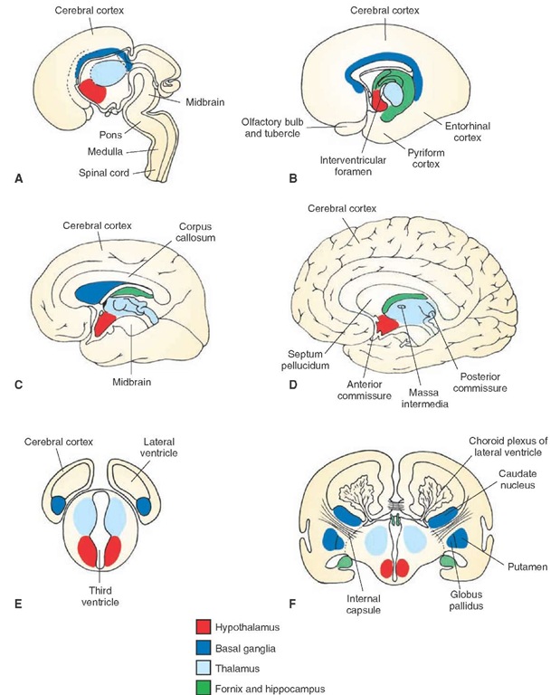
Diencephalon. The diencephalon appears as swellings of the lateral aspect of the neural canal. In this region, the canal originally had a large lumen. The lumen is diminished with the emergence of the swellings forming the thalamus, dorsally, and hypothalamus, ventrally (Fig. 2-5). The derivation of the diencephalon is somewhat controversial. However, it has been suggested that it develops mainly from the alar plate because the basal plate appears to be absent in this region. Likewise, the diencephalon does not appear to contain a floor plate but does retain a roof plate, which differentiates into the choroid plexus after becoming attached to the pia mater.
The thalamus displays the greatest amount of growth and becomes the largest component of the diencephalon. The cells will differentiate into many cell groups, forming the varied nuclei of the thalamus. In fact, the rapid growth of the thalamus is such that a small bridge is often formed between the two sides called the massa intermedia (or interthalamic adhesion) (Fig. 2-5D).
As the walls of the third ventricle expand ventrally, they do so to a much smaller extent than at dorsal levels. This region will become the hypothalamus and will contain a smaller number of anatomically well-defined nuclei than the thalamus does. However, other structures will become associated with the hypothalamus. These structures include the optic chiasm, which is formed at a rostral level of the hypothalamus and is the product of the growth of retinal fibers; rounded bodies called the mammillary bodies; the tuber cinereum; and the infundibulum (infundibular stalk), all of which are located on the ventral surface of the hypothalamus.The infundibulum gives rise to a pituitary stalk and neural hypophysis (posterior lobe of pituitary). The anterior lobe of the pituitary is derived from an ectodermal diverticulum called Rathke’s pouch, which is in contact with the infundibulum. Rathke’s pouch ultimately develops into the anterior lobe of pituitary (adenohypophysis). The rostral limit of the third ventricle is formed by the lamina terminalis, in front of which lies the telencephalon.
FIGURE 2-5 Development of the forebrain. (A-D) Medial views of four different stages of development of the cerebral hemispheres and their fusion with the diencephalon. Note the C-shaped arrangement of the telencephalic structures, including the hippocampus and fornix as well as the basal ganglia complex formed later in development. (E and F) Cross-sectional diagram taken from levels indicated in panels A and D illustrating how the cerebral hemispheres are formed at different stages of development. Note that, as shown in panel F, the choroid plexus develops from the roof of the ventricles.
Telencephalon. After the growth of the telencephalic vesicles of the telencephalon from the dorsal aspect of the fore-brain, primitive sac-like structures form within each cerebral hemisphere in the developing brain. These structures will become the lateral ventricles, which are continuous with the third ventricle through a small channel known as the interventricular foramen (of Monro) (Fig. 2-5B).
The cerebral cortex is formed by the continued growth of the cerebral hemispheres in both anterior and dorsal directions during the third and fourth months of development (see Fig. 2-2B). The anterior and dorsal expansion results in the formation of the frontal lobe. Expansion laterally and dorsally results in development of the parietal lobe, and growth in a posterior and ventral direction results in the development of the temporal and occipital lobes. As immature neurons within the cortex begin to differentiate, they form different cell groups. Much of the cerebral cortex contains six histologically distinct layers, which are present in higher vertebrates; this form of cortex is referred to as neocortex. One cell type that is formed, the pyramidal cell, gives rise to axons that will grow out into other regions of cortex and form the internal capsule (see discussion in "Internal Capsule" section). Thus, this cell type constitutes the principal means by which the cerebral cortex communicates with other regions of the CNS, including the spinal cord. Other cell types are also formed; the most common is the granule cell, which receives input principally from different regions of the thalamus.
As the cerebral hemispheres display growth in rostral and dorsal directions, the roof plate becomes fused with the pia mater, which contains tissue of vascular mesodermal origin to form the choroid plexus of the lateral ventricle (Fig. 2-5F). With continued growth, the choroid plexus becomes most extensive within the lateral ventricles. As the cerebral hemispheres continue to expand, the lateral ventricles also appear to be carried along with them, thus contributing to their continued growth and elongation.
Basal Ganglia. Major components of the basal ganglia, which include the caudate nucleus, putamen, and globus pal-lidus, are formed when immature neurons within the floor of the telencephalon and situated lateral to the interven-tricular foramen begin to proliferate. With continued growth of the basal ganglia, differentiation is noted. One group of cells, the caudate nucleus, comes to occupy a dor-somedial position. A second group of cells arise from the same general region but migrate ventrally to form the amygdaloid complex.Other groups of cells, the lentiform or lenticular nucleus (i.e., putamen and globus pallidus), display considerable growth and development and are displaced in a ventrolateral position relative to that of the caudate nucleus. The putamen assumes a position directly lateral to that of the globus pallidus (Fig. 2-5F).
The main body of the caudate nucleus (i.e., the region that will become the head and body of the caudate nucleus) displays little change from its original position, whereas the posterior aspect (i.e., the part that will become the tail of the caudate nucleus) becomes elongated by virtue of the growth of the hemisphere. The resulting effect is that the tail of the caudate follows the growth pattern of the lateral ventricle, which, in turn, is directed by the rapid growth of the cerebral hemispheres relative to that of the dien-cephalon. In this manner, the tail of the caudate is first pulled backward toward the occipital pole, then downward, and, finally, somewhat anteriorly together with the inferior horn of the lateral ventricle. The trajectories of both structures, therefore, basically follow the contour of the posterior aspect of the thalamus.
Internal Capsule. Neurons associated with the cerebral cortex give rise to axons that are directed caudally to the basal ganglia, thalamus, brainstem, and spinal cord. These developing neurons form the internal capsule and pass between the thalamus and the lentiform nucleus (i.e., globus pallidus and putamen [Fig. 2-5F]). Neurons associated with much of the frontal lobe contribute to the formation of the anterior limb of the internal capsule. Neurons located in portions of the cortical region that will develop into the precentral and postcentral gyri as well as other parts of the parietal lobe contribute to the formation of the posterior limb of the internal capsule. Those fibers situated in the temporal lobe are called the sublenticular component of the internal capsule. In addition, the internal capsule is also formed by fibers arising in the thalamus that grow toward the cerebral cortex and innervate different regions of the cortex.
Hippocampal Formation and Related Structures. The hippocampal formation (archipallium) arises from the medial surface of the telencephalic vesicle. The entorhinal and pyriform cortices (paleopallium) arise from the ventral surface of the telencephalon and are further directed in a ventrome-dial direction where they become situated on the medial and ventral surfaces of the temporal lobe adjacent to the hippocampal formation in which the entorhinal cortex lies caudal to the pyriform cortex (Fig. 2-5B). With the growth of the temporal neocortex, the hippocampal formation is pulled in a caudal direction that follows the course of the inferior horn of the lateral ventricle. Axons of the hippocampal formation form a major pathway called the fornix that is directed in a dorsomedial direction to the level of the anterior commissure, at which point it then passes downward and caudally until it makes contact with hypothalamic nuclei (Fig. 2-5D). The anterior aspect of the forebrain becomes enlarged to form structures directly associated with olfactory functions, which include the olfactory bulb and a region located near the ventral surface of the anterior aspect of the fore-brain called the olfactory tubercle and (Fig. 2-5B).
Commissures. Several prominent commissures can be identified within the forebrain. These include the corpus cal-losum (Fig. 2-5C), anterior commissure (Fig. 2-5D), and the posterior commissure (Fig. 2-5D). The corpus callo-sum is the largest and most extensive of the commissures. It grows out of the dorsal aspect of the lamina terminalis and extends caudally beyond the level of the posterior aspect of the thalamus. The corpus callosum arises from pyramidal cells of the cerebral cortex and extensively connects homotypical regions of both sides of the brain.
The anterior commissure passes through the lamina ter-minalis and provides a connection between the temporal lobes, olfactory cortices, and olfactory bulbs on both sides of the brain. The posterior commissure is located on the border between the midbrain and diencephalon. The posterior commissure is located on the border between the midbrain and diencephalon and connects the pretectal area and neighboring nuclei on both sides of the rostral midbrain.
Myelination in the Central Nervous System
Myelination within the CNS is essential for efficient and rapid transmission of signals. It begins at approximately the fourth month of fetal development at cervical levels of the spinal cord. But myelination within the spinal cord is not completed until after the first year of birth. In the brain, myelination begins at approximately the 6th month of gestation and is generally limited to the region of the basal ganglia. This is followed by myeli-nation of ascending fiber systems, which extends into the postnatal period. Surprisingly, much of the brain remains unmyelinated at birth. For example, the corti-cospinal tract begins to become myelinated by the 6th month after birth and requires several years for myelina-tion to be completed. Other regions of the brain may not be fully myelinated until the beginning of the second decade of life.


