Excitability of neurons depends on the short-term history of the membrane potential (e.g. refractoriness and accommodation). However, properties of neural cells in normal, no-learning conditions (the rare presentation of indifferent stimuli) can be described by the all-or-none principle [815], which means that the threshold for AP generation does not depend on the magnitude and variety of the input signal, and after threshold attainment the neuron generates an AP of maximal amplitude. A model of a neuron possessing such properties was named the formal neuron. Nevertheless, it turns out that excitability is a changeable parameter and depends on long-term neuron history [428, 255].
During learning, the reactions of many neurons to signals change in correspondence with animal behavior. Theoretically, a neuron may change its reactions either passively or it may acquire new properties that are not consistent with the all-or-none principle. Generally though not always, responses to biologically significant stimuli (for example, conditioned ones) increase and responses to insignificant stimuli (for example, habitual ones) decrease. How does a neuron decide which stimulus is more important in the current behavior? An elementary explanation is that synaptic efficacy changes for the specific input: the synaptic input corresponding to a more important signal becomes stronger. However, there is no evidence that during normal learning, such as habituation as well as classical or instrumental conditioning, the efficacy of the synapse changes independently from the activation of other synapses.
Change in synaptic efficacy is not the exclusive manner of storing information in the brain. Control of excitability may serve, too, as the means for modulation of neuronal activity. Excitability usually increases [1258, 1355] during augmentation of the biological importance of the signal during conditioning and decreases when it falls during habituation [1266, 1267, 641, 1272] and during extinction of conditioned reflexes [1259]. During instrumental learning, excitability also changes, but in a complex manner [881, 1270, 1351]. Learning also leads to changes in a post-spike afterhyperpolarization [24, 865], the AP amplitude [723, 1266], the AP duration [641, 547, 713, 245], input resistance of neurons [348, 54] and the conduction velocity in the axon [1314, 1351]. The more important the stimulus, the more powerful an AP it evokes. Plasticity of excitable membrane demonstrates a postsynaptic mechanism of learning with its powerful biochemical machinery. Thus, a neural cell might take on a role of self-contained unit within the brain. During development of visual circuits, plasticity of excitable membrane also plays a central role [992].
In the last half of twentieth century, the paradigm of the formal neuron appeared strong. It was substantiated by physiological achievements in the investigation of giant axon of squid, which produced an analytical description of the behavior of an excitable membrane [548] that was so fine that this till this present time it is still the basic means for analysis of membrane currents. Besides that, theoretical generalizations of the neural network theory have lead to the advent of a new branch of science with its adherents, journals, conferences and accomplishments. However, investigation of excitable membrane plasticity did not develop on the back of experiments devoted to synaptic plasticity, particularly, LTP. Instead, easily reproducible phenomena that, as expected, are related to learning and memory have become the firm scientific background of synaptic plasticity. Work on excitable membrane plasticity, in contrast has been seen as sporadic attempts to explain a few data that are contradictory to conventional thinking and relate to vague and previously unexplored chemical events in neural cells. In many cases, researcher published their results dedicated to excitable membrane plasticity and never returned to the problem, without enlarging scientific knowledge, confirmation or refutation [1309, 135, 1157, 641, 752, 982, 524, 348, 547, 1419]. Excitable membrane plasticity has remained the concern of only a few researchers, who continued investigations during many years [1253, 1355, 24].
Nevertheless, leading physiologists over and over again returned to the idea of a connection of brain functions with the individual, goal-directed behavior of neural cells and with the modulation of neuronal membranes by means of chemical processes, which might or might not have electric exhibitions [1126, 18, 19, 661, 181, 43]. Now we satisfactorily understand the LTP phenomenon, but memory comprehension is till rudimentary. At present, the role of neurons in signal processing has evolved conceptually from that of a simple integrator of synaptic inputs until a threshold is reached and an output pulse is initiated, to a much more sophisticated process with mixed analog-digital logic and highly adaptive synaptic elements [647]. This enriches calculating function of a neuron, but very likely does not allows for the hypothesis of memory as a reorganization of the neural network. Versatile complexity of neuronal chemical interactions is still underestimated.
Recently a new interest in the plasticity of the excitable membrane has flourished [713, 71, 1166, 54, 1024, 291, 1278, 1339, 1367, 255]. Voltage threshold of an AP and other membrane parameters can be spontaneously changeable [71, 482]. A preliminary change in neuronal excitability by blocking of subunits of potential-dependent K + channels impairs complex learning, but more simple learning was found to be normal [468]. Such enhanced sensitivity of complex learning to various harmful influences, as we had described earlier, is a general property of the learning mechanism and a weak influence of any impact to simple learning does not undermine the significance of this impact. Sluggish change in electrical activity, neuronal environment and chemical disturbance lead to a homeostatic change in excitability [15, 672, 14, 1386, 296, 791], which supplements the homeostatic forms of synaptic plasticity [887, 966] is the property of living beings, which regulates its internal state in order to maintain a stability). Synaptic plasticity and plasticity of the excitable membrane can mutually assist reorganization of the neu-ronal reaction. Change in the ion currents of an excitable membrane of a neuron has been supposed to influence postsynaptic potentials affecting the same neuron and to sharpen the input specificity [1185, 1351, 33, 54, 1331, 997].
One consequence of experience is to alter the biophysical and integrative properties of neurons [1004, 14], which can respond to changing inputs by adjusting their firing properties, through the modification of voltage-gated channels [672]. It is important to note that besides homeostatic forms of reorganization of excitability, further development receives investigation of excitability during learning [670, 865, 713, 1100, 1004, 762] and LTP [33, 15, 54, 414, 1367]. Difference in excitability could also play a role as a means for maintenance of inborn specificity in image recognition [1308, 1339].
The threshold, as one expects and as usually observed, is independent of the input signal. Therefore threshold specificity in respect to different signals, as a rule, is not examined at all. In a majority of cases, change in excitability during learning is considered as an unspecific parameter and as a steady alteration concerning any input of the given neuron. The unselective form of excitable membrane plasticity cannot be the basis for the selectivity of neu-ronal reactions [48, 818] and such unselective phenomenon might influence plasticity but cannot explain the main particularities of learning. Nevertheless, sometimes the specific, in relation to the input, modifications of threshold were described [1258, 1259, 291, 414, 997, 1339], although there are different explanations for this non-standard phenomenon: the selective change in excitability looks like an unconventional result.
The various pieces of discovered data concerning plasticity of the excitable membrane sometimes seem contradictory, but they are clearly dependent on the methods of measurement of excitability. Measurement of characteristics associated with excitability is not a simple task. Spike amplitude is the easiest controlled factor, even in the course of extracellular recording, but changes in AP amplitude are usually small, with infrequent exceptions. AP duration, afterhyperpolarization and membrane potential are accessible during intra-cellular recording. Nevertheless AP duration and afterhyperpolarization are formed after the AP generation has been completed and, hence, role of excitability in the given spike generation is already completed, too, and may affect only the follow APs. Membrane potential is a non-specific parameter and moreover it is usually stable during learning. AP threshold is the most important factor directly connected with actual spike generation and it is necessary to discuss methods of how to find the threshold in experiments.
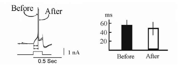
Threshold is sometimes an ill-defined value. There is a difference between voltage threshold (the voltage value at which the membrane is able to generate sufficient current to drive the AP without further applied current) and current threshold (the minimal amount of sustained current needed to generate a spike). A fast and powerful input signal is an appropriate condition for calculation of the voltage threshold, while a sustained and small input for current threshold. Sustained and small input signals generate a spike at a voltage that is smaller than voltage threshold [646]. Frequently used intracellular current injection [1100, 54, 14, 414, 1367] corresponds more closely to the conditions under which current threshold is observed, at the same time that synaptic current delivered to the soma has a short duration and corresponds to conditions appropriate for the voltage threshold. Therefore, measurement of excitability by the intracellular current pulse does not correspond to the thresholds that are essential for synaptic responses. Examination of the change in threshold of mollusk neurons after classical conditioning by means of an intracellular depolarizing current pulse in our experiments revealed only a weak change in excitability (Fig. 1.12).
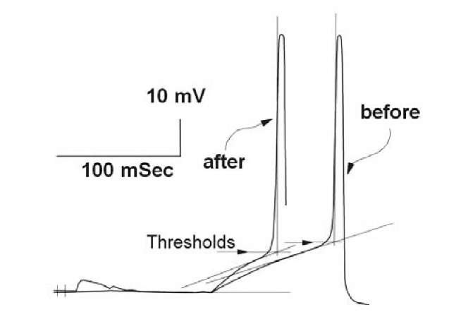
In general, it is natural to examine a change in excitability using tests for unspecific excitability, such as by the response to the current pulse. However, in order to evaluate specific change in excitability within the given response, a threshold ought to be measured within this response. Excitability within responses has been determined by measurements of levels of membrane depolarization immediately prior to AP generation. The simplest method for this purpose is evaluation of the given rate of depolarization, which one chooses to be rather high, so that it is observed only during an AP generation [71, 413, 672, 1308]. This method is acceptable, but depends on an arbitrary choice of the ultimate rate of depolarization, which itself may alter during learning and, besides, the evaluation is rather rough. The threshold also can be found as a maximum curvature at the leading front of an AP (Fig. 1.13), to be exactly the depolarization necessary to activate an action potential [1258, 1259, 54, 1339, 1367]. We determined maximum curvature (Fig. 1.13) according to the points of intersection of tangential lines at the points of inflections of the EPSP and at the leading edge of an AP at the intersection of two tangents [620]. Both methods are appropriate [1043]. An AP waveform cannot be analyzed when a neuron failed to generate an AP.
Fig. 1.12. Response of the Helix neuron LPa3 to current pulse (0.5 nA, 150 ms) before and after acquisition (at the left). Excitability within the current pulse was evaluated by means of AP latency in response to the current pulse. At the right, mean change of AP latency in response to the current pulse for 13 neurons (means and standard errors).
A perfect illustration of the importance of choice of the method of excitability measurement is the investigation of LTP in the rat hippocampus. When LTP was induced by pairing subthreshold synaptic stimulation with backpropagation of AP into the dendrites, current threshold in response to current injection increased, voltage threshold of spontaneous APs (measured as the voltage where the first derivative of the action potential waveform exceeded 10 V/s) remained stable, while voltage threshold of spikes arising from the potentiated EPSP was lowered up to 2 mV and voltage threshold of unpo-tentiated inputs was less increased [374]. Thus, we may conclude that voltage-threshold from potentiated and unpotentiated inputs differs with respect to threshold within spontaneous APs, but these data cannot be compared with the results of measurements of current threshold.
The selective form of excitable membrane plasticity is characterized by different excitability corresponding to different signals (for example, conditioned and discriminated, habitual and novel). The levels of AP generations shifted to de- or hyperpolarization side during training and t his result is not fully consistent with the all-or-none principle of neural cell function.
Fig. 1.13. The first action potential generated within responses to the CS+ before and after classical conditioning in snail neuron. The method of the level of spike generation determination, as the level of intersection of two tangents is shown (explanations in the text). Threshold levels are shown by the pointers. Responses were aligned in the points of EPSP beginning. Short vertical lines at the start of frame are times of tactile stimulus presentations.
We first discovered [1253] that spike amplitudes evoked in mitral cells (pyramidal neurons) of the olfactory bulb of frog by different stimuli may be different. The effect was large enough and visible by a naked eye (Fig. 1.14), since action potential duration was 15-20 ms, ten times longer than in mammalian cells. For our knowledge, we made the first intracellular recording from neurons of the olfactory bulb of frog and firstly it was difficult to believe that so elongated large positive intracellular potentials are really spikes. Therefore, we tried to examine whether these were conventional action potentials and whether they follow the all-or-none principle. At the time of discovery, classical neuron paradigm was so strong that nobody wasted efforts to measure alteration of spike amplitude in the experiment. So, when spike amplitude turns out to be dependent on neuronal input and this had been published, certain colleagues (for example, the theorist V. Dunin-Barkovsky) considered this as a joke. Plasticity of excitable membrane seemed to contradict to the basic principles of neuroscience.
After stimulation of epithelium by the smell, the AP generated in response to olfactory nerve stimulation decreased by 10-30% and this inhibition went on for approximately 15 sec without perceptible change in the membrane potential. During this period, spontaneous spikes and spikes induced by repeated adequate stimulus have normal magnitudes. The character of spike amplitude inhibition in the responses to stimulation of olfactory nerve after adequate stimulus corresponds to the inhibition of the evoked potential recorded from the surface of olfactory bulb in the same response (Fig. 1.15). We never observed so large an effect in other objects (see further) and later it was always necessary to undertake labor consuming measurements.
Fig. 1.14. Decrease in amplitude of spike induced by stimulation of olfactory nerve of frog after irritation of olfactory epithelium by the smell. At the left control response to the nerve stimulation (pointer). At the right – the nerve stimulation soon after presentation of smell evokes low amplitude spike (second spike), while spontaneous spike was normal (left spike). Calibration 50 mV and 100 ms.
Fig. 1.15. Inhibition in the olfactory bulb of frog after adequate stimulus. 1) Amplitude of spikes in response to olfactory nerve stimulation after presentation of smell; 2) evoked potential in olfactory bulb after smell; 3) the same in the embryonic olfactory cortex of forebrain; 4) evoked potential in the olfactory cortex after stimulation of olfactory tract. Control, before adequate stimulation 100%.
A decrease of the spike amplitude probably inhibits activity of the next neuron, since it was accompanied by reduction of the evoked potential in the olfactory cortex. Probably, the small somatic spike had incomplete amplitude also in the axon and this leads to decreased postsynaptic potentials in the cortical neurons. At the same time, the evoked potential in the olfactory cortex in response to stimulation of the olfactory tract (axons of mitral cells) does not undergo inhibition (Fig. 1.15). Inhibition of the evoked potential in the cortex under these conditions was more pronounced (Fig. 1.15). Thus, neurons were inhibited exclusively for one stimulus, but not for another. This is a very parsimonious inhibition that extends the possibilities of neurons and selectively blocks the neuron from some types of activity but leaves it free for other types of activity. This parsimonious inhibition, evidently, does not require supporting stimulation, like postsynaptic inhibition, and it is difficult to explain by the classical processes on the neuron membrane. We supposed [1253] that this phenomenon relates to neuronal plasticity and some types of events in intracellular organelles are essential here.
Similar phenomenon we further discovered in the general cortex of the turtle forebrain when we investigated habituation to light flashes [1267]. During habituation, the amplitude of the AP evoked by a habitual stimulus falls when compared with spontaneous APs and an AP in the response to a rare stimulus (Fig. 1.16).
The results of these experiments suggest that plasticity is connected with a selective change in the state of the neuron, which is manifested only during the action of a particular stimulus. Habituation of turtle cortex neurons to light flashes resulted in a selective decrease in the AP amplitude measured from the point of maximal curvature at the start of the AP to the top of the AP. The result of intracellular measuring corresponded to the results obtained during extracellular recording (Fig. 1.17) when the AP amplitude can easily be measured without any element of subjectivity and without utilizing the point of maximal curvature [1266]. Thus, AP waveform selectivity for the different responses evidently is not an epiphenomenon. After training, neurons can reveal different excitability within different responses. Change in the amplitude of the first extracellular AP in the response to habitual stimulus was as small as 3-5%.
General cortex is the higher neural center of the turtle and therefore we cannot determine whether the decrease in amplitude of an AP is connected with impairment of the transmission of excitation to the next neuron. It is impossible to rule out that a slightly decreased somatic AP generates a normal reaction in the target neuron. On the other hand, the value of the EPSP and the threshold of an AP are directly connected with the output signal of the neuron. It is important to reveal the principles governing changes in EPSP and the level of an AP threshold during habituation. We have investigated this problem in neurons of the mollusk Helix.
Habituation of identified neurons for defensive closure of the pneumostome in the Helix mollusk to tactile stimuli produced decrease in amplitude of the first synaptic AP in the response to habitual stimulus [1272]. At the same time, an AP in the response to the rare stimulus (another tactile stimulus, directed to other point of the body) did not change. The response was restored after a long interruption in stimulation and after a change in the parameters of the stimulus. The rate of habituation increased with an increase in the frequency of presentation and during a repeated series of habituation. No significant shifts of membrane potential were noted during habituation.
Fig. 1.16. The effect of habituation on the neuronal responses in the turtle general cortex. At the left upper beam, recording of the evoked potential from the surface of the cortex; lower beam intracellular recording from the basic cortex neuron. The number of stimuli is pointed out at the left. The moment of presentation of the habitual stimulus is pointed out by the negative-positive line (shorter than APs). R response to the new (rare) light stimulus. Pointer the place of generation of the short-latent AP and levels for its measure (maximum curvature at the AP front). At the right change in AP amplitudes; parts of the recording, presented at the left, were augmented. In each frame, at the left, first spike – spontaneous AP, second evoked short-latent AP. The Fig. 1.16 was redrawn in accordance with the data [1267]
A decrease in AP amplitude during habituation proceeds in parallel with an increase in the threshold of the same AP [1272]. The cause of the blockade of an AP during repetitive stimulation was the combined action of two factors: a rise in threshold and a fall of the EPSP. Change in the threshold was also selective, since threshold within a response to the rare stimulus did not increase. The selective raising of the threshold relative to the ordinary stimulus indicates that the neuron can identify the stimulus and change the state of its excitable membrane rapidly.
The fall in amplitude of the AP was very small in terms of absolute magnitude. When averaged for all identified neurons it amounted to 3-5% of the mean AP. The threshold of generation of the first synaptic AP rose during repetition of the stimulus on an average of 20% after the habituation series.
Fig. 1.17. Neuronal responses of the general cortex of turtle to repeated presentation of light flashes and rare stimuli. At the left upper, evoked potentials, lower extracellular activity. Numbers of habitual stimulus are pointed out, R response to the rare stimulus (another light stimulus). The points indicate APs, which are shown at the right: in each frame, at the left- spontaneous AP, at the right evoked AP. The Fig. 1.17 was redrawn in accordance with the data [1266].
Neuron excitability can both selectively decrease (during habituation) and selectively increase (during classical conditioning). Selective decrease in threshold we observed in land snails Helix during classical conditioning [1258, 1259]. A typical example of neuronal activity during classical conditioning is presented in Fig. 1.18. Tactile stimulation of the foot was used as the conditioned stimulus (CS+). A similar tactile stimulus directed to another point on the foot served as the discriminated stimulus (CS-). Cutaneous electrical stimulation of the foot caudal area served as the unconditioned stimulus.
During acquisition, the number of AP in response to the CS+ in both recorded neurons increased from 1 to 2-3 AP (combinations of CS+ and US number 2, 20 and 30). AP threshold in response to the CS+ during acquisition decreased. At the same time, the response to the CS- during acquisition revealed a small change in the number of APs as well as in the threshold value (trials 1, 21, 27). At the end of the acquisitions, the threshold in the response to the CS- exceeded the threshold in the response to the CS+. During an extinction series, the number of APs in both the response to the CS+ and the response to the CS- decreased and the AP threshold in these responses increased. After a 20-minute break, the number of APs in the response to the CS+ recovered (reacquisition), but did not reach the amplitude found at the end of acquisition. Response to the CS- did not increase after the break. An enlarged version of some traces in Fig. 1.18 demonstrates selective change in AP threshold in response to the CS+ compared with the thresholds in the response to the CS-.
Fig. 1.18. Representative intracellular recordings of neuronal activities during acquisition (letter A), extinction (letter E) and reacquisition (letter R) of the neuronal analog of a classical conditional reflex. In each frame, at the top – activity of the neuron LPa3, at the bottom – RPa3. The arrows indicate thresholds. The threshold was measured from the membrane potential level to the point of maximal curvature at the leading front of the AP. The number of CS+ at each exposure is indicated. For the responses to the CS" the number of the preceding CS+ is indicated (in the brackets – the ordinal number of the CS"). Sections marked in A CS+ 20, A CS" 21, E CS+ 4, E CS" 6, R CS+ 1 and R CS" 2 were magnified (X 1.8) and are presented at the end of acquisition, extinction and reacquisition. The real amplitudes of the APs are shown only in the magnified responses. CS+ – ellipsis, US vertical rectangles, CS" – horizontal rectangles. Calibration, 10 mV (trial A CS" 1). The Fig. 1.18 was redrawn in accordance with the data [1259] Methods. The training procedure consisted of the elaboration of a neuronal analog of classical conditioning. Acquisition consisted of 25-35 combinations of the CS+ and US and 8-15 presentations of the CS". The interval between the CS+ and US was 1s. The CS" was never paired with the US and was presented every one to four combinations over the course of training. An extinction series (after a 5-10 min break) consisted of 15-20 presentations of the CS+ and 6-12 presentations of the CS". Reacquisition (after a 20 min break) consisted of 5-15 combinations of the CS+ and US and 2-6 presentations of the CS" . Properties of our neuronal model of learning were similar to the properties of a well-known behavioral conditioned reflex of the defensive closure of the pneumostome in Helix [810].
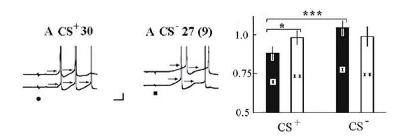
There is peculiarity of thresholds during classical conditioning. We examined whether the decrease in the first AP threshold coincided with the decrease in the second AP threshold in the same response. The above-mentioned selective change in the AP waveform which we use for determination of the threshold applied in general only to the first AP (Fig. 1.19). During the last part of acquisition, the threshold of the first AP in response to a CS+ differed significantly from the threshold of the first AP in response to the CS-. At the same time, the threshold of the first AP in response to the CS+ was smaller than the threshold of the second AP in the same response (p < 0.05). Thresholds of the second AP in responses to the CS+ and CS- did not differ significantly. A change in AP threshold during habituation is also an integral factor of the first AP in the response [1272].
Fig. 1.19. The change in the first and second AP thresholds (are indicated by the arrows) during the last part of acquisition. Examples of response to the CS+ (at the left) and CS- (middle). At the right, averaged changes in the first (I) and second AP (the latency 50-200 ms, II) thresholds in the responses to the CS+ (left) and CS- (right) during acquisition (trials 21-30). The thresholds of the second APs were normalized according to the average threshold of the first AP in the same neuron. Asterisks indicate a significant difference (Student t-test, *p < 0.05, < 0.001). Calibration 10 mV, 0.1 s.
Thus, the decrease in the threshold of the first AP only poorly promotes the generation of the next AP in the response and hence, change in excitability within the response is short-term and transient. Thus, specific change in excitability concerns just the first AP in the response. This corresponds to general observations that only short latency responses selectively change during learning, compared with discriminated signals. Current changes in excitability of a neuron in time are probably linked with the degree of the real participation of given neuron in current behavior.
The AP threshold, latency, EPSP slope and amplitude (Fig. 1.20A-D) changed in a rough correspondence to the number of AP changes (Fig. 1.20E). During acquisition the number of APs in response to the CS+ on average increased and in response to the CS~ decreased, while during extinction both responses decreased. During the reacquisition, the number of APs in the responses to the CS+ increased rapidly.
During training, a change in spike threshold, spike latency and postsynap-tic potential slope and amplitude after pairing also displayed selectivity for responses to the significant and insignificant stimuli (Fig. 1.20A-D). All these characteristics correlated with the efficacy of the corresponding stimulus for the spike generation. The properties of the AP threshold, EPSP and the number of APs in the response correspond to well-known properties of behavioral classical conditioning, but in spite of the existence of a roughly inverse relationship between AP threshold and EPSP (Fig. 1.20), there is a significant difference between the dynamics of the EPSP and the threshold at the end of acquisition and at the beginning of extinction. The cause-and-effect relationship between the strength of the presynaptic volley and the excitability was evidently weak4.


![The effect of habituation on the neuronal responses in the turtle general cortex. At the left upper beam, recording of the evoked potential from the surface of the cortex; lower beam intracellular recording from the basic cortex neuron. The number of stimuli is pointed out at the left. The moment of presentation of the habitual stimulus is pointed out by the negative-positive line (shorter than APs). R response to the new (rare) light stimulus. Pointer the place of generation of the short-latent AP and levels for its measure (maximum curvature at the AP front). At the right change in AP amplitudes; parts of the recording, presented at the left, were augmented. In each frame, at the left, first spike - spontaneous AP, second evoked short-latent AP. The Fig. 1.16 was redrawn in accordance with the data [1267] The effect of habituation on the neuronal responses in the turtle general cortex. At the left upper beam, recording of the evoked potential from the surface of the cortex; lower beam intracellular recording from the basic cortex neuron. The number of stimuli is pointed out at the left. The moment of presentation of the habitual stimulus is pointed out by the negative-positive line (shorter than APs). R response to the new (rare) light stimulus. Pointer the place of generation of the short-latent AP and levels for its measure (maximum curvature at the AP front). At the right change in AP amplitudes; parts of the recording, presented at the left, were augmented. In each frame, at the left, first spike - spontaneous AP, second evoked short-latent AP. The Fig. 1.16 was redrawn in accordance with the data [1267]](http://what-when-how.com/wp-content/uploads/2011/06/tmp2316_thumb_thumb.jpg)
![Neuronal responses of the general cortex of turtle to repeated presentation of light flashes and rare stimuli. At the left upper, evoked potentials, lower extracellular activity. Numbers of habitual stimulus are pointed out, R response to the rare stimulus (another light stimulus). The points indicate APs, which are shown at the right: in each frame, at the left- spontaneous AP, at the right evoked AP. The Fig. 1.17 was redrawn in accordance with the data [1266]. Neuronal responses of the general cortex of turtle to repeated presentation of light flashes and rare stimuli. At the left upper, evoked potentials, lower extracellular activity. Numbers of habitual stimulus are pointed out, R response to the rare stimulus (another light stimulus). The points indicate APs, which are shown at the right: in each frame, at the left- spontaneous AP, at the right evoked AP. The Fig. 1.17 was redrawn in accordance with the data [1266].](http://what-when-how.com/wp-content/uploads/2011/06/tmp2317_thumb_thumb.jpg)
![Representative intracellular recordings of neuronal activities during acquisition (letter A), extinction (letter E) and reacquisition (letter R) of the neuronal analog of a classical conditional reflex. In each frame, at the top - activity of the neuron LPa3, at the bottom - RPa3. The arrows indicate thresholds. The threshold was measured from the membrane potential level to the point of maximal curvature at the leading front of the AP. The number of CS+ at each exposure is indicated. For the responses to the CS" the number of the preceding CS+ is indicated (in the brackets - the ordinal number of the CS"). Sections marked in A CS+ 20, A CS" 21, E CS+ 4, E CS" 6, R CS+ 1 and R CS" 2 were magnified (X 1.8) and are presented at the end of acquisition, extinction and reacquisition. The real amplitudes of the APs are shown only in the magnified responses. CS+ - ellipsis, US vertical rectangles, CS" - horizontal rectangles. Calibration, 10 mV (trial A CS" 1). The Fig. 1.18 was redrawn in accordance with the data [1259] Methods. The training procedure consisted of the elaboration of a neuronal analog of classical conditioning. Acquisition consisted of 25-35 combinations of the CS+ and US and 8-15 presentations of the CS". The interval between the CS+ and US was 1s. The CS" was never paired with the US and was presented every one to four combinations over the course of training. An extinction series (after a 5-10 min break) consisted of 15-20 presentations of the CS+ and 6-12 presentations of the CS". Reacquisition (after a 20 min break) consisted of 5-15 combinations of the CS+ and US and 2-6 presentations of the CS" . Properties of our neuronal model of learning were similar to the properties of a well-known behavioral conditioned reflex of the defensive closure of the pneumostome in Helix [810]. Representative intracellular recordings of neuronal activities during acquisition (letter A), extinction (letter E) and reacquisition (letter R) of the neuronal analog of a classical conditional reflex. In each frame, at the top - activity of the neuron LPa3, at the bottom - RPa3. The arrows indicate thresholds. The threshold was measured from the membrane potential level to the point of maximal curvature at the leading front of the AP. The number of CS+ at each exposure is indicated. For the responses to the CS" the number of the preceding CS+ is indicated (in the brackets - the ordinal number of the CS"). Sections marked in A CS+ 20, A CS" 21, E CS+ 4, E CS" 6, R CS+ 1 and R CS" 2 were magnified (X 1.8) and are presented at the end of acquisition, extinction and reacquisition. The real amplitudes of the APs are shown only in the magnified responses. CS+ - ellipsis, US vertical rectangles, CS" - horizontal rectangles. Calibration, 10 mV (trial A CS" 1). The Fig. 1.18 was redrawn in accordance with the data [1259] Methods. The training procedure consisted of the elaboration of a neuronal analog of classical conditioning. Acquisition consisted of 25-35 combinations of the CS+ and US and 8-15 presentations of the CS". The interval between the CS+ and US was 1s. The CS" was never paired with the US and was presented every one to four combinations over the course of training. An extinction series (after a 5-10 min break) consisted of 15-20 presentations of the CS+ and 6-12 presentations of the CS". Reacquisition (after a 20 min break) consisted of 5-15 combinations of the CS+ and US and 2-6 presentations of the CS" . Properties of our neuronal model of learning were similar to the properties of a well-known behavioral conditioned reflex of the defensive closure of the pneumostome in Helix [810].](http://what-when-how.com/wp-content/uploads/2011/06/tmp2318_thumb_thumb.jpg)

