Introduction: What Is Electroneuropathology?
Neuropathological studies can reveal a great deal about the appearance of cells and structures within the nervous system during the course of a disease, but they cannot determine which neurons were working properly when the samples were taken or in the period before death. Neurons may look normal, but are they still able to integrate incoming synaptic information, to generate and propagate action potentials, and lay the foundations of new memories? The answers to these questions are central to the understanding of the relationships between neuropathological abnormalities and particular signs or symptoms. Prion diseases also affect laboratory animals, and, as a consequence, it is possible to study them in great detail, from the presymptomatic to the terminal stages. Of course, mice do not report symptoms, but it is possible to assess their health status by using scoring systems such as those described by Irwin (1), and behavioral tests can, to some extent, model psychiatric disturbances. The crucial benefit is that the functioning of neurons can be assessed in a pathological and behavioral context. The possibility thereby exists to relate abnormalities in the electrophysiological properties of neurons, or groups of neurons, to particular neuropathological or behavioral changes. We have called this approach "electroneuropathology" (Fig. 1) (2).
Electrophysiological techniques have been used to study the effects of prion disease in whole animals, and in slices of living brain. Experiments in whole animals, including humans, have used electroencephalographic techniques. When applied to rats, they have indicated that electroencephalogram changes occur early in the disease, and have consistent onset times in a wide range of different strains of rats (3). These changes reflect alterations in the properties of groups of neurons, and have been reviewed elsewhere (4). The majority of studies have used slices of living brain, and it is on these that this topic concentrates.
Fig. 1. Electroneuropathology. Application of electrophysiological techniques to study diseased or behaviorally abnormal neuronal tissues.
Experiments Using Slices of Living Brain
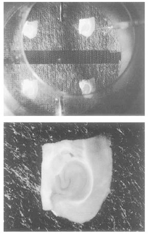
Living slices of brain have been used for electrophysiological experiments over many years (Fig. 2) (5). Workers in the prion field have tended to use similar methods for brain-slice preparation and maintenance. Our own methods have been described in detail elsewhere (6,7), but, briefly, they comprise the following: Under blind conditions, a diseased or matched control mouse is selected, anesthetized, and decapitated. The brain is removed, and horizontal slices of hippocampal formation, 400 ^m thick, are then made on a vibroslice (Camden Instruments). Slices are transferred to an interface-type recording chamber, where they are superfused with an artificial cerebrospinal fluid at 36°C. The slices are given 2 h to recover from the preparation procedure, before starting the experiments, and, in general, they remain useful for a further 6 h. The quality of the recordings made in this type of experiment depends on the effectiveness of slice preparation, and this in turn depends on many variables, including the speed and delicacy of dissection, the thickness of slices and method of slicing, and the temperature and composition of the fluid used during dissection and superfusion. Poor technique may result in slices that contain a higher proportion of damaged neurons, which then produce unreliable recordings, and this may be particularly so in diseased tissue.
The same general method can be used to investigate brain tissue of different origins. Experiments have been made on slices from mice with the ME7 strain of scrapie (8-12), from hamsters with the Sc237 strain (13), and from trans-genic mice with the hamster prion protein gene that have been infected with the Sc237 strain (14). In general, these studies have examined neurons in the hippocampal formation, but the neocortex (13) and the lateral geniculate nucleus (9) have also been studied. The electrophysiological techniques employed have in the main been a mixture of extracellular field potential recording and intracellular current-clamp recording.
Fig. 2. Brain slice recording chamber. The top panel shows four horizontal slices of mouse hippocampal formation in an interface-type recording chamber. The slices lay on strips of lens-cleaning tissue that are about 1 cm wide, which in turn rest on a mesh of tensioned stocking. They are surrounded by a humidified mixture of 95% oxygen and 5% carbon dioxide. The artificial cerebrospinal fluid (ACSF) enters via the stainless steel tubes seen at the top and the bottom of the picture, and is carried away, by the capillarity of the lens tissue, to an annulus that surrounds the stocking at a lower level. From here, it is sucked from the bath. Flow rates in this chamber are typically 0.1 mL/ min (for each side), but are adjusted to ensure that pools of ACSF do not collect around the slice or on the lens tissue. The ACSF is warmed before entering the chamber, which itself sits above a thermostatically controlled water bath. The slices are covered by a lid, which has an aperture to admit the recording (and in some experimental situations, a stimulating) electrode. In this example, the recording electrode is entering at the top right of the picture, and its tip enters the slice in the subiculum. The bottom panel demonstrates that, even in a slice of living, unstained brain 400 ^m thick, the subregions of the hippocampal formation can be distinguished easily.
A central question is whether the symptoms of prion disease are as a result of accumulation of the abnormal protein or a consequence of the loss of functioning of the normal protein. To address this question, experiments have also been conducted in slices from mice that lack the prion protein gene, and therefore presumably lack the activity normally bestowed by the prion protein (assuming that a replacement protein does not compensate) (15-20). The experiments using null mice have also made wider use of whole-cell and voltage-clamp techniques.
The literature describing the electrophysiological consequences of scrapie infection or prion gene knockout contains contradictions and some confusion. There are valid reasons for differences in results, and these should be borne in mind throughout. They include differences in the species that have been studied, and differences in the strains of scrapie that have been used. The diseases produced by different combinations of host species and scrapie strain have different time-courses and different neuropathological profiles. Of the two chief combinations studied electrophysiologically, ME7 in mice results in marked neuronal loss, and Sc237 infection in hamsters produces no substantial neuronal loss, and has a shorter incubation period.
However, differences in experimental methods are also important. There are undoubtedly subtle differences in the criteria that different researchers use, either consciously or subconsciously, to decide whether or not to include particular neurons in their studies. A brain slice contains millions of neurons, but only a handful will be recorded during any one session. Achieving a representative sample from a control group, which can be compared with another sample that is representative of a scrapie group, is difficult but fundamental. It is necessary to account for the fact that the neuronal population is not homogeneous, and that many neuronal subtypes, with distinct electrophysiological characteristics, exist. It is crucial to compare like with like, so that the only variable between the samples is the presence of disease. In the first instance, all experiments should be done blind to disease status. It is then necessary to keep an open mind about what to expect. By the very nature of the experiments, some of the recorded neurons are sick, and their records will look very abnormal; indeed, they may look like bad recordings from healthy neurons, but these neurons should not be excluded arbitrarily. Acceptance criteria for continuing with a recording should be kept as relaxed as possible, and the setting of minimum acceptable membrane potentials, and so on, should be avoided. The pragmatic view is to accept any neuron in which it is thought likely that the planned experiments can be conducted and interpreted. Finally, when complex and relatively poorly understood phenomena have been investigated, the precise details of the experimental methods employed are likely to have a significant influence on the results obtained. When such matters of experimental design or conduct are thought to be relevant to discrepancies in the literature, they are highlighted.
Effects on Basic Membrane Properties
The basic membrane properties that have been examined are principally the resting membrane potential and the cell input resistance. Studies that have addressed this issue in ME7 mice (8,10,11) have found the resting membrane potential to be more depolarized (more positive) in neurons from infected mice. In contrast, in Sc237 hamsters, the neurons are not depolarized, compared to controls (13). When a depolarization has not been found, this may be because depolarized neurons were excluded by the application of predetermined acceptance criteria. Other things being equal, the depolarized neurons would be closer to action potential threshold, and therefore more easily excited. Indeed, increases in the proportion of spontaneously active hippocampal neurons in ME7 mice (8) and epileptiform activity in transgenic Sc237 mice (14) have been reported. This change in the pattern of action potential discharge may then lead to accumulation of calcium, activation of enzymes or ion channels, and excitotoxic effects. Although the functioning of high-threshold voltage-gated Ca2+ channels has been examined specifically, and found not to be compromised in ME7 mice (11), this does not exclude the possibility that, in scrapie infection, the resting calcium concentration is elevated.
Effects on the Parameters of Action Potential
In ME7 mice, the electrophysiological properties of neurons in area CA1 of the hippocampus have been examined at different, but predominantly later, stages of the disease. The amplitude and the rise time of the action potentials were unaffected, but there was a significant decrease in the fall time (8), which represents an increase in the rate of repolarization.
The so-called "after-hyperpolarization" (AHP), divided into three components, has also been examined. The early AHP occurs at the end of the repolarization phase of a single action potential (Fig. 3); the middle and late AHPs, which are mediated by a range of different voltage- and calcium-activated potassium currents (17), occur after a sequence of action potentials, and can be difficult to compare accurately among studies. In ME7 scrapie, an increase in the amplitude of the early AHP is reported, and, although not examined in detail, it has been suggested that there is also an increase in the medium AHP (8).
In area CA1 neurons from Sc237 hamsters, there were also no changes in action potential amplitude or rise time. However, the early AHP was unaffected, rather than increased, and the medium and late AHP were attenuated, rather than increased (13). These two sets of results are difficult to reconcile.
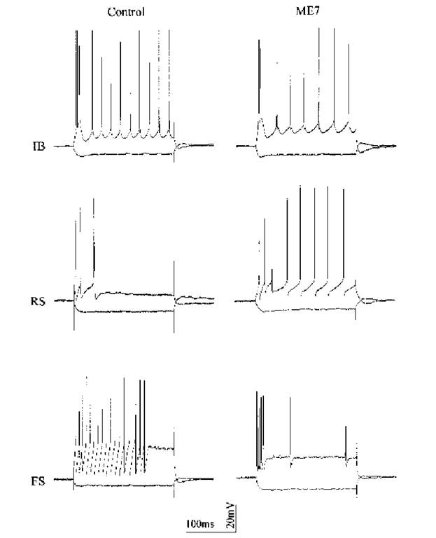
Fig. 3. Types of neuron encountered in control and ME7 mice. Intracellular recordings were made in the subiculum of mice injected, 21 wk previously, with either normal brain homogenate or with ME7 homogenate. The records show, superimposed, the response of each neuron to a depolarizing current pulse (0.4 nA except for the control FS neuron, where it was 0.8 nA) and a hyperpolarizing current pulse (-0.4 nA). Intrinsically burst-firing (IB) and regular-spiking (RS) subtypes of pyramidal neuron are shown. Examples of fast-spiking neurons are given also. It was confirmed morphologically that these were nonpyramidal neurons, but no attempt was made to subtype them. The purpose of this figure is to demonstrate that the same types of neuron can be recorded and distinguished in ME7 as in control mice, and to suggest that failure to account for differences between subtypes could confound a comparison between control and scrapie mice. This figure is not intended to provide a summary of the differences postulated to be present between control and prion affected neurons.
The discordant results for the medium AHPs may reflect differences in the level of analysis, and interpretation should perhaps be left until studies of equal depth have been performed. However, the difference in the early AHP does warrant some consideration.
There is an important philosophical point to consider in this type of experiment, which may be particularly pertinent here: When is a neuron an abnormal example of a particular subclass, and when is it a normal example of a different subclass? The rodent hippocampal formation contains pyramidal neurons that fire single action potentials, these are termed "regular-spiking" (RS) neurons. It also contains neurons that fire bursts of action potentials, riding on single depolarizing waves, and these are termed "intrinsically burst-firing" (IB) neurons (7,21). Examples of the different classes of neurons recorded in control and ME7 mice are given in Figure 3.
RS neurons have a more obvious early AHP (7). Thus, if the sample from a scrapie group contained more RS neurons than did a corresponding sample from a control group, the false impression might be gained that the AHP was increased in amplitude in the scrapie group. Indeed, there is some weak evidence, discussed below, to suggest that hippocampal RS neurons may be less susceptible, and thus perhaps more likely to be recorded. Before the issue regarding the amplitude of the early AHP can be settled, further work is needed in ME7 mice that distinguishes clearly between subtypes of neurons.
As well as differences in the shapes of individual action potentials, changes in the patterns of multiple action potential discharges have also been reported. There was an increase in the proportion of spontaneously active neurons in area CA1 neurons in ME7 mice. Also in ME7 mice, there was a reduction in spike frequency adaptation following depolarizing current pulses, and thus a tendency to fire more action potentials when stimulated in this way (8). In the Sc237 hamsters, synaptic stimulation led to double action potential discharges, rather than to the single action potentials seen in controls (13). In both cases, the changes in the pattern of action potential discharge were consistent with a reduction in the AHP.
Effects on Synaptic Transmission
Extracellular Field Potentials
Extracellular field potential recording techniques have been used to record the integrated response of many cells to an artificial stimulus. In the hippocampal formation, recording usually takes place in the stratum radiatum, and follows stimulation of the Schaffer collaterals, some 500 ^m away. The amplitude and waveform of the recording are influenced by the number of axons successfully stimulated and the number of neurons contributing to the recorded signal. In Sc237 hamsters, the field potential recorded in the hippocampal formation, at different stages of the disease, was no different from controls. In contrast, in ME7 mice, there was a marked reduction, and, in the terminal stages of the disease, it was impossible to record (8). The most likely reason for this discrepancy lies in the pathological consequences of infection in the two cases. In ME7 mice, there is marked cell loss, but this is not the case in Sc237 hamsters. A lower density of neurons may well produce a decrease in the field potential. These differences emphasize the importance of placing the electrophysiologi-cal data in their proper pathological context: the essence of electroneuropathology, in fact.
Intracellular Recordings
Experiments have been conducted in which the recording electrode is placed intracellularly, and the responses of single neurons to spontaneous or evoked synaptic stimulation are assessed. Although the electrode is recording the response of a single neuron, that neuron is in contact with other cells, and should not be regarded as in any way isolated. The only synaptic components that have been examined in any detail in scrapie are the excitatory postsynaptic events mediated by glutamate acting at N-methyl-d-aspartate (NMDA) and non-NMDA receptors, and by inhibitory postsynaptic events mediated by y-amino-butyric acid (GABA) acting at GABAA and GABAB receptors. Two forms of recording technique have been employed. In so-called "current clamp," events are recorded as changes in membrane potential, and are referred to as excitatory postsynaptic potential (EPSP) and inhibitory postsynaptic potential (IPSP). In voltage clamp, the membrane potential is kept constant, and events are recorded as changes in membrane current; thus, they are referred to as excitatory or inhibitory postsynaptic currents (EPSC and IPSC, respectively). Differences in experimental technique can be important when comparing apparently disparate sets of results.
It is possible to record a near-normal EPSP in ME7 mice, but only if the stimulus intensity is turned higher than is used in controls (12). This increase in stimulus intensity is probably needed to compensate for the neuronal loss associated with ME7 in mice. In Sc237 hamsters, in which there is no marked neuronal loss, normal EPSPs were recorded without any need to increase the stimulus strength (13). These results imply that any change in the EPSP results from a change in the density of neurons being stimulated, rather than from a specific effect of prion infection on the postsynaptic machinery that underpins the EPSP. There is less agreement about IPSPs. Although not studied in detail in ME7 mice, evoked IPSPs appeared to be attenuated (8). On the other hand, in Sc237 hamsters, the GABAA-mediated IPSP, assessed using multiple criteria, was unchanged (13). More detailed studies of the IPSP in ME7 mice are needed to resolve this issue.
Long-Term Potentiation
Long-term potentiation (LTP) is the phenomenon, first described in detail by Bliss and Lomo (22), in which repeated use of a synapse strengthens the functional connection across that synapse. The reasons why those studying a dementing illness are drawn to a phenomenon described as a synaptic model of memory (23) are obvious. In the experimental setting, synaptic potentiation that persists for more than 1 h is usually regarded as LTP, but there are other forms of synaptic potentiation that are less long-lived. In ME7 mice at the later stages of the disease, but before there is frank neuropathological change or loss of dendritic spines, the ability to maintain LTP is lost. In these mice, although the stimulus protocol failed to produce LTP, it continued to produce short-term potentiation (12). These results have been interpreted as a PrPSc-induced loss of the ability to change short-term potentiation into LTP. The locus of this effect is likely to be postsynaptic, but occurs before there are detectable changes in the numbers of dendritic spines (12).
Electrophysiological Changes in Prion-Null Mice
It is important to know if mice that lack the prion protein gene are electro-physiologically normal or not. If they are, this suggests that loss of the functioning of the normal prion protein is not of great importance, and that the symptoms of prion disease stem from the presence of the abnormal protein. Abnormal knockouts suggest that loss of the functioning of the normal protein is important. Furthermore, any such abnormalities, similar to those seen in animals with prion disease, support the view that loss of the normal is important, rather than accumulation of the abnormal. It is prudent to keep in mind the caveat that the function of the missing proteins may have been taken over by different proteins. Unfortunately, electrophysiological experiments using knockout mice have yielded a range of different results. The passive membrane properties of null mice are unchanged (15,19), and this is at odds with the results obtained in ME7 mice (8,10), but in agreement with those from Sc237 hamsters (13). The medium AHP is reduced in Sc237 hamsters (13), but not in null mice (17). However, the late AHP is reduced both in null mice (17) and in Sc237 hamsters (13), and this suggests that prion protein is in some way involved in the calcium-activated potassium current that mediates the late AHP (17). Thus, for passive membrane properties, and for the late AHP, there is concordance between the data derived from prion-null mice and Sc237 hamsters, but not between prion-null mice and ME7 mice.
In prion-null mice, attenuation of evoked, GABAA-mediated IPSCs has been reported (15), but not replicated (19). Synaptic transmission has also been investigated using somewhat different techniques in thin slices (150 ^m, rather than the 400 ^m used in other experiments) of cerebellum (18). In these experiments, spontaneous, rather than evoked, IPSCs were measured and found to be normal. Some degree of caution is required when comparing these two results, because they represent different phenomena. A spontaneous IPSC is likely to be the result of a discharge from a single inhibitory neuron; an evoked IPSP may result from several neurons. It has also been argued that, because spontaneous IPSCs are small, they would have been difficult to measure accurately (13). On the other hand, inhibitory events mediated by GABAA receptors may be measured more accurately as currents, using voltage-clamp techniques, because very small changes in the resting membrane potential can have profound effects on the amplitude of the IPSP.
In these same experiments, the EPSC, elicited in cerebellar neurons by stimulation of the climbing fibers, was also investigated and found to be normal (18), and this result does agree with previous studies that have reported the EPSC to be qualitatively normal (15). Thus, there is agreement that the EPSC is unchanged in both infected and knockout animals. However, regarding the IPSPs, although there is agreement among some studies derived from prion-null mice and Sc237 hamsters, data from prion-null mice and ME7 mice do not agree.
Investigations of synaptic plasticity in relation to prion disease are particularly fraught with mistakes. LTP was reported to be abnormal in the null mice (15). A letter describing a replication of this finding was published the following year by a different group (16). However, a subsequent study by a leading LTP laboratory failed to find any differences in LTP between prion-null and control mice, under blind conditions (19). It was suggested at the time that this discrepancy resulted from differences in the genetic backgrounds of the knockouts. This seems unlikely, given that LTP was apparently normal in knockouts with three different backgrounds (19). Thus, on balance, LTP is probably normal in prion-null mice.
The difficulty of using LTP to probe the functions of the prion protein lies primarily in the lack of consensus about the mechanisms that underlie LTP. It is generally agreed that a rise in the postsynaptic calcium concentration is necessary, and occurs as a consequence of calcium entry through NMDA receptor channels that open in response to membrane depolarization (23), but there has been much debate surrounding the primary locus of LTP. If the locus is at least in part presynaptic (23), this would point to a role for prion protein in transmitter release, and this possibility is supported by the results of experiments in cerebellar Purkinje cells (18). If, as much current thinking favors (24), the locus is predominantly postsynaptic and involves the unmasking of silent synapses by the translocation of glutamate receptors it is possible that prion protein has a role in the membrane trafficking of such receptors.
Ironically, just as attention is focused on a postsynaptic locus for LTP, recent results suggest that normal prion protein is concentrated presynaptically (25).
A presynaptic effect of PrP is also suggested by the results of a small number of experiments in cerebellar Purkinje cells. It has been reported that the sum amplitude of spontaneous IPSCs was reduced by the application of copper ions in prion-null, but not in wild-type mice (20).The LTP literature also counsels on the difficulty of making valid comparisons between sets of results, when different methodologies have been used to obtain them (26). Factors that are likely to affect results, not only of the LTP experiments, but of many of the experiments described in this topic, include the age of the animals, the brain region, the presence (or not) of intact synaptic inhibition, the details of the recording technique, the temperature, composition of bathing and electrode filling solutions, and the measurement of EPSP amplitude, rather than slope. This does not mean that all conflicting results are equally reliable and have valid explanations. As far as prion proteins and LTP are concerned, it is too soon to tell.
Thus, overall, the electrophysiological studies have not yet establish conclusively whether it is loss of normal protein or accumulation of abnormal protein that is important to the generation of signs of disease.
Differential Vulnerability Among Neuronal Subtypes
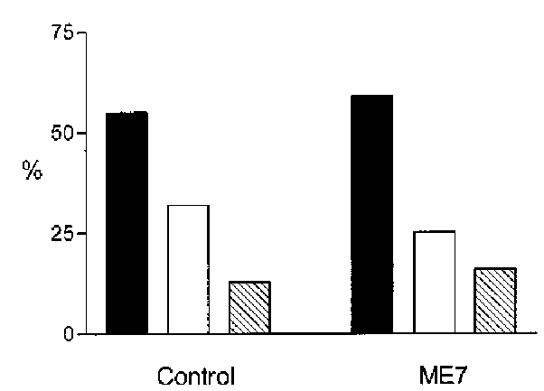
Whether or not some neurons are more vulnerable to the effects of PrPSc, be they direct or indirect, is an important question. If the answer is yes, and the vulnerability resides in a pharmacologically accessible aspect of the neuron, it may be possible to design drugs that remove that vulnerability. In our studies on ME7 mouse subiculum, no evidence was found of a differential loss of nonpyramidal neurons (note that nonpyramidal neurons were not subtyped), or indeed a differential loss of one subtype of pyramidal neuron over another (Fig. 4).
In the neocortex, differences in the action potential in Sc237 mice are present in RS, but not IB neurons (13). In ME7 mice, even at late stage, some electro-physiological and morphologically normal lateral geniculate neurons remain (9). In studies in the subiculum, membrane depolarization was present in IB, but not RS, neurons (10). Thus, the electrophysiological data suggest that particular neurons are more vulnerable than others. The explanations for this are more elusive, but could lie in the anatomy of neuronal circuits. It has been suggested that, in subiculum, IB and RS neurons receive different inputs, and have different projection targets (7,27). It may simply be that the abnormal protein reaches IB neurons more quickly, because of differences in the length of circuits or in the numbers of interposed synapses. However, in the rat sub-iculum, IB and RS neurons do have different pharmacological properties, and these could relate to vulnerability. RS neurons contain the neuronal form of nitric oxide synthase, but IB cells do not (28). IB neurons respond much more strongly to the neuropeptide, somatostatin (6).
Fig. 4. Proportion of neuronal subtypes recorded in control and ME7 mice. Recordings were made in the subiculum: 22 neurons were recorded from three control mice that had received intrahippocampal injections of normal brain homogenate 21 wk previously, and 32 neurons from four matched mice that had been injected with ME7 homogenate. Solid bars indicate the proportion of neurons that were of IB pyramidal subtype, open bars, the RS pyramidal subtype; and the hatched bars, the proportion of interneurons.
IB neurons also show a much more prominent sag in the voltage response to hyperpolarizing current pulses (21), and this is known to be true also in guinea pig (29) and mice (10). This sag is thought to be mediated by a mixed cation current, referred to as Ih . Potassium is one of the charge carriers involved in Ih (30), and possible selectivity for neurons that express much sag adds support to the view (11) that potassium currents are involved in scrapie infection.
Abnormalities of Neuronal Morphology
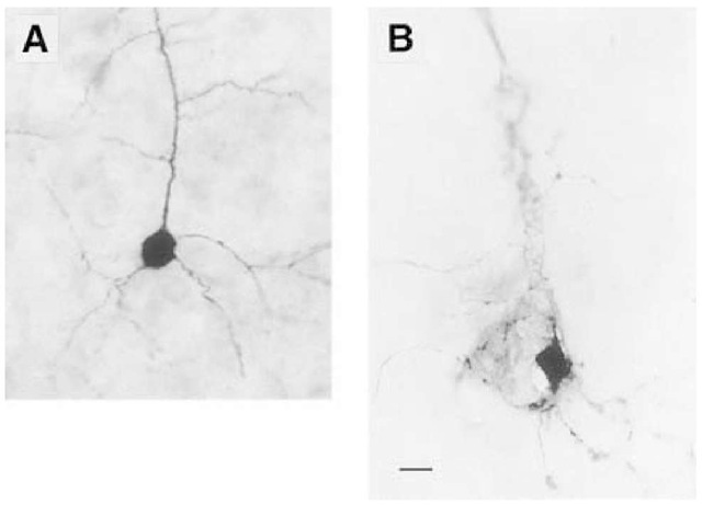
A number of studies have been made in which the electrophysiologically characterized neurons have also been filled with an intracellular label, such as Neurobiotin (Vector), and examined morphologically. These studies have revealed a spectrum of abnormalities. In a small study in ME7 mice, there was a loss of dendritic spines and the presence of a small number of membrane diverticuli (8). We found more severe morphological abnormalities in ME7 mice (10). This may be because very relaxed acceptance criteria were used, and we may therefore have sampled from more severely affected neurons. Neurons were found where only the apical dendrite remained, and branching was very much less extensive. Neurons were also found in which the label has apparently been excluded from some regions of the soma and apical dendrite; An example of this is given in Figure 5. The localization and the scale of these filling defects suggests that they are caused by the presence of (unlabeled) some regions of the soma and the apical dendrite, giving a honeycombed appearance.
Fig. 5. Morphological changes in neurons from ME7 mice. Two subicular pyramidal neurons (IB subtype), that have been filled with Neurobiotin during electrophysiological characterization, are shown. (A) is from a control mouse, and has the typical appearance of a pyramidal (projection) neuron. There is flask-shaped soma with prominent apical dendrite, a skirt of basal dendrites, and the presence of some dendritic spines, note that the intracellular dye is evenly distributed. (B) This neuron is from a scrapie-infected mouse, note that the neuron is distended, and that the intracellular dye is displaced from some regions of the soma and the apical dendrite, giving a honeycombed appearance. The scale bar is 10 ^m.
The scale bar is 10 ^m. coalescences of mitochondria or lysosomes, which have been reported to occur in scrapie infection (31,32). In general, the morphological abnormalities found in electrophysiologically characterized neurons from ME7 mice are similar to those reported using more traditional techniques. Golgi impregnation and confo-cal microscopy in ME7 mice reveals that the first morphological abnormality is a loss of dendritic spines (33), and that swelling of the dendrites, intraneuronal vacuolation, and extensive neuronal loss follow.
In contrast, in a detailed study of electrophysiologically characterized neurons from terminal-stage Sc237 hamsters, there were only mild morphological changes, consisting of an increase in the arborization of the basal dendrites, and no change in the numbers of dendritic spines (13). This may be a sampling issue, but it may also be that those neurons with severe morphological abnormalities are those that are destined to be lost, and these would be expected to be of greater number in ME7 mice, in which neuronal loss is a feature, than in Sc237 hamsters, in which it is not.
Conclusions and Future Directions
It is clear that the electrophysiological effects seen in prion-affected mice are sensitive to the combination of scrapie strain and animal species employed. Different combinations produce distinct neuropathological changes, and it is likely that the underlying pathology determines the electrophysiological changes that are seen. Electrophysiological studies point to changes in potassium currents and alterations in intracellular calcium concentrations. There is also evidence to suggest that the locus of effect may be both pre- and postsyn-aptic. At present, it appears that not all neurons are affected equally, but the basis of any difference in vulnerability is not understood. Nor is it possible to say with confidence if the electrophysiological effects seen result from the direct effects of PrPSc, to a loss of functioning of PrP, or indeed to mediators released by the host in response to the presence of PrPSc. Previously, prion infection has been shown to be associated with an inflammatory response (33-37). It is known that some mediators of inflammation in the brain have their own elec-trophysiological effects (38,39), and it is possible that these contribute to the overall picture seen in acute slices from diseased animals. Experiments are needed in which the effects of the normal and the abnormal forms of the prion protein are assessed by applying them directly to neurons in an experimental environment that excludes the complication of an inflammatory response.
At first sight, the contradictions in the literature are annoying: Some may result from differences in methodology, but others may have a solid biological basis. After intracerebral inoculation, both ME7 mice and Sc237 hamsters develop gross neurological symptoms and die from their disease in a predictable fashion. However, in these two models, the neuropathological characteristics are different. In ME7 mice, there is marked neuronal loss; in Sc237 hamsters, neuronal loss and neuropathology are much less apparent (at least in the brain regions that have been studied), but the hamsters die anyway (13). One interpretation of these results is that, in the Sc237 hamsters, electrophysiological changes (and signs of disease) are caused directly by alterations in the prion protein whereas in the ME7 mice, in addition to these effects, there are electrophysiological abnormalities related directly to the neuropathological changes that occur. In this respect, the ME7 model may be closer to the human condition. If death really can occur without extensive neuropathological involvement, this has implications for identifying potential therapeutic targets. These issues must be clarified, and this can be achieved by the application of the electroneuropathological approach, in which electrophysiological changes are placed in the context of the pathological status of the individually characterized neurons themselves.


