The general organization of the postcranial soft tissues does not vary appreciably among mammals. Factors that may influence the relative proportions or positions of organs and organ systems include phylogeny and adaptations to a particular environment or trophic level.
This article provides a “road map” that orients a prosector to the organs and organ systems of marine mammals. For comparative purposes, we focus on the California sea lion (Zalo-phus California mis), Florida manatee (Trichechus manatus latirostris), harbor seal (Phoca vitulina), and common bot-tlenose dolphin (Tursiops truneatus). Our descriptions are at the gross anatomical level.
To recognize variations on a theme, one must first recognize the theme. Although there is 110 “typical” mammal, we shall use our own species and the domestic dog as the norms against which to make comparisons. To appreciate human and clog anatoinv, we suggest Hollinsliead and Rosse (1985) and Evans (1993), respectively. Anatomy of internal organs of domestic mammals is covered by Schummer et al. (1979). For discussions of the anatomy of various types of marine mammals, consult Fraser (1952)’, Green (1972), Herbert (1987), Howell (1930), King (1983), Murie (1872, 1874). Pabst et al. (1999), von Schulte (1916), Slijper (1962), and St. Pierre (1974).
Wherever possible, anatomical terms follow the Nomina Anatomica Veterinaria as illustrated by Schaller (1992).
I. Mammalian Postcranial Landmarks
Marine mammals are generally dissected either ventrally or laterally, but some large, stranded animals must be examined in whatever position they are found. For consistency, we provide figures that describe anatomy in terms of a lateral view, and we discuss organs and organ systems in the order in which they are revealed during necropsy. Although this approach may take some getting used to if one is accustomed simply to the ventral approach, the lateral orientation approximates the living condition more closely.
A. The Diaphragm
The diaphragm of most marine mammals is generally similar in orientation to that of the diaphragm in both the human and the dog. It lies in a transverse plane and provides a musculotendinous sheet to separate the heart and its major vessels, the lungs and their associated vessels and airwavs, the thyroid, thymus, and a variety of lymph nodes (all located cranial to the diaphragm) from the major organs of the digestive, excretory, and urogenital systems (all typically caudal to the diaphragm). The diaphragm is generallv confluent with the transverse septum (a connective tissue separator between the heart and liver) and, thus, attaches medially at its ventral extremity to the sternum.
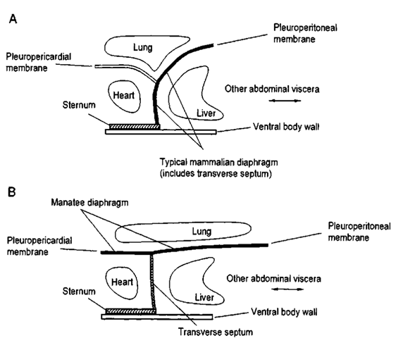
Although the diaphragm separates the heart and lungs from the other organs of the body, the diaphragm is traversed bv nerves and other structures, such as the aorta (crossing in a dorsal and medial position), the vena cava (crossing more ventrally than the aorta, and often slightly right of the midline, although appearing to approximate the center of the liver), and the esophagus (crossing slightly right of the midline, at roughly a mid-horizontal level). This approximately transverse orientation exists in most marine mammals, although the orientation of the diaphragm may be more or less diagonal, with the ventral portion being more cranial than the dorsal portion (Fig. 1A).
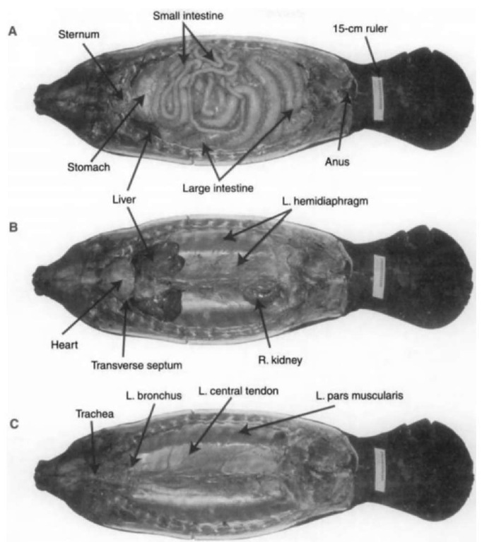
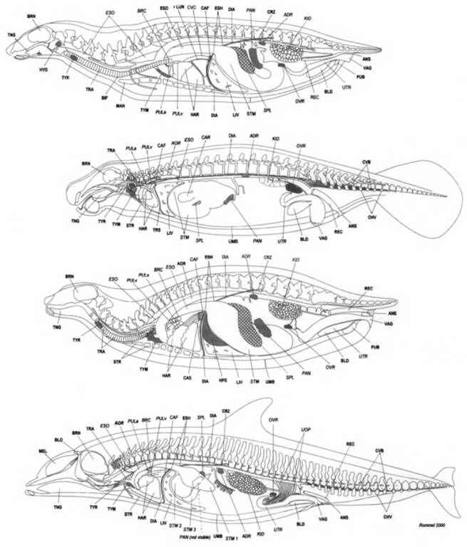
The West Indian manatees diaphragm differs from this general pattern of orientation and attachment. The diaphragm and the transverse septum are separate, with the septum occupying approximately the “typical” position of the diaphragm and the diaphragm itself occupying a horizontal plane extending virtually the entire length of the body cavity (Fig. IB). This apparently unique orientation contributes to buoyancy control (Rommel and Reynolds, 2000). Additionally, there are two separate hemidi-aphragms in the manatee (Figs. 2B and 2C). The central tendons attach firmly to the ventral aspects of the thoracic vertebrae, producing two isolated pleural cavities. The position of the manatee diaphragm stands in contrast with the curved, oblique diaphragms (DIA, Fig. 3) of the sea lion, seal, and dolphin.
Figure 1 Schematic arrangements of mammalian diaphragms. Modified after Rommel and Reynolds (2000). (A) The typical mammalian diaphragm extends ventrally from the dorsal midline to attach to the sternum. The typical diaphragm is a separator between the heart and lungs in the front and the liver and other abdominal organs in the back. (B) The manatee diaphragm extends dorsally to the heart and does not touch the sternum. There is a mechanical barrier between the heart and the liver and other abdominal organs, but it is a relative/ weak barrier called the transverse septum.
Figure 2 Ventral views of the Florida manatee. Modified after Rommel and Reynolds (2000). The rider is 15 cm long. (A) After removal of the ventral skin, fat, and musculature, the small and large intestines are exposed; the large intestine (with contents) may account for 10% of the total body weight and can measure 20 m long. Portions of the stomach and ventral margins of the liver are visible caudal to the sternum. (B) Removal of the Gl tract reveals the heart, transverse septum, liver, hemidiaphragms, and right kidney (the left kidney was removed to expose that portion of the hemidiaphragm). (C) The two central tendons of the hemidiaphragms attach medially to the ventral aspects of the vertebral column. The diaphragm mtiscles attach laterally to the ribs. The lungs are flattened, elongate structures dorsal to the hemidiaphragms; when fully inflated, the lungs extend almost the entire length of the region dorsal to the hemidiaphragms. Note the junctions of the central tendon and the pars muscularis of each hemidiaphragm; this approximates the lateral margin of each lung.
B. Regions and Structures Cranial to the Diaphragm
The region cranial to the diaphragm is typically compartmentalized into three sections: (1) the pericardium (containing the heart), (2) the pleural cavities (containing the lungs), and (3) the mediastinum (Figs. 3 and 4).
The pericardium is a fluid-filled sac surrounding the heart (HAR, Fig. 3); in manatees, it often contains more fluid than is found in the pericardia of the typical mammal or in those of other marine mammals. The heart occupies a ventral position in the thorax (immediately dorsal to the sternum), making it easy to see when the overlying muscles, ribs, and sternum are removed. The heart lies immediately cranial to the central portion of the diaphragm (or just the transverse septum in the manatee). Some lungs may embrace the caudal aspect of the heart, separating the heart from the diaphragm. As do the hearts of all other mammals, marine mammal hearts have four chambers, separate routes for pulmonary and systemic circulation, and the usual arrangements of great vessels (venae cavae, aorta, coronary arteries, pulmonary vessels). Cardiac fat is commonly found in manatees but is typically absent in pinnipeds and cetaceans.
The pleural cavities and lungs of mammals are generally found dorsally and laterally to the heart and are separated along the midline by the heart and mediastinum (see later). In the manatee, the lungs are unusual in that they extend virtually the length of the body cavity and remain dorsal to the heart (Rommel and Reynolds, 2000). Lungs of some marine mammals (cetaceans and sirenians) tend to be unlobed. The size of the lungs of marine mammals varies according to each species’ diving proficiency. Marine mammals that make deep and prolonged dives (e.g., elephant seals, Miroungn spp) tend to have smaller lungs than expected (based on allometric relationships), whereas shallow divers (e.g., sea otters, Enhydra lutris) tend to have larger than expected lungs.
The mediastinum is typically considered to be the area between the lungs, excluding the heart and pericardium. The mediastinum contains the major vessels leading to and emanating from the heart, nerves (e.g., the phrenic nerve to the diaphragm), and lymph nodes. The thymus, which is larger in younger individuals, is found on the cranial aspect of the pericardium (sometimes extending caudally to embrace almost the entire heart) and may extend into the neck in some species. The thyroid gland is located in the cranial part of the mediastinum along either side of the distal part of the trachea, cranial to its bifurcation into the bronchi (in sea lions, but not in other marine mammals, the bifurcation is cranial to the thoracic inlet).1 In most ‘The thoracic inlet is the cranial opening of the thoracic cavity and is bounded by the vertebral and sternal ribs and sternum.
Marine mammals, the mediastinum is generally not remarkable; in the manatee, however, the unusual placement of the lungs and the unique diaphragm change how one must define the mediastinum (Rommel and Reynolds, 2000).
One additional structure, located on the cranial aspect of the diaphragm in seals and sea lions, is an atypical mammalian muscular feature associated with the heart. This is the caval sphincter (CAS, Fig. 3), which can regulate the flow of oxygenated2 blood in the large venous hepatic sinus to the heart during dives (Eisner, 1969).
C. Structures Caudal to the Diaphragm
Easy to find landmarks caudal to the diaphragm include a massive liver and the various components of the gastrointestinal (GI) tract. The urogenital organs are generally found only after removal of the GI tract (note that the exception is the uterus of a pregnant female).
1. The Liver Typically, the liver is located immediately caudal to the diaphragm. It is a large, brownish, multilobed organ positioned so that most of its volume/mass is to the right of the midline of the body. Although marine mammal livers are generally similar to the livers oi other mammals, in manatees, the organ is displaced somewhat to the left and dorsal relative to its location in most other mammals. The size, color, and “sharpness” of the liver margins can be used to assess the nutritive state and health of individual animals. Bile mav be stored in a gallbladder (often greenish in color) located ventrally between the lobes of the liver, although some species (e.g., cetaceans, horses, and rats) lack a gallbladder. Bile enters the duodenum to facilitate the chemical digestion of fats.
“Diving mammals with abundant arteriovenous anastomoses (shunts between arteries and veins before capillary beds) can have high blood pressure and highly oxygenated blood in their veins. One such venous reservoir of oxygenated venous blood is the hepatic sinus of seals (King, 1983).
Figure 3 Left lateral illustrations of the superficial internal structures and “anatomical landmarks” of the California sea lion, Florida manatee, harbor seal, and common bottlenose dolphin with the skeleton (minus the distal appendictdar elements) superimposed for reference. Our view is a left lateral view, focused on relatively superficial internal structures (labeled in bold) visible from that perspective; the other important bony or soft “landmarks” are not necessarily visible from a left lateral view but they are useful for orientation and are labeled in italics. Skeletal elements are included for reference, but not all are labeled. Each drawing is scaled so that there are equivalent distances between the shoulder and the hip; thus, the thoracic and abdominal cavities are roughly equal in length. The shoulder joints are aligned. The left kidney (not visible from this vantage in the manatee) is illustrated. The relative sizes of the lungs represent partial inflation—full inflation would extend margins to distal tips of ribs (except in the manatee). The follotving abbreviations are used as labels (structures on the midline are in bold, those off-midline are not): ANS, anus; BLD, urinary bladder; BLO, blowhole of dolphin; VIA, diaphragm, midline extent (except manatee); EYE, eye (note small size in manatee); HAR, heart; 1LC, iliac crest of the pelvis; INT, intestines, note the large diameter of the large intestines in the manatee; KID, left, kidney (not visible from this vantage in the manatee); LIV, liver; LUN, lung (note that in this illustration, the lung extends under the scapula except in the seal); MEL, melon, dolphin only; OLE, olecranon; OVR, left ovari/; PAN, pancreas (in this view visible only in seal and sea lion); PAT, patella; PEL, pelvic vestige; KEC, rectum; SCA, scapula; SPL, spleen; STM, stomach; TRA, trachea (not visible in this view of the manatee); TYM, thymus gland; TYR, thyroid gland; UMB, umbilical scar; UOP, uterovarian plexus in dolphins; UTR, uterine horn; VAC, vagina. Copyright S. A. Rommel.
2. The GI Tract Most of the volume of the cavity caudal to the diaphragm (the abdominal cavity) is occupied by the various components of the GI tract: the stomach, the small intestine (duodenum, jejunum, and ileum), and the large intestine (cecum, colon, and rectum). The proportions and functions ol these components reflect the feeding habits and trophic levels of the different marine mammals. Therefore, the gastrointestinal tracts of marine mammals vaiy considerably.
Food and water travel from the mouth, through a muscular pharynx, and into the esophagus. As noted earlier, the latter pierces the diaphragm to join the stomach, which is typically a single, distensible sac. The distal end of the stomach (the pylorus) is marked by a strong sphincter before it connects with the small intestine (duodenal ampulla in cetaceans). The separation between jejunum and ilium of the small intestine is difficult to distinguish grossly although the two sections are different microscopically. The junction of the small and large intestines is often (but not in cetaceans) marked by the presence of a cecum (homologous to the human ). In manatees, the midgut cecum has two blind pouches called cecal horns. In some marine mammals, the large intestine, as its name implies, has a larger diameter than the small intestine.
The gastrointestinal tracts of pinnipeds and other marine mammal carnivores follow the general patterns outlined earlier, although the intestines can be remarkably long in some species. Cetaceans, however, have some unique specializations (Gaskin, 1978). Cetaceans can have two or three stomachs (usually three), depending on the species being examined. The multiple stomachs of cetaceans function in much the same way as the single stomach found in most other mammals. The first stomach of cetaceans, called the forestoinach (essentially an enlargement of the esophagus), is muscular and very distensible, and it acts much like a bird crop, i.e., as a receiving chamber. The second or glandular stomach is the primary site of chemical breakdown among the stomach compartments; it contains the same types of enzymes and hydrochloric acid that characterize a “typical” stomach. Finally, the “U-shaped” third or pyloric stomach ends in a strong sphincteric muscle that regulates the flow ol digesta into the duodenum (the duodenal ampulla is sometimes mistakenly called a fourth stomach) of the small intestine. The cetacean duodenum is expanded into a sac-like ampulla. The only other remarkable feature at the gross level is the lack of a cecum, which makes it difficult to tell where the small intestine ends and the large intestine begins. The intestines of some cetaceans may be extremely long (especially in the sperm whale, Phijsetcr macrocephalus; Slijper, 1962), but they are not especially long in many other marine mammal species.0
Among marine mammals, sirenians have the most remarkably developed gastrointestinal tract. Sirenians are herbivores and hindgut digesters (similar to horses and elephants) so the large intestine (specifically the colon) is extremely enlarged, enabling it to act as a fermentation vat (see Marsh et al, 1977; Reynolds and Rommel, 1996). In horses, the cecum is the region of the large intestine that is enlarged, but in sirenians, the cecum is relatively small and has two “horns.” The sirenian stomach is single chambered and has a prominent accessory secretory gland (the cardiac gland) extending from the greater curvature. The duodenum is capacious and has two obvious di-verticulae projecting from it. The GI tract and its contents can account for more than 20% of a manatee’s weight.
The length and mass oldie gastrointestinal tract are impressive and create three-dimensional relationships that can be complex. Simplifying the organization is the fact that tough sheets of connective tissue called mesenteries suspend die organs from the dorsal part of the abdominal cavity and shorter bands of connective tissue (ligaments)4 hold organs close to one another in predictable arrangements (e.g., the proximal spleen is always found
”Assessing the length of intestines is fraught with potential bias because it is extremely difficult not to stretch the intestines to unnatural lengths after they are freed from the mesenteries and straightened. Linear measurements of gastrointestinal tract are, therefore, highly subjective.
4Liganient has several meanings in anatomy: a musculoskeletal element (e.g.. the anterior [cranial] cruciate ligament), a vestige of”a fetal artery or vein (e.g.. the round ligament of the bladder), the margin of a fold in a mesentery (e.g., broad ligament), and a serosal fold between organs (e.g.. the gastrolienal ligament).
Figure 4 A view slightly to the left of the midsagittal plane illustrates the circulation, body cavities, and selected organs of the California sea lion, Florida manatee, harbor seal, and common bottlenose dolphin, with the skeleton for reference. The left lung is removed. Note that the diaphragm separates the heart and lungs from the liver and other abdominal organs. Each drawing is scaled so that there are equivalent distances between the shoulder and the hip: thus, the thoracic and abdominal cavities are roughh/ equal in length. The shoulder joints are aligned. Note that the manatee’s diaphragm is unique and that the distribution of organs and the separation of thoracic stnwtures from abdominal structures require special consideration in these beasts. The following abbreviations are used as labels (structures on the midline are in bold, those off midline are not): ADR, adrenal gland; ANS, anus; AOR, aorta; BLD, urinan/ bladder: BLO, blowhole; BRC, bronchus; BRN, brain; CAF, caval foramen; CAR. cardiac gland, in manatee only; CAS, caval sphincter, surrounding the vena cava in the seal and sea lion; CHV, chevron bones; CRZ, crus (plural crura) of the diaphragm; CVB, caudal vascular bundle, in manatee and dolphin; D1A, diaphragm, cut at midline, extends from crura dor-sally to sternum ventrally (except in manatees); ESH, esophageal hiatus; ESO, esophagus (to the left of the midline cranially, on the midline caudally); HAR, heart; HPS, hepatic sinus within liver, in seals only; KID. right kidney; LTV, liven cut at midline; LVN, lung, right lung between heart and diaphragm; MEL, melon, dolphin only; PAN, pancreas; PUB, pubic symphysis (seals and sea lions only); PULa, pulmonary artery, cut at hilus of lung: PULv, pulmonary vein, cut at hilus of lung; REC, rectum, straight pari of terminal colon; SPL, spleen; STM1, forestoinach: STM2, main stomach (STM in noncetaceans); STM3. pyloric stomach; STR, sternum, sternabrae; TNG, tongue; TRA, trachea; TRS, transverse septum; TYM, thymus gland; TYR, thyroid gland: VMB, umbilicus; UOP, right uterovarian vascular plexus in dolphin; UTR. uterus, VAG, vagina. Copyright S. A. Rommel along the greater curvature of the stomach and is connected to the stomach by the gastrolienal, or gastrosplenic, ligament). Also suspended in the mesenteries are numerous lymph nodes and fat.
Accessory organs of digestion include the salivary glands (small in most marine mammals but very large in die manatee), pancreas, and liver (where bile is produced and then stored in the gall bladder). The pancreas is sometimes a little difficult to locate because it can be a rather diffuse organ and it decomposes rapidly; however, a clue to its location is its proximity to the initial part of the duodenum, into which pancreatic enzymes flow. Another organ that is structurally, but not functionally, associated with the GI tract is the spleen, which is suspended by a ligament, generally from the greater curvature of the stomach (the first stomach in cetaceans) on the left side of the body. The spleen may be a single organ accompanied by accessory spleens in some species. The spleen is bluish in color and varies considerably in size among species; in manatees and cetaceans it is relatively small, but is more massive in some deep-diving pinnipeds (Za-pol et al., 1979) and acts as a storage region for red blood cells.
3. Urogenital Anatomy The kidneys lie in a retroperitoneal position, typically against the musculature of the back (epaxial muscles) at or near the dorsal midline attachment of the diaphragm (crura). In the manatee, the unusual placement of the diaphragm means that the kidneys lie against the diaphragm, not against the epaxial muscles. All mammals have metanephric kidneys (i.e., containing cortex, medulla, calyces). In many marine mammals, the kidneys are specialized as reniculate (mul-tilobed) kidneys, where each lobe (renule) has all the components of a complete metanephric kidney. Why marine mammals have reniculate kidneys is uncertain, but the fact that some large terrestrial mammals also have reniculate kidneys has led to speculation that they are an adaptation associated simply with large body size (Vardy and Biyden, 1981).
The renal arteries of cetaceans enter the cranial poles of the kidneys, whereas in other marine mammals, they enter the hilus (typical of most mammals). Additionally, in manatees, there are accessor}’ arteries on the surface of the kidney. The kidneys are drained by separate ureters, which carry urine to a medially and relatively ventrally positioned urinary bladder. The urinary bladder lies on the floor of the caudal abdominal cavity and, when distended, may extend as far forward as the umbilicus in some species. The pelvic landmarks are less prominent in fully aquatic mammals. In the manatee, the bladder can be obscured by abdominal fat.
Pabst et al. (1999) noted that the reproductive organs tend to reflect phylogeny more than adaptations to a particular niche. If one were to examine the ventral side of cetaceans and sire-nians before removing the skin and other layers, one would discover that positions of male and female genital openings are different, permitting rather easy determination of sex without dissection. In all marine mammals, the female urogenital opening is more caudal than the opening for the penis in males. One way to approach dissection of the reproductive tracts is to follow structures into the abdomen from their external openings.
The position and general form of the female reproductive tract in marine mammals are generally similar to those of the female reproductive tracts in terrestrial mammals. The vagina opens cranial to the anus and leads to the uterus, which is biconiuate in marine mammal species. The body of die uterus is found on die midline and is located dorsally to the urinary bladder (die ventral aspect of the utems rests against the bladder). Although the body of the uterus lies along the midline, it has bilaterally paired, relatively large diameter projections called uterine horns (cornua), which extend laterally. The relatively small-diameter oviducts conduct eggs from the ovaries to the uterine horns where implantation of the fertilized egg and subsequent placental development occur. The dimensions of the uterine homs vary with reproductive history and age. Often die fetus may expand the pregnant horn to the point that it fills a substantial portion of the abdominal cavity. The horns terminate abruptly, narrowing and extending as uterine tubes (fallopian tubes) to paired ovaries. The uterus and the uterine horns are held in place in the abdominal cavity by the broad ligaments. Uterine and ovarian scarring may provide information about the reproductive history of the individual.
The ovaries of mature females may have one or more white or yellow-brown scars, called corpora albicantia and corpora lutea, respectively. Although ovaries are usually solid organs, in sirenians they are relatively diffuse.
Mammary glands are ventral, medial, and relatively caudal in most marine mammals, but they are axillary in sirenians. Many marine mammals have a single pair of nipples, sea lions and polar bears, Ursus maritimus, (DeMaster and Sterling, 1981), have two pairs of nipples, and cetaceans have mammary slits (note that some male cetaceans have distinct mammary slits).
The male reproductive tracts of marine mammals have the same fundamental components as the tracts in “typical” mammals, but positional relationships are significantly different. This difference is due to the testicond (ascrotal) position of the testes in most marine mammal species [sea otters are scrotal (J. Bodkin personal communication); polar bears are seasonally scrotal (I. Stirling personal communication); sea lion testes become scrotal when temperatures are elevated]. The testes of some marine mammals are intraabdominal, but in phocids, for example, they lie outside the abdomen, partially covered by the oblique muscles and blubber. The position of marine mammal testes creates certain thermal problems because spermatozoa do not survive well at body (core) temperatures; in some species, these problems are solved by the circulatory adaptations mentioned later.
The penis of marine mammals is retractable and it normally lies within the body wall. The general structure of the penis relates to phylogeny (see Pabst et al, 1999).
4. Adrenal Glands The term “suprarenal gland” is often used interchangeably with “adrenal gland.” Although the suprarenals often lie immediately atop or very close to the kidneys of terrestrial mammals, adrenals of marine mammals may lie several centimeters cranial to the kidneys, along either side of the median. Adrenal glands can be confused with lymph nodes, but if one slices the organ in half, an adrenal gland is easv to distinguish grossly by its distinct cortex and medulla.
5. Circulatory Structures Basically, blood vessels are named for the regions they feed or drain. Thus, the fully aquatic marine mammals (cetaceans and sirenians) lack femoral arteries that supply the pelvic appendage. However, most organs in marine mammals are similar to those of terrestrial mammals so their blood supply is also similar. Therefore, readers who want to learn details of typical circulatory anatomy should consult one of the anatomy references cited earlier. The thoracic aorta leaves the heart and lies ventral to the vertebral column, giving off segmental arteries to the vertebrae and epaxial muscles (and in the case of cetaceans and manatees to the thoracic re-tia). The aorta continues through the aortic hiatus of the diaphragm (between the crura) and into the abdomen as the abdominal aorta and lumbar aorta, which give off several paired (e.g., renal, gonadal) and unpaired (e.g., celiac, mesenteric) arteries. The caudal aorta follows the ventral aspect of the tail vertebrae. In the permanently aquatic marine mammals, there are robust ventral chevron bones that form a canal in which the caudal aorta, its branches, and some veins are protected.
Some of the diving mammals (e.g., seals, cetaceans, and sirenians) have few or no valves in their veins (Rommel et al, 1995); this adaptation simplifies blood collection.5 Other exceptions to the general pattern oi mammalian circulation are associated with thermoregulation and diving. Countercurrent heat exchangers abound, and extensive arteriovenous anastomoses exist to permit two general objectives to be fulfilled: (1) regulating loss of heat to the external environment, while keeping core temperatures high; and (2) permitting cool blood to reach specific organs (e.g., testes, uteri, spinal cord) that cannot sustain exposure to high body temperatures (see rev iews by Rommel et al, 1998: Pabst et al, 1999).
In mammals, several paths for supplying blood to the brain exist; via the internal carotid, the external carotid, and the vertebral/basilar arteries. Some species use only one, others use two, and manatees use all three pathways. In cetaceans, the path for supplying blood to the brain is unique. The blood destined ior the brain first enters the thoracic rete. a plexus of convoluted, small diameter arteries in the dorsal thorax. Blood leaves the thoracic rete and enters the spinal rete where it surrounds the spinal cord and enters the base of the skull (Mc-Farland et al, 1979). There are two working explanations for this convoluted path of blood to the brain: (1) the elasticity of the retial system allows mechanical damping of the blood pulse pressure wave (McFarland et al, 1979) and (2) the juxtaposition of the thoracic retia to the dorsal aspect of the lungs may provide thermal control of the blood entering the spinal retia. Combined with cooled blood in the epidural veins, the spinal retia may provide some temperature control of the central nervous system (Rommel et al 1993, paper presented at the Tenth Biennial Conference on the Biology of Marine Mammals).
II. Overview
Marine mammal postcranial soft tissue anatomy is, in many regards, similar to that of “typical” mammals. However, the relative proportions of and, to some extent, the positions of organs may be somewhat different from the norm.
5The near absence of valves in the veins of seals and dolphins allows two-way flow to occur, increasing the blood available when venipuncture is used; in contrast, sea lions have numerous valves in their hind flipper veins.
We close with a reminder about orientation: namely the orientation of the prosector relative to the orientation of the specimen and the orientation of the specimen to the orientation of that animal when it was alive. The position of animals during necropsy may be belly-up. obviously not the usual position of the living animals. Thus, gravitational forces make the positional relationships we may observe during necropsy somewhat artificial; we assess “dead anatomy” rather than “living anatomy.” We suggest that people examining marine mammal postcranial anatomy bear this fact in mind and try to constantly picture how the structures being observed during necropsy might be arranged in a free-ranging animal. The more the latter perspective can be maintained, the easier it will be to envision dynamic relationships among organs and systems and to relate function (physiology) to structure (anatomy).


![tmpBA-23_thumb[1] tmpBA-23_thumb[1]](http://lh5.ggpht.com/_NNjxeW9ewEc/TNFYFUGiAjI/AAAAAAAAOZo/kgHIaofgmOY/tmpBA23_thumb1_thumb2.jpg?imgmax=800)

