Ethos
Serology is defined as the study of the composition and properties of the serum component of blood.
Despite this inadequate description of what has colloquially become known as forensic serology, many individuals and laboratories use this rubric to describe a practice that more accurately could be described as forensic biology or biochemistry. ‘Forensic serology’, then, is the application of immuno-logical and biochemical methods to identify the presence of a body fluid or tissue sample encountered in connection with the investigation of a crime, and the possible further genetic characterization of the sample with a view to determining likely donors thereof. For the purposes of this article the genetic characterization involves polymorphic cellular antigens and proteins (i.e. those that exhibit variable forms in the population) but does not include DNA genetic markers.
The probative significance of biological evidence transfer
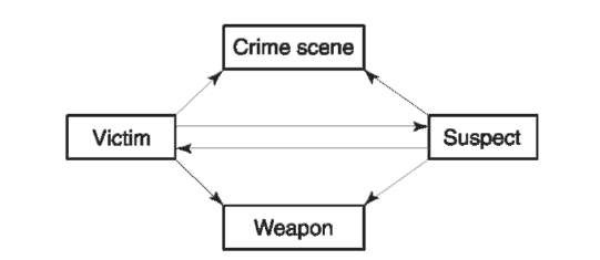
The perpetration of a violent crime often results in a number of different types of biological material being transferred in a unidirectional or bidirectional manner between the victim, the perpetrator, the crime scene or the weapon (Fig. 1). A genetic analysis of such biological material by the forensic biologist may yield important legal evidence that may associate, or exclude, a particular individual with the crime in question. Such analysis also may aid in the reconstruction of the sequence of events which occurred before, during or after the commission of the crime. Based upon the circumstances of the case, it is incumbent on the investigators (whether law enforcement officials or forensic scientists) to evaluate the potential ‘probativeness’ of biological evidence that may have been transferred. By ‘probativeness’ is meant the degree of meaningfulness of the potential information gleaned by examination of a particular piece of biological evidence as it relates to establishing a relevant fact which may be at issue. Examples of good probative evidence would include the finding of the presence of the perpetrator’s semen in the vagina of the rape victim or the presence of the deceased’s blood on the perpetrator’s clothing. Examples of evidence that would possess low or almost nonexistent probative value would include the finding of blood from a deceased person in immediate proximity to the bloodied body itself, or the finding of semen on the inside of a rape suspect’s underpants. The probative value of the evidence greatly increases if there is a demonstrated bidirectional transfer. An example of such transfer would be the transfer of the rape victim’s menstrual blood on to the rape perpetrator’s underwear concomitant with the assailant’s semen being deposited on the victim. In some circumstances the analysis of the nature and distribution of the stain is more important than the genetic characterization of it. A tiny spot of blood consistent with a low-velocity blood spatter pattern on an individual’s clothing may belie an attempt to explain the presence of the blood as a result of a Good Samaritan act. Thus it is important to evaluate the circumstances of each crime in order to make a rational judgment as to which evidence to remove for analysis.

Figure 1 Directionality of biological evidence transfer.
Importance of Communication Between Law Enforcement and Laboratory Personnel
Communication of the circumstances of the case to the appropriate laboratory staff is essential in order that an appropriate analytical scheme can be developed. Failure to do so could result in the inadvertent omission of certain tests that should have been conducted. An example of this would include an altercation that may have been precipitated as a result of one individual spitting at another. If this information is not communicated to the laboratory, it is unlikely that testing for saliva would be performed and an important set of extenuating circumstances might not be corroborated. This is so because testing of clothing items for the presence of saliva is not normally performed in the forensic laboratory unless there is a specific reason to do so.
Importance of Laboratory Search Activities
Evidence submitted to the laboratory must be processed and searched for the presence of probative physiological stains and for other trace materials. Searching takes place under a variety of lighting conditions, both with and without the aid of magnification. This general search phase is critical for locating materials of evidential value, recording information about the nature and location of these materials and collecting and preserving them for later analysis. The general search phase often employs the use of a number of so-called presumptive chemical or biochemical tests for the presence of particular body fluids such as blood or semen. These preliminary tests allow the scientist to screen items efficiently before the use of more specific confirmatory tests.
Identification of Body Fluids and Tissues
Physiological material must first be identified as such before genetic analysis is performed. The most commonly encountered body fluids are blood, semen and saliva, although there may be instances in which the identification of vaginal secretions, fecal material and urine is necessary.
Blood
Blood consists of hematopoietic lineage cells (red blood cells, white blood cells and platelets) in a proteinaceous fluid known as plasma. Serum is the fluid exuded from blood once it has clotted and thus comprises the plasma minus the proteins (principally fibrinogen and fibrin) responsible for the clotting process.
Presumptive catalytic screening tests The principal function of blood is to transport oxygen to the tissues and remove carbon dioxide therefrom. The hemoglobin molecule (Hb), which constitutes most of the protein content of the erythrocyte, or red blood cell, is principally responsible for this function, and binding of the gas molecules takes place via the nonpro-tein prosthetic heme group of Hb. The heme group is a nitrogenous planar structure, comprising a proto-porphyrin IX ring and conjugated ferrous atom, which happens to possess an associated peroxidase activity: H202 – H20+i02.
The peroxidase-induced reduction of hydrogen peroxide can be coupled to the oxidation of a number of colorless (reduced) dyes, such as phenolphthalein (Kastle-Meyer reagent), leuco-malachite green (LMG), o-toluidine and tetramethyl benzidine, to form their respective colored moieties. Alternatively luminol (3-amino-phthalhydrazide) can be oxidized to a product that luminesces and this is used to screen large areas for the presence of blood. In this case the area to be searched is sprayed with luminol reagent and the heme-induced luminescence is detected in the dark. The catalytic color tests are extremely sensitive to minute amounts of blood but can produce false-positive results in the presence of a number of substances, including chemical oxidants and catalysts and plant sources containing the enzyme peroxidase. The presence of chemical oxidants and catalysts can normally be excluded by performing a two-step test in which the colorless reduced dye is added first to check for the presence of heme-independent oxidation prior to the addition of the hydrogen peroxide substrate.
Confirmatory tests Subsequent to a positive presumptive test, the presence of blood is confirmed by the immunological identification of Hb or serum proteins, which also serves to identify the blood as being of human (more strictly, primate) origin. Alternatively a positive crystal test for the presence of certain heme derivatives, or the demonstration of the characteristic visible absorption spectrum of hemoglobin, is regarded as conclusive proof of the existence of blood.
Immunological identification of blood and determination of species origin The precipitin reaction occurs when a precipitating antibody combines with its conjugate antigen to produce an insoluble proteinac-eous immune complex, which can be detected under appropriate oblique lighting conditions by the naked eye or by the use of general protein stains such as Coomassie Blue or Amido Black. Polyclonal and monoclonal antibodies to serum proteins from humans and a range of domesticated animals are commercially available, as are antibodies to human hemoglobin. The antibody-antigen reaction is either allowed to take place in an inert agar gel by diffusion of the antigen and antibody molecules towards one another (the 0uchterlony reaction or double diffusion in two dimensions) or is facilitated by an electric field (counterimmunoelectrophoresis or crossed-over electrophoresis). An immunochromatographic method, initially developed for clinical use for occult blood, has begun to gain widespread acceptance. In this method a dye-linked mobile monoclonal antibody to human hemoglobin is allowed to react with the sample. If human hemoglobin is present in the sample, it combines with the antibody-dye complex and moves along a membrane until, in a reaction zone, it meets an immobilized polyclonal antibody to human hemoglobin and is concentrated. A positive reaction is indicated by the formation of a dye front. A control zone consists of immobilized anti-IgG that concentrates any unbound mobile monoclonal antibody-dye complex and forms a second dye front.
Confirmatory crystal tests for the presence of blood The two main confirmatory crystal tests are the Teichmann and Takayama tests, named after their developers who initially described the reactions in 1853 and 1912, respectively. These tests rely on the formation of certain heme derivatives. The Teich-mann test involves the formation of hematin (heme in which the ferrous ion has been oxidized to the ferric state) halide crystals, whereas the Takayama test induces the formation of salmon pink pyridine hemochromogen crystals. Positive crystal tests, while specific for blood, provide no information regarding its species origin.
Identification of blood by visible spectrophotometry The heme moiety of hemoglobin has a characteristic absorption spectrum which, if present in a sample, is often regarded as conclusive evidence for the presence of blood. Although different heme derivatives produce different spectra, they all possess the Soret band at 400-425 nm. Oxyhemoglobin produces two absorption bands at 538 nm and 575 nm and a shoulder peak at 610 nm, whereas hemochromogen displays a sharp peak at 550-560 nm. This technique is confounded by the presence in bloodstains of a broad region of absorption at 500-600 nm that tends to obscure the diagnostic heme derivative peaks.
Fetal blood Normal adult hemoglobin is a tetramer consisting primarily of two a and two p polypeptide chains (a2p2). However, during development of the embryo and subsequently the fetus, s and y polypeptide chains are respectively expressed instead of the p chain. Fetal blood can thus be distinguished from adult blood by the presence of the fetus-specific y subunit, which is still detectable up to 6 months after birth. This can be accomplished immunological-ly with the use of antisera specific to fetal hemoglobin (HbF) or by separation in an electrical field. Specificity problems with many anti-HbF preparations for determining the Hb status in dried and aged stains have prevented its widespread use in forensic laboratories.
Menstrual blood Menstrual blood may be transferred from a female victim of rape or assault to the assailant, and under certain circumstances the identification of it is of some investigative use. Menstrual flow is comprised of endometrial tissue, mucus and blood. Usually not more than 50-60 ml of blood is lost during the uterine cycle and it has the characteristic property of being unable to clot due to extensive degradation of the clotting factor fibrino-gen (or its product fibrin). Fibrinogen degradation products are present in relatively high concentration in menstrual blood and can be detected immunologi-cally and normalized against total protein to distinguish menstrual from venous blood. Alternatively, the isoenzymes of lactate dehydrogenase (LDH) can be used to distinguish venous from menstrual blood. Isoenzymes are structurally distinct forms of enzymes that have equivalent catalytic specificities. LDH is a tetrameric protein, the polypeptide chains of which can be of two types, H and M, thus giving rise to five possible isoenzymes H4,H3M, H2M2,HM3,M4 or LDH 1, LDH 2, LDH 3, LDH 4, LDH 5. Venous blood consists primarily of the three isoenzymes LDH 1, LDH 2 and LDH 3, whereas menstrual blood additionally contains elevated levels of LDH 4 and LDH 5. Other tissues possess varying amounts of the LDH isoenzymes and, although the is oforms are readily separated by electrophoresis, difficulties with the presence of body fluid mixtures have limited the efficacy of this technique.
Semen
Semen principally comprises the germ cells (spermatozoa) suspended in a complex mixture of fluids secreted by various accessory glands of the male reproductive tract, including the prostate, seminal vesicles, Cowper’s glands and the glands of Littre. Spermatozoa make up approximately 10-25 % of the volume of the semen and normal sperm density ranges from 60 to 100 million per milliliter. The ejaculate volume typically ranges from 1 to 6 ml, with an average of 3 ml, and is dependent upon the time interval since the last ejaculation, the metabolic activity of the glands and the presence or absence of partial ductal obstruction.
Screening tests Screening tests comprise the classical crystal tests for the presence of spermine (Barberio test) and choline (Florence test) and the more commonly used seminal acid phosphatase (SAP).
The Barberio test relies on the formation of sperm-ine phosphate or picrate crystals upon the suspected stain extract’s reaction with appropriate anions. The Florence test detects the presence of choline periodide crystals when a semen extract is treated with a solution of iodine in potassium iodide.
SAP is an enzyme present in high concentration in semen; as a nonspecific orthophosphoric monoester phosphohydrolase, it cleaves a variety of organic phosphates, including p-nitrophenyl-, a-naphthyl-and thymolphthalein monophosphates. As implied in its name, it is active at acid pH (4.9-5.5). Although SAP is a sensitive test it is not specific for semen owing to its presence in a number of other tissues, including in particular, vaginal fluid.
Confirmatory tests The presence of semen can be confirmed microscopically by the presence of spermatozoa or by the presence of the semen-specific protein p30.
Spermatozoa Spermatozoa have a distinct and characteristic appearance as viewed under the microscope. They are approximately 50-60 um in length and comprise a flattened ovoid head (dimensions, 4.6 x 2.6 x 1.5 um) and a 50 um tail. However, owing to the lability of the tail-head junction, dried stains often possess sperm heads without tails. The head structure, which is principally comprised of a nucleus surrounded by a thin layer of cytoplasm, contains at its anterior end a secretory vesicle known as the acrosome. This appears as a caplike structure and can be differentially stained by standard histochemical stains. The spermatozoa are the principal sources of DNA in semen.
P30 protein P30 protein or prostate specific antigen (PSA) is a protein that is synthesized in the prostate and is an important clinical indicator of malignancy. Its normal range in semen is 300-4200 ugmr1 with a mean of 1200 ugml-1. However, it is found in breast, lung and uterine cancers and it may function as an endogenous antiangiogenic protein. Commercial antibodies to PSA are readily available and standard immunochemical techniques can be applied to detect it, including crossed-over electrophoresis, 0uchterlony double diffusion, immunochromatogra-phy and enzyme-linked immunoassay (ELISA). The immunochromatographic and ELISA techniques are sensitive to 1 in 105-106 dilutions of semen (i.e. approximately 1ngml_1) and care must be taken in the interpretation of weak results. For example, it may be possible to get false-positive reactions from postejaculate urine, and urine from adult males, as PSA is present at a mean level of 260 ng ml ~~1 therein.
Stability of semen components Dried seminal stains on clothing and bedding can generally exhibit some or all of the semen factors months or even years after deposition. Washing will tend to remove any seminal material, although there have been reports of spermatozoa persistence after machine washing. However, persistence in the postcoital vaginal canal is a different matter and the differential stability of p30, SAP and spermatozoa can be used to assess how long has passed since the last act of sexual intercourse. Semen is lost from the vagina of the living victim due to drainage, dilution with vaginal fluid and phagocytosis of spermatozoa by neutrophilic lymphocytes and mononuclear cells. However, significant levels of p30 tend to be lost within 24 h of deposition in the vaginal vault (as measured by immunodiffusion or crossed-over electrophoresis), SAP is normally lost 48 h postcoitus and spermatozoa do not normally persist after 72 h. In deceased individuals these semen components can last for several days, depending upon the environmental conditions and the rate of atrophy of the body tissues.
Vaginal secretions
Although vaginal secretions are often encountered in postcoital vaginal swabs and stains, there is no definitive test for their presence. The squamous epithelial cells lining the vaginal tract are glycogen-rich and some investigators have used staining with Lugol’s iodine to try and distinguish these from other epithelial cells, such as those from the oral cavity. DNA extracts from stains containing a mixture of vaginal epithelial cells and spermatozoa can be differentially enriched for both components.
Saliva
Saliva is a secretion that acts as a digestive aid and contains secretions from the salivary gland. There is currently no definitive test for the positive identification of saliva, although there are a number of substances present in higher concentration in saliva than elsewhere. These include the inorganic anions thio-cyanate and nitrite and the enzymes alkaline phos-phatase and a-amylase. The presence of significant levels of a-amylase is strongly indicative of the presence of saliva and the detection of a-amylase is the most commonly used test for it.
a-Amylase is produced by two different genetic loci, AMY1 and AMY2. The enzyme hydrolyses the a(1,4) glycosidic bonds of glucose polymers such as glycogen or starch. AMY1 encodes the salivary form of the enzyme, which is found in saliva, breast milk and perspiration, whereas AMY2 encodes the pancreatic isoform, which is expressed in semen, vaginal secretions, urine and feces. AMY1 and AMY2 can be distinguished by differential inhibition with wheat seed lectin (WSL) and kidney bean extract (KBE), in that WSLand KBE produce greater inhibitory effects on AMY1 and AMY2, respectively. The two most commonly used methods for a-amylase detection are radial diffusion and dyed starch substrates.
Radial diffusion A stain extract from a suspected saliva stain is placed into a well of an agar gel, which also has starch incorporated therein, and allowed to diffuse into the gel. If the extract contains saliva the diffusing a-amylase will hydrolyze the starch and this can be detected by the classical starch-iodine reaction. Starch will give a characteristic purple reaction with iodine, in contrast to a circular clear area where the starch polymer has been hydrolyzed by the a-amylase. A semilogarithmic relationship exists between the diameter of the clear circle and the amount of a-amylase present.
Dyed starch substrates Starch is covalently linked to a dye such as cibachron blue or procion red to form an insoluble complex. Subsequent to a-amylase activity the dye is released into solution and can be measured, for example by spectrophotometry. This forms the basis of the often-used Phadebas test which uses starch-cibachron blue tablets as the substrate.
Urine
Urine contains a variety of inorganic ions, such as sulfate, phosphate and chloride, that can be identified by the formation of their barium, magnesium, ammonium and silver salts, respectively. However, urine also contains a significant number of amines,including urea and creatinine, and a positive chemical reaction for the presence of amines is regarded as presumptive evidence for the presence of urine.
Urea If the enzyme urease is added to a urine stain it will catalyze the breakdown of any urea present and produce ammonia, which can be detected using a variety of acid base indicators:
![]()
Creatinine Urine will give a bright-red coloration in the presence of picric acid and a weak base, the so-called Jaffe reaction for the presence of creatinine.
Amines The general detection of amines is possible by reaction with p-dimethylaminocinnamaldehyde (DMAC), which gives a dark-pink/red coloration upon Schiff’s base formation.
Fecal material
Fecal material can be identified by a combination of microscopy and testing for the presence of urobilin (which gives feces its characteristic color).
Microscopy Microscopical identification of fecal material relies on the presence of various undigested fibrous food residues, such as meats, fish and vegetables, and enterobacteriaceae such as Escherichia coli.
Urobilin In a test known as the Edelman test, uro-bilinogen (a precursor of urobilin) is oxidized to urobilin by alcoholic mercuric chloride. Subsequent addition of alcoholic zinc chloride produces a green fluorescence, which is due to the formation ofa stable zinc-urobilin complex.
Genetic Marker Analysis
Classical genetic markers are inherited biochemical substances that exhibit variation (polymorphism) in the population. Questioned biological stains from the crime scene are typed in various genetic market systems and compared to reference samples obtained from individuals who may be possible donors of the stain. Based upon the results obtained, it is possible either to exclude an individual as being the stain donor or to include that person as belonging to a class of individuals who cannot be excluded as having been the stain donor. Alternative forms of a particular genetic marker are known as alleles, and polymorphic genetic loci are ones for which the most common allele frequency is < 0.95 (or < 0.99). At each of these genetic loci it is possible to assign each individual in the population to one of a small number of possible types, the frequency of which varies according to the alleles present and the subpopulation to which the individual belongs. If there are n alleles then there are a possible n(n + 1)/2 possible (geno)-types. Classical genetic marker systems normally possess 2, 3 or 4 alleles, thus giving rise to 3, 6 or 10 subtypes, respectively. Importantly, the more genetic markers tested in a particular crime scene sample, the smaller the proportion of individuals who would possess the constellation of alleles found, and the more probative the evidence. Classical genetic markers can be classified into cellular antigens and extracellular proteins and intracellular isoenzymes.
Cellular antigens
The classical blood groups comprise a diverse set of polymorphic cell surface molecules that are mostly erythrocyte-tethered glycolipids with carbohydrate moieties defining an antigenic specificity that can be detected with an appropriate antiserum. At least 30 different blood group loci exist but only a small subset of these have been successfully employed in forensic serology. These include the ABO, Rhesus and Lewis systems.
ABO blood groups In 1901 Landsteiner discovered that certain combinations of red blood cell suspensions from different people, mixed with blood serum from other people, reproducibly produced a cell clumping or agglutination reaction, whereas other combinations produced no such reaction. Individuals could be classified into four distinct groups, which were named A, B, O and AB, that occur with a frequency of approximately 42%, 8%, 47% and 3%, respectively, in the population. The agglutination reaction takes place because there is recognition of the A or B agglutinogens (antigens) on the cell surface by corresponding isoagglutinins (antibodies). Uniquely, the ABO isoagglutinins are naturally occurring and found in all individuals who are type A (who possess B isoagglutinins), type B (who possess A isoagglutinins) and type O (who possess both A and B isoagglutinins). AB individuals possess neither A nor B agglutinins. A liquid blood sample is typed by firstly separating the red blood cells from the serum and then using commercially obtained monoclonal anti-A and anti-B antisera to test for agglutination of the red blood cells. Confirmatory reverse typing is carried out by testing the sample serum for the presence of isoagglutinins by its reaction with A or B cells. Although the three common ABO alleles (A, B and O) give rise to six different genotypes, AA, AO, BB, BO, AB, OO, only the four types (A, B, O and AB) are distinguishable by liquid typing owing to genetic dominance effects of the A and B alleles over the 0 allele. Two common subtypes of A exist, namely A1 and A2. Hence there are four common alleles at the AB0 locus giving rise to ten different genotypes A1A1,A1A2,A2A2,A10, A20, BB, B0, A1B, A2B, 00.
Secretors and nonsecretors The agglutinogens of red blood cells and the endothelial cells of the cardiovascular system consist of alcohol-soluble glycosphingo-lipids and, with rare exceptions, all individuals possess these. However, the mucous secretions from the gastrointestinal tract, vaginal secretions and semen of certain individuals contain water-soluble glycopro-tein blood group substances with the same antigenic specificity as their red blood cell agglutinogens. These individuals are known as ‘secretors’ and approximately 80% of the population belong to this group, whereas the remaining 20% of individuals who do not secrete blood group substances into their body fluids are classified as ‘nonsecretors’. The secretor/nonsecre-tor dichotomy has important forensic implications. For example, it is expected that most body fluids recovered from a crime scene (80%) can be ABO typed. In addition, nonsecretor individuals can be excluded as having been the body fluid donor even if the individual’s AB0 type is the same as that found in the stain, although aberrant secretors exist and each case has to be examined on its own merits.
ABO typing of stains Stains can be typed for the presence of AB0 agglutinogens by absorption-elution, absorption-inhibition or mixed agglutination methods, or for isoagglutinins by the Lattes crust method.
1. Absorption-elution Blood-stained threads from a questioned stain are affixed by a suitable adhesive to three separate locations on a solid surface. A drop of anti-A is added to the first thread, anti-B to the second thread and anti-H is added to the third thread. If the cognate antigen is present in the bloodstain the antibody (or lectin) will be bound (‘absorbed’) to the stained thread, whereas if the antigen is absent no such absorption will take place. Upon heating to 56°C, any absorbed antibody or lectin will be ‘eluted’ and be available for agglutinating red blood cell suspensions of the appropriate type.
2. Absorption-inhibition Blood-stained threads are placed in separate test tubes and are able to ‘absorb’ the cognate antisera, as in the absorption-elution method described above. An aliquot of each anti-sera supernatant is then removed and tested for its ability to agglutinate red blood cell suspensions of the appropriate type. A reduction in titer of the antisera due to absorption with its cognate antigen results in an inability to agglutinate the red blood cells. Thus ‘inhibition’ of agglutination of a particular red blood cell type signals the presence of the cognate antigen in the bloodstain.
3. Mixed agglutination In this method the antibody is allowed to absorb on to blood-stained threads, as before. Red blood cell suspensions of the cognate antigen are allowed to come into contact with the threads. Any absorbed antibody is detected microscopically by the presence of a layer of red blood cells coating the thread.
4. Lattes crust Separate portions of a blood-stained crust are allowed to react with A, B and 0 red blood cell suspensions. Microscopical observation of agglutination indicates the presence of the cognate isoagglutinin.
Rhesus The clinically important rhesus system consists of six antigens C, c, D, d, E and e, for which appropriate commercial antisera are readily available (except for the d antigen). Rhesus typing of bloodstains can be performed using the absorption-elution methodology but is less sensitive than the AB0 system.
Lewis The Lewis system is most commonly used to confirm the secretor status of an individual. Individuals whose red blood cells are Le (a —b+) are secretors, whereas Le (a+b —) individuals are nonse-cretors. The rarer Le (a— b— ) type is noninformative with respect to secretor status determination.
Extracellular proteins and intracellular enzymes
Genetic variation in the structure of proteins, including enzymes, is mainly due to amino acid substitutions, and at least a third of these are expected to result in charge differences in the protein. In the case of isoenzymes (enzymes which possess the same catalytic specificity but are structurally distinct), such variants are known as allozymes, and they can be electrophoretically separated according to size and/or charge in an electrical field using sieving media such as starch, agarose and polyacrylamide. After electro-phoretic separation, isoenzymes are detected using appropriate substrates which produce a color change due to the transfer of electrons from a donor such as NADPH or NADH to a dye such as MTT tetrazo-lium, thus forming the colored, insoluble MTT-tetra-zolium complex. In the case of nonenzyme proteins, the variants are detected postelectrophoretically using a variety of methods, including immuno-fixation with human protein-specific antibodies subsequent to capillary transfer of the separated proteins on to an inert membrane (Western blotting). A number of polymorphic isoenzymes and extracellular proteins have been commonly used for forensic purposes. The efficacy of genetic marker systems for individual discrimination can be mathematically ranked by comparing their discriminatory potentials (DP). The DP of a genetic marker system is defined as the probability of discriminating two randomly chosen, biologically unrelated individuals. Mathematically, DP =1 — Hpi2, where pi is the frequency of the ith phenotype. Table 1 lists some commonly used genetic marker systems and their DP values for a US African American population.
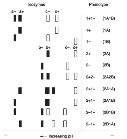
An illustrative example of a genetic marker is the enzyme phosphoglucomutase 1 (PGM1) which is an important ‘housekeeping’ enzyme involved in the metabolism of glucose. There are four common alleles PGM1*2A(PGM12+), PGM1*2B(PGM12—), PGM1*1A(PGM11+) and PGM1*1B(PGM11 —), as well as at least 30 rare variants. The enzyme is detected by means of a substrate overlay (zymogram), as shown in Fig. 2, where the underlined reagents are those that are added to the ‘reaction mix’. The allozymes are separated by isoelectric focusing, which is an electrophoretic technique in which separation is effected in a pH gradient according to differences in the proteins’ isoelectric points (the pH at which the net charge on the molecule is zero). The ten common phenotypes are depicted in Fig. 3.
Significance of Genetic Marker Typing Data
A typical case involves the genetic marker testing of a body fluid sample from a crime scene and reference samples from the victim(s) and suspect(s). Subsequent to the analysis, conclusions are drawn with respect to possible donor(s) of the identified body fluid. As an illustrative example consider the following genetic marker data obtained from a violent assault, whereby a bloodstain found at the crime scene was typed, as was that of the victim and two suspects:

Table 1 Commonly used protein genetic markers
| Genetic marker | Protein type | Discrimination potential (DP) |
| ACP1 | Isoenzyme | 0.67 |
| ADA | Isoenzyme | 0.06 |
| AK | Isoenzyme | 0.25 |
| CAM | Isoenzyme | 0.25 |
| ESD | Isoenzyme | 0.27 |
| GL01 | Isoenzyme | 0.57 |
| PEPA | Isoenzyme | 0.28 |
| GC | Extracellular | 0.69 |
| HP | Extracellular | 0.62 |
| AHSG | Extracellular | 0.54 |
![Pathway for phosphoglucomutase reaction. Reagents underlined are those that are added to the reaction mix. PMS = phenazine methosulfate; MTT = 3-[4,5-dimethylthiazol-2-yl] 2,5-diphenyltetrazolium bromide. Pathway for phosphoglucomutase reaction. Reagents underlined are those that are added to the reaction mix. PMS = phenazine methosulfate; MTT = 3-[4,5-dimethylthiazol-2-yl] 2,5-diphenyltetrazolium bromide.](http://lh6.ggpht.com/_1wtadqGaaPs/TFHE1p9S6iI/AAAAAAAAMro/VX3Hr8mlBS4/tmp213_thumb_thumb.jpg?imgmax=800)
Figure 2 Pathway for phosphoglucomutase reaction. Reagents underlined are those that are added to the reaction mix. PMS = phenazine methosulfate; MTT = 3-[4,5-dimethylthiazol-2-yl] 2,5-diphenyltetrazolium bromide.

Figure 3 Isolectric focusing patterns of the 10 common PGM1 phenotypes. Black bands indicate increased intensity.
Note that it is not uncommon for some genetic markers to yield negative results, as can be seen for the AHSG and HP genetic markers in this case. This can be due to a variety of factors, such as sensitivity of the system and protein stability. Notwithstanding these negative results, what conclusions can be drawn from the above? Firstly, the victim and suspect 1 are excluded as the source of the crime scene bloodstain. Secondly, suspect 2 is included as a possible donor of the bloodstain. What is the significance of this evidence? To evaluate this we ascertain the frequency of each genetic marker type in different population groups from published tables of genetic marker frequencies:
| Caucasian | African American | Hispanic | |
| ACP1 B | 0.39 | 0.57 | 0.52 |
| PGM1 1 + | 0.36 | 0.44 | 0.29 |
| ESD 1 | 0.79 | 0.85 | 0.77 |
| GC1S | 0.31 | 0.04 | 0.22 |
| Combined | 0.034 or | 0.008 or | 0.026 or |
| approximately approximately 1 | approximately | ||
| 1in29 | in 120 | 1in39 | |
For example, the ACP1 B type occurs with a frequency of 39%, 57% and 52% in the Caucasian, African American and Hispanic populations, respectively. The probability of obtaining the composite phenotype (i.e. ACP1 B, PGM1 1+, ESD 1, GC 1S) is determined by use of the product rule, by simply multiplying together each system’s phenotype frequency. The report would state that suspect 2 could not be excluded as the source of the crime scene bloodstain and that approximately 1 in 29, 1 in 120 and 1 in 39 of the Caucasian, African American or Hispanic population cannot be excluded as possible donors. Both the victim and suspect 1 are excluded as possible sources of the blood.
Sexual Assault Investigations
Conventional genetic marker analysis can aid sexual assault investigations but misinterpretation of the data, whether intentional or not, can occur and a discussion of the issues involved may be useful. A typical sexual assault case involves detection of semen stains on the victim’s vaginal samples taken from the victim shortly after the incident. Also as a result of postcoital drainage, seminal stains are often found on the victim’s underpants, and such stains can be a good source of seminal material. For the purposes of this exercise let us assume that a woman (victim, V) is raped by an individual (perpetrator, P) but shortly before this she has had consensual intercourse with her husband/boyfriend (B). This scenario must not be uncommon. Before consideration of the scientific data generated from the genetic marker results, a number of mutually exclusive events may have in actuality occurred:
1. Semen is present and only comes from P.
2. Semen is present and only comes from B.
3. Semen is present and comprises a mixture from P and B.
4. No semen is present from either P or B.
From this analysis it is clear that the absence of semen from P does not mean that P did not rape V (due to the possibility of events (2) and (4) above). Other factors may confound the analysis of genetic marker data. For example, semen stains on vaginal swabs (and often on underpants) will be mixed with vaginal epithelia/ secretions and therefore testing will identify the genetic factors from V. Although sperm are lost from the vaginal vault (normally not present after 72 h) and seminal stains on underpants are stable (months or years), the vaginal swab best represents semen deposited at the time of rape. The genetic profiles of V, P and B may have alleles in common and masking of one component by another is possible. There is often no scientifically reliable method of determining the number of semen donors using conventional methods. From the foregoing it should be obvious that the interpretation of conventional genetic marker testing data in sexual assault evidence is multifactorial and requires a case-by-case consideration of the meaning of the data generated. The use of highly discriminating DNA typing systems has helped alleviate some, but not all, of these problems. For example, it may be a facile matter to determine the number of semen donors in certain cases with the use of Y chromosome short term repeat (STR) markers.
