Correlating with the system function
The method proposed above is extremely effective in reducing blurring and artifacts in images, as will be shown later. However, this method emphasizes the signal from the isolated signal source that exists at the center of each FFP, and assumes that the signal generated from that circumference is an unnecessary interference. This is a problem when adjacent signal sources (MNPs) exist, as the intrinsic signal generated from these sources is recognized as an unnecessary interference to each other.
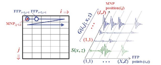
In order to overcome this unexpected effect, a method of reconstructing the exact spatial distribution of MNPs is proposed. Using the same method proposed by Weizenecker et al. (2009), the waveform generated from a MNP at each FFP is measured as a system function, but the correlation between this system function and a waveform generated by the unknown MNPs’ distribution at each FFP is calculated without any inverse matrix operation. More specifically, the estimation of the MNPs’ distribution is based on the correlation between the observed signal S(x, z) from the unknown MNPs’ distribution and the system function (i.e., point-spread function) G(i, j; x, z), which is a space-variant system determined by the interaction of the magnetic field and the MNPs’ distribution. As shown in Fig. 9, this system function can be determined by measuring the waveforms at FFP points of (x, z) when a MNP is set at each point (i, j) within the FOV, and by connecting all these measured waveforms as one-dimensional data, which is sequentially arranged in an array of rows and columns (i, j). Consequently, the MNP distribution in the x-z plane is reconstructed using equation (5).
Fig. 9. Calculation of correlation coefficients: the system function G(i, j; x, z) is defined as a space-variant system. It can be determined by measuring the waveforms at FFP points (x, z) when a MNP is set at each point (i, j) within the FOV. The MNP distribution is calculated by correlation with S(x, z), which is the waveform measured by each FFP of (x, z) and arranged in a one-dimensional array.
Numerical simulation
Simulation methods
In order to examine the validity of the proposed method using the higher harmonic components appropriately, a numerical analysis using the system model shown in Fig. 2 was conducted. In this examination, two Maxell coil pairs (diameter: 50 mm, opposite distance: 50 mm) and a receiver coil (diameter: 16 mm, number of turns: 200) were used, and the FOV was set as 9 x 9 mm2 with a matrix size of 64 x 32. A magnetic field distribution with a gradient magnetic field of about 5 T/m formed in the z direction at the MNPs with a particle diameter of 20 nm was applied as an FFP using this Maxell coil pair. In addition, an alternating magnetic field of 20 mT was applied in the same direction.
Simulation results
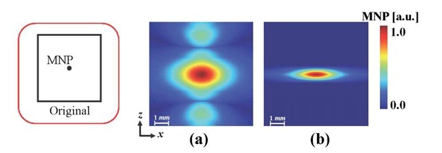
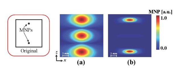
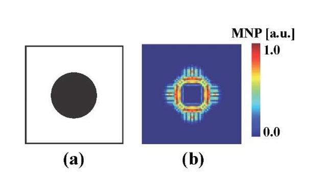
Figure 10 shows the reconstructed images of the MNP placed at the center of the FOV using the conventional method with equation (2) and the proposed method based on equation (3). The magnetic field was distributed over the region where the MNPs were positioned owing to the influence of the FFP formed as a result of the imperfection of the local magnetic field distribution. Therefore, because the alternating magnetic field was applied only in the z-direction, image blurring was observed particularly along the z-direction in the image reconstructed using the conventional method (Fig. 10(a)). On the other hand, such image blurring was reduced by suppressing the even harmonics on the basis of equation (3) and by using optimized parameters (Nh = 7, a = 0.19, ft = 0.12, k = 1.39) (Fig. 10(b)). Furthermore, it was confirmed that a drastic reduction in the image artifact compared to the conventional method could be achieved by using the proposed method shown in Fig. 11 by differentiating between the interference of the signal from the MNPs placed outside and inside the region on the basis of equation (4) using the optimized parameters (ca = 10). Although this proposed method enables us to improve image resolution by suppressing the interference signal due to the even harmonic components generated from the MNPs and also to improve the sensitivity by emphasizing the odd harmonic components, the outer part of the reconstructed image was weighted excessively, as in a high-pass-filter effect. For example, the perimeter region is emphasized when the MNPs indicate continuous distribution, as shown in Fig. 12 (a) while there are fewer blurring and artifacts compared with the conventional method shown in Figs. 10 and 11 when the MNPs are separated.
Fig. 10. Improvement of image resolution by adjusting the harmonic components based on equation (3). Overall image blurring due to the imperfection of the local magnetic field distribution formed as FFP is observed on the image reconstructed by the conventional method (a). The spread of the distribution along the z-direction was suppressed by using the proposed method with equation (3) (b).
Fig. 11. Improvement of image resolution by suppressing the interference signals on the basis of equation (4). When using the conventional method, an image artifact was formed at the FFP between the nanoparticles placed symmetrically on either side of the FFP (a). With the proposed method, this image artifact was eliminated, and the spread of the field distribution in the z-direction was suppressed on the basis of equation (4) (b).
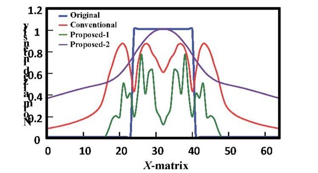
Consequently, the efficiency of the reconstruction method based on the newly proposed equation (5) was evaluated. Figure 13 illustrated the reconstructed results for the original image (Fig. 13(a)) using the conventional method (Fig. 13(b)), the method based on equations (3) and (4) (Fig. 13(c)), and the method based on equation (5) (Fig. 13(d)). With the conventional method, the reconstructed distribution was spread around the region where the MNPs were actually positioned, and an image artifact was observed. On the other hand, with the reconstruction method based on equation (5), although some image blurring is observed in the peripheral part of the image, a more exact reconstruction image with fewer artifacts is obtained, compared to other methods. Furthermore, it is confirmed from Fig. 14, which shows the profile of the central section of these images, that the sensitivity of this method based on equation (5) was high — about 20% compared with other methods.
Fig. 12. The reconstruction result for a continuous distribution of MNP. The outer part of the reconstructed image is excessively emphasized, as in the effect of a high pass image filter.
Fig. 13. Reconstruction results of each method. (a) Original image. (b) With the conventional method, significant image blurring and an artifact appear. (c) With the proposed method based on equations (3) and (4); although the image is less blurred, the edges are emphasized excessively. (d) With the proposed method based on equation (5); although the image is somewhat blurred, the original image is reconstructed more accurately than with the other methods.
Fig. 14. Profile of a reconstructed image. The proposed method based on equation (5) reconstructs the MNP distribution more accurately than the other methods. In addition, this method has excellent sensitivity.
Experiment
Materials and methods
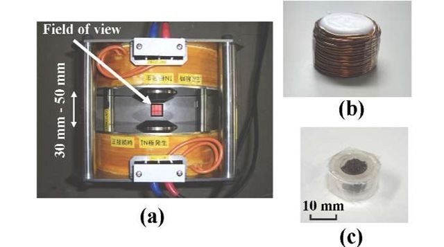
In order to confirm the validity of the proposed methods and numerical computation, the influence of the interference originating from the magnetization response waveform generated outside the target region was estimated by a fundamental experiment. The prototype Maxell coil pair (diameter: 180 mm, number of turns: 285 each, and opposite distance: 30-50 mm) (Toyojiki industry Co. Ltd., Niiza, Japan) used for the experiment is shown in Fig. 15.
Detecting magnetization response
When an alternating magnetic field was applied, the higher harmonic component contained in the magnetization response detected from the MNPs was evaluated, and the validity of the numerical analysis was confirmed. In this experiment, an alternating magnetic field with an amplitude of about 90 mT was generated in the center of the coil by applying an alternating current (frequency of 35 Hz, and amplitude 10.6 A) in the same direction to each coil. As a measuring object, a 2.0-g dry particle of iron oxide (nominal diameter of 10 nm), which has polar surface properties (EMG1500, Ferrotec Corp., Chiba, Japan)) was enclosed in the container, as shown in Fig. 15 (b).
The magnetization response waveform generated from the MNPs at the center of the Maxwell pair coil was detected using a receiver coil (diameter: 35 mm, number of turns: 40) which surrounds the phantoms. Here, in order to reduce a nonlinear error, intrinsic to the system, which originates from imperfections in the power supplies and the Maxell coil pair, difference processing between the signals observed with and without the existence of the measuring object was carried out.
Fig. 15. Experimental modules for signal detection. A Maxwell coil pair (diameter: 180 mm, turns: 285 each, distance between coils: 30-50 mm) was used in the z-direction for generation of an FFP and an alternating magnetic field. The magnetization response from the dry particle of iron oxide enclosed within the container (c) was detected by a receiver coil (diameter: 35 mm, turns: 40) (b).
Differentiating the waveforms obtained from inside and outside of the FFP
Next, it was evaluated by the experiment that the interference of the magnetization response signal generated from MNPs outside an FFP can be decreased with the proposed method based on equations (3) and (4). In this experiment, an alternating magnetic field with an amplitude of about 20 mT was generated in the center of the coil by applying an alternating current (frequency of 80 Hz, and amplitude 4.7 A) in the same direction to each coil. At the same time, a direct current of 9.4 A was applied to each coil in the opposite direction and a gradient magnetic field of about 1.2 T/m was generated, and as a result, an FFP was generated around the center of the coil gap.
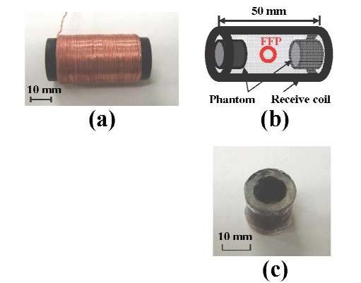
A 0.5-cc hydrophilic colloidal solution of superparamagnetic iron oxide (concentration about 500 mM/liter), which is coated with carboxydextran (Ferucarbotran, Fujifilm RI Pharma Co., Ltd., Tokyo, Japan), enclosed in the container shown in Fig. 16 (c) was used as a phantom. The magnetization response waveform generated from the MNPs at the center of the Maxwell coil pair was detected using a receiver coil (diameter: 35 mm, number of turns: 40) (Fig. 16 (a)) that surrounds the phantoms. In order to correspond to the numerical analysis (Fig. 11), the two phantoms were coaxially arranged, 20 mm apart, between the opposite coils (Fig. 16 (b)).
The FFP was set at the center between the coils, at an equal distance from the phantoms, and the magnetization response waveform generated from the phantoms was detected using the receiver coil (diameter: 23 mm, number of turns: 400) that surrounded the phantoms. The ideal waveforms (the system function) were determined by arranging a phantom at the center between the coils and measuring the signal, in order to calculate the correlation with the detected signal.
Fig. 16. Experimental modules for evaluation of the signal interference. The magnetization signal from the MNPs (MRI contrast agent: Ferucarbotran) (c), which are placed in two containers in order to reproduce the conditions of numerical analysis (b), was observed using a receiver coil (diameter: 23 mm, turns: 400) (a).
Experimental results
Detecting magnetization response
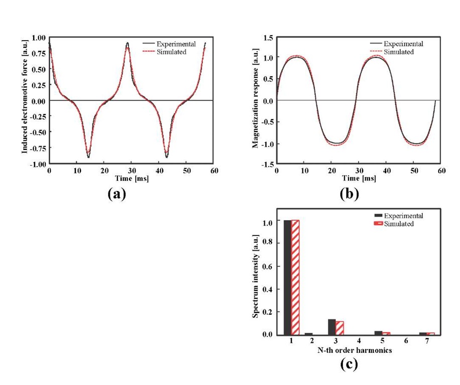
Figure 17 (a) shows the electromotive force induced by the receiver coil when applying the alternating magnetic field to the MNPs. By integrating over this electromotive force wave, the magnetization response waveform of the particles was obtained (Fig. 17 (b)) and is shown in Fig. 17 (c) with the result of the Fourier transform. These experimental results confirmed that the method of our numerical analysis is accurate.
Detecting magnetization response
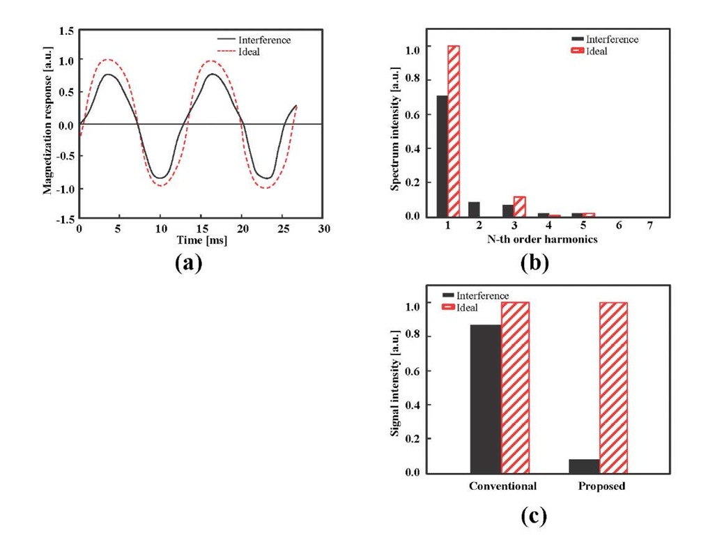
The magnetization response waveform detected by the receiver coil and the ideal waveforms are shown in Fig. 18 (a). The influence of interference is reflected in the detected magnetization response waveform, and a similar wave shape to that shown in Fig. 7 was observed. In order to confirm that the interference of the magnetization response waveform generated outside the FFP region can be suppressed by using the proposed method, the conventional method based on equation (2) and the proposed method based on equations (3) and (4) were applied to the magnetization response waveform obtained in the experiment. Here, the correction coefficients used in equations (3) and (4) were determined based on the characteristics of the ideal waveforms (Nh = 7, a = 0.10, p = 0.05, k = 1.22, Ca = 5.0). The signal strength reconstructed by each method is shown in Fig. 18 (c). For the conventional method, the signal strength, which reflects the interference of the magnetization response waveform generated from outside the FFP, (which corresponds to the interference signal detected at the center of the Maxwell coil pair) was about 79.0% of the ideal waveform signal. On the other hand, it was confirmed that this interference signal can be suppressed to about 9.0% by our proposed method based on equations (3) and (4). The results of this fundamental experiment were well in agreement with the results of the numerical analysis, confirming the efficiency of the proposed methods and the computational process.
Fig. 17. Detected signal from MNPs. The signal from the MNPs is detected as electromotive force induced by the receiver coil (a). A magnetization response is obtained by integrating this signal (b), and the harmonics are computed by the Fourier transform of this response (c).
Fig. 18. Suppression effect of interference signal. It is confirmed that by using the proposed method, the interference signal detected from the center of the Maxwell coil (a) can be suppressed to less than 9.0% (c).
Conclusion
In MPI, interference of the magnetization signal generated from the MNPs outside the boundary of an FFP due to the nonlinear responses, results in degradation of the signal sensitivity. Although we proposed an image reconstruction method that suppresses the interference component while emphasizing the signal component using the property of the higher harmonic components generated from the MNPs, the perimeter of the reconstructed image was over-emphasized due to the high-pass-filter effect when using this method. We therefore proposed a new method based on the correlation information between the observed signal and a system function, and performed a numerical analysis. As a result, the image blurring was still visible, but we clearly showed that the detection sensitivity can be improved without the inverse-matrix operation used by the conventional image reconstruction method. In addition, although such proposed methods and numerical analyses could be demonstrated by a basic experiment, the reconstruction of an image by means of a phantom experiment should be evaluated in the future.

