INTRODUCTION
Heart-related pathologies are among the most frequent health problems in western society. Symptoms that point towards cardiovascular diseases are usually diagnosed with angiographies, which allow the medical expert to observe the bloodflow in the coronary arteries and detect severe narrowing (stenosis). According to the severity, extension, and location of these narrowings, the expert pronounces a diagnosis, defines a treatment, and establishes a prognosis.
The current modus operandi is for clinical experts to observe the image sequences and take decisions on the basis of their empirical knowledge. Various techniques and segmentation strategies now aim at objectivizing this process by extracting quantitative and qualitative information from the angiographies.
BACKGROUND
Segmentation is the process that divides an image in its constituting parts or objects. In the present context, it consists in separating the pixels that compose the coronary tree from the remaining “background” pixels.
None of the currently applied segmentation methods is able to completely and perfectly extract the vascula-ture of the heart, because the images present complex morphologies and their background is inhomogeneous due to the presence of other anatomic elements and artifacts such as catheters.
The literature presents a wide array of coronary tree extraction methods: some apply pattern recognition techniques based on pure intensity, such as thresholding followed by an analysis of connected components, whereas others apply explicit vessel models to extract the vessel contours.
Depending on the quality and noise of the image, some segmentation methods may require image preprocessing prior to the segmentation algorithm; others may need postprocessing operations to eliminate the effects of a possible oversegmentation.
The techniques and algorithms for vascular segmentation could be categorized as follows (Kirbas, Quek, 2004):
1. Techniques for “pattern-matching” or pattern recognition
2. Techniques based on models
3. Techniques based on tracking
4. Techniques based on artificial intelligence
5. Main Focus
This section describes the main features of the most commonly accepted coronary tree segmentation techniques. These techniques automatically detect objects and their characteristics, which is an easy and immediate task for humans, but an extremely complex process for artificial computational systems.
Techniques Based on Pattern Recognition
The pattern recognition approaches can be classified into four major categories:
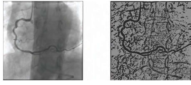
Figure 1. Regions growth applied to an angiography
Multiscale Methods
The multiscale method extracts the vessel method by means of images of varying resolutions. The main advantage of this technique resides in its high speed. Larger structures such as main arteries are extracted by segmenting low resolution images, whereas smaller structures are obtained through high resolution images.
Methods Based on Skeletons
The purpose of these methods is to obtain a skeleton of the coronary tree: a structure of smaller dimensions than the original that preserves the topological properties and the general shape of the detected object. Skeletons based on curves are generally used to reconstruct vascular structures (Nystrom, Sanniti di Baja & Svensson, 2001). Skeletonizing algorithms are also called “thinning algorithms”.
The first step of the process is to detect the central axis of the vessels or “centerline”. This axis is an imaginary line that follows each vessel in its central axis, i.e. two normal segments that cross the axis in opposite sense should present the same distance from the vessel’s edges. The total of these lines constitutes the skeleton of the coronary tree. The methods that are used to detect the central axes can be classified into three categories:
Methods Based on Crests
One of the first methods to segment angiographic images on the basis of crests was proposed by Guo and Richardson (Guo & Ritchardson, 1998). This method treats angiographies as topographic maps in which the detected crests constitute the central axes of the vessels.
The image is preprocessed by means of a median filter and smoothened with non-linear diffusion. The region of interest is then selected through thresholding, a process that eliminates the crests that do not correspond with the central axes. Finally, the candidate central axes are joined with curve relaxation techniques.
Methods Based on Regions Growth
Taking a known point as seed point, these techniques segment images through the incremental inclusion of pixels in a region on the basis of an a priori established criterion. There are two especially important criteria: similitude in the value, and spatial proximity (Jain, Kasturi & Schunck, 1995). It is established that pixels that are sufficiently near others with similar grey levels belong to the same object. The main disadvantage of this method is that it requires the intervention of the user to determine the seed points.
O’Brien and Ezquerra (O’Brien & Ezquerra, 1994) propose the automatic extraction of the coronary vessels in angiograms on the basis of temporary, spatial, and structural restrictions. The algorithm starts with a low-pass filter and the user’s definition of a seed point. The system then starts to extract the central axes by means of the “globe test” mechanism, after which the detected regions are entangled through the graph theory. The applied test also allows us to discard the regions that are detected incorrectly and do not belong to the vascular tree.
Methods Based on Differential Geometry
The methods that are based on differential geometry treat images as hypersurfaces and extract their features using curvature and surface crests. The points of hypersurface’s crest correspond to the central axis of the structure of a vessel. This method can be applied to bidimensional as well as tridimensional images; angiograms are bidimensional images and are therefore modelled as tridimensional hypersurfaces.
Examples of reconstructions can be found in Prinet et al (Prinet, Mona & Rocchisani, 1995), who treat the images as parametric surfaces and extract their features by means of surfaces and crests.
Correspondence Filters Methods
The correspondence filter approach convolutes the image with multiple correspondence filters so as to extract the regions of interest. The filters are designed to detect different sizes and orientations.
Poli and Valli (Poli, R & Valli, 1997) apply this technique with an algorithm that details a series of multiorientation linear filters that are obtained as linear combinations of Gaussian “kernels”. These filters are sensitive to different vessel widths and orientations.
Mao et al (Mao, Ruan, Bruno, Toumoulin, Col-lorec & Haigron, 1992) also use this type of filters in an algorithm based on visual perception models that affirm that the relevant parts of the objects in images with noise appear normally grouped.
Morphological Mathematical Methods
Mathematical morphology defines a series of operators that apply structural elements to the images so that their morphological features can be preserved and irrelevant elements eliminated. The main morphological operations are the following:
• Dilatation: Expands objects, fills up empty spaces, and connects disjunct regions.
• Erosion: Contracts objects, separates regions.
• Closure: Dilatation + Erosion.
• Opening: Erosion + Dilatation.
• “Top hat” transformation: Extracts the structures with a linear shape
• “Watershed” transformation: “Inundates” the image that is taken as a topographic map , and extracts the parts that are not “flooded”.
Eiho and Qian (Eiho & Qian, 1997) use a purely morphological approach to define an algorithm that consists of the following steps:
1. Application ofthe “top hat” operator to emphasize the vessels
2. Erosion to eliminate the areas that do not correspond to vessels
3. Extraction of the tree from a point provided by the user and on the basis of grey levels.
4. Slimming down of the tree
5. Extraction of edges through “watershed” transformation
MODEL-BASED TECHNIQUES
These approaches use explicit vessel models to extract the vascular tree. They can be divided into four categories: deformable models, parametric models, template correspondence models, and generalized cylinders.
Figure 2. Morphological operators applied to an angiography
Deformable Models
Strategies based on deformable models can be classified in terms of the work by McInerney and Terzopoulos (McInerney & Terzopoulos, 1997).
Algorithms that use deformable models (Merle, Finet, Lienard, & Magnin, 1997) are based on the progressive refining of an initial skeleton built with curves from a series of reference points:
• Root points: Starting points for the coronary tree.
• Bifurcation points: Points where a main branch divides into a secundary branch.
• End points: Points where a tree branch ends.
These points have to be marked manually.
Deformable Parametric Models: Active Contours
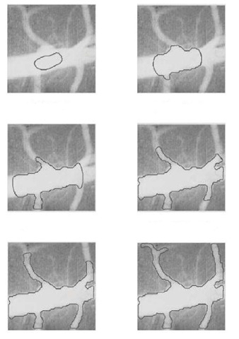
These models use a set of parametric curves that adjust to the object’s edges and are modified by both external forces, that foment deformation, and internal forces that resist change. The active contour models or “snakes” in particular are a special case of a more general technique that pretends to adjust deformable models by minimizing energy.Klein et al. (Klein, Lee & Amini, 1997) propose an algorithm that uses “snakes” for 4D reconstruction: they trace the position of each point of the central axis of a skeleton in a sequence of angiograms.
Deformable Geometric Models
These models are based on topographic models that are adapted for shape recognition. Malladi et al. (Malladi, Sethian & Vemuri, 1995) for instance adapt the “Level Set Method” (LSM) by representing an edge as a level zero set of a hypersurface of a superior order; the model evolves to reduce a metric defined by the restrictions of edges and curvature, but less rigidly than in the case of the “snakes”. This edge, which constitutes the zero level of the hypersurface, evolves by adjusting to the edges of the vessels, which is what we want to detect.
Propagation Methods
Quek and Kirbas (Quek & Kirbas, 2001) developed a system of wave propagation combined with a backtracking mechanism to extract the vessels from an-giographic images. This method basically labels each pixel according to its likeliness to belong to a vessel and then propagates a wave through the pixels that are labeled as belonging to the vessel; it is this wave that definitively extracts the vessels according to the local features it encounters.
Approaches based on the correspondence of de-formable templates:
This approach tries to recognize structural models (templates) in an image by using a template as context, i.e. as a priori model. This template is generally represented as a set of nodes connected by a segment. The initial structure is deformed until it adjusts optimally to the structures that were observed in the image.
Petrocelli et al. (Petrocelli, Manbeck, & Elion, 1993) describe a method based on deformable templates that also incorporates additional previous knowledge into the deformation process.
Parametric Models
These models are based on the a priori knowledge of the artery’s shape and are used to build models whose parameters depend on the profiles of the entire vessel; as such, they consider the global information of the artery instead of merely the local information. The value of these parameters is established after a learning process.
The literature shows the use of models with circular sections (Shmueli, Brody, & Macovski, 1983) and spiral sections (Pappas, & Lim, 1984), because various studies by Brown, B. G., (Bolson, Frimer, & Dodge, 1977) (Brown, Bolson, Frimer & Dodge, 1982) show that sections of healthy arteries tend to be circular and sections with stenosis are usually elliptical. However, both circular and elliptical shapes fail to approach irregular shapes caused by pathologies or bifurcations.
This model has been applied to the reconstruction of vascular structures with two angiograms (Pellot, Herment, Sigelle, Horain, Maitre & Peronneau, 1994), which is why both healthy and stenotic sections are modeled by means of ellipses. This model is subsequently deformed until it corresponds to the shape associated to the birth of a new branch or pathology.
Figure 3. “Snakes” applied to a blood vessel.
Generalized Cylinder Models
A generalized cylinder (GC) is a solid whose central axis is a 3D curve. Each point of that axis has a limited and closed section that is perpendicular to it. A CG is therefore defined in space by a spatial curve or axis and a function that defines the section in that axis. The section is usually an ellipse. Tecnically, GCs should be included in the parametric methods section, but the work that has been done in this field is so extense that it deserves its own category.
The construction of the coronary tree model requires one single view to build the 2D tree and estimate the sections. However, there is no information on the depth or the area of the sections, so a second projection will be required.
ARTERIAL TRACKING
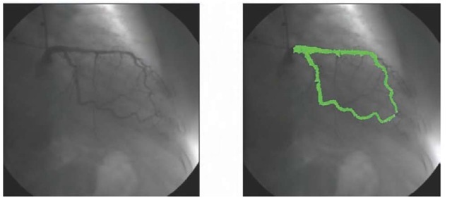
Contrary to the approaches based on pattern recognition, where local operators are applied to the entire image, techniques based on arterial follow-up are based on the application of local operators in an area that presumibly belongs to a vessel and that cover its length. From a given point of departure the operators detect the central axis and, by analyzing the pixels that are orthogonal to the tracking direction, the vessel’s edges. There are various methods to determine the central axis and the edges: some methods carry out a sequential tracking and incorporate connectivity information after a simple edge detection operation, other methods use this information to sequentially track the contours. There are also approaches based on the intensity of the crests, on fuzzy sets, or on the representation of graphs, where the purpose lies in finding the optimal road in the graph that represents the image.
Figure 4. Tracking applied to an angiography
Lu and Eiho (Lu, Eiho, 1993) have described a follow-up algorithm for the vascular edges in angiog-raphies that considers the inclusion of branches and consists of three steps:
1. Edge detection
2. Branch search
3. Tracking of sequential contours
The user must provide the point of departure, the direction, and the search range. The edge points are evaluated with a differential smoothening operator in a line that is perpendicular to the direction of the vessel. This operator also serves to detect the branches.
TECHNIQUES BASED ON ARTIFICIAL INTELLIGENCE
Approaches based on Artificial Intelligence use high-level knowledge to guide the segmentation and delineation of vascular structures and sometimes use different types of knowledge from various sources.
One possibility (Smets, Verbeeck, Suetens, & Oosterlinck, 1988) is to use rules that codify knowledge on the morphology of blood vessels; these rules are then used to formulate a hierarchy with which to create the model. This type of system does not offer any good results in arterial bifurcations or in arteries with occlusions.
Another approach (Stansfield, 1986) consists in formulating a rules-based Expert System to identify the arteries. During the first phase, the image is processed without making use of domain knowledge to extract segments of the vessels. It is only in the second phase that domain knowledge on cardiac anatomy and physiology is applied.
The latter approach is more robust than the former; but it presents the inconvencience of not combining all the segments into one vascular structure.
FUTURE TRENDS
It cannot be said that one technique has a more promising future than another, but the current tendency is to move away from the abovementioned classical segmentation algorithms towards 3D and even 4D reconstructions of the coronary tree.
Other lines of research focus on obtaining angio-graph images by means of new acquisition technologies such as Magnetic Resonance, Computarized High Speed Tomography, or two-armed angiograph devices that achieve two simultaneous projections in combination with the use of ultrasound intravascular devices. This type of acquisition simplifies the creation of tridimensional structures, either directly from the acquisition or after a simple processing of the bidi-mensional images.
KEY TERMS
Angiography: Image of blood vessels obtained by any possible procedure.
Artery: Each of the vessels that take the blood from the heart to the other bodyparts.
Computerized Tomography: Exploration of X-rays that produces detailed images of axial cuts of the body. A CT obtains many images by rotating around the body. A computer combines all these images into a final image that represents the bodycut like a slice.
Expert System: Computer or computer program that can give responses that are similar to those of an expert.
Segmentation: In computer vision, segmentation refers to the process of partitioning a digital image into multiple regions. The goal of segmentation is to simplify and/or change the representation of an image into something that is more meaningful and easier to analyze. Image segmentation is typically used to locate objects and boundaries (structures) in images, in this case, the coronary tree in digital angiography frames.
Stenosis: A stenosis is an abnormal narrowing in a blood vessel or other tubular organ or structure. A coronary artery that’s constricted or narrowed is called stenosed. Buildup of fat, cholesterol and other substances over time may clog the artery. Many heart attacks are caused by a complete blockage of a vessel in the heart, called a coronary artery.
Thresholding: A technique for the processing of digital images that consists in applying a certain property or operation to those pixels whose intensity value exceeds a defined threshold.


