Neuroendocrine theory
The neuroendocrine system, which consists of several endocrine glands under the control of the central nervous system, coordinates an animal’s physiology.
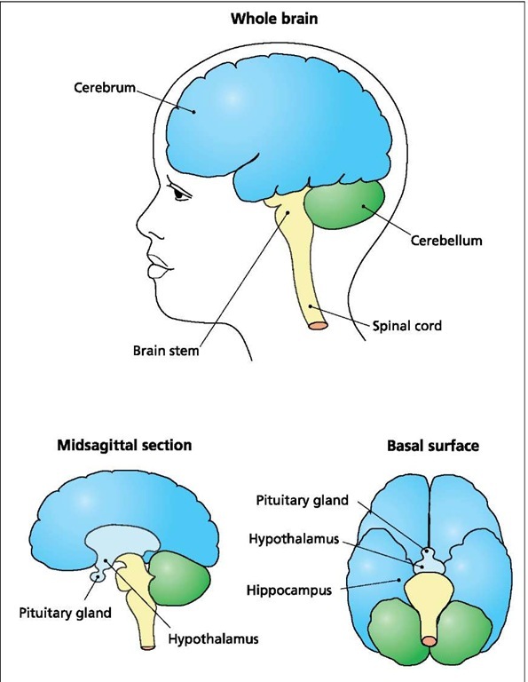
The human central nervous system. The human brain consists of the cerebrum, the cerebellum, and the brain stem, which is continuous with the spinal cord. The brain and spinal cord are called the central nervous system (CNS). The pituitary gland, a crucial part of the neuroendocrine system, is connected to the hypothalamus at the base of the brain (mid-sagittal section). The hippocampus, located on the basal surface of the brain, coordinates memory functions.
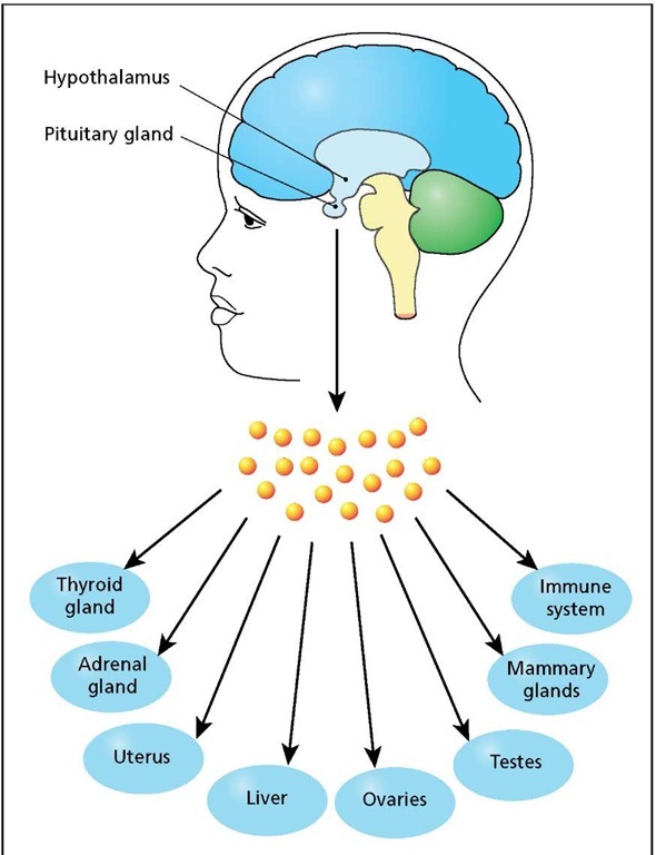
The human endocrine system is controlled by the hypothalamus, which regulates the production and release of various hormones from the pituitary gland. The pituitary hormones, in turn, regulate other glands, tissues, and organs of the body.
This system consists of a command center located in the hypothalamus; a master endocrine gland called the pituitary, which is connected directly to the hypothalamus; and a variety of secondary endocrine glands located in various parts of the body. The hypothalamus controls the pituitary by releasing hormone messengers that pass directly to the gland, where they stimulate or inhibit the release of pituitary hormones.
The pituitary gland, also known as the master gland of the vertebrate body, is located at the base of the brain and is about the size of a cashew nut. Despite its small size, this gland is in charge of producing all of the hormones that coordinate the many physiological processes occurring in animals and humans. There are 10 different kinds of pituitary cells, all constructed of cuboidal epithelium. Each cell type specializes in the synthesis and release of a different hormone, and is named after the hormone it produces. Thus, growth hormone-producing cells are known as GH cells or somatotrophs, and thyroid hormone-producing cells are known as thyrotrophs.
The cells that produce hormones that stimulate the gonads (ovaries and testes) to develop are known as gonadotrophs. Other pituitary cells produce hormones that stimulate the adrenal glands (ad-renocorticotrophs) and lactation (lactotrophs). The three remaining cell types are involved in the production of oxytocin, vassopressin, and melanocyte-stimulating hormone (MSH). Oxytocin is a hormone that causes milk ejection from the breasts and contraction of the uterus during birth. Vassopressin is a hormone that regulates salt and water balance by stimulating the kidneys to retain water. MSH stimulates melanin synthesis in human melanocytes and is very important in lower vertebrates, such as lizards and amphibians, where its release can stimulate a rapid change in skin color. The pituitary hormones, released into the blood, control the activity of other glands, such as the adrenal and thyroid glands, as well as organs such as the ovaries, testes and liver. All of the hypothalamic messengers and the pituitary hormones are small proteins. Overall, the system is responsible for regulating reproductive cycles, growth, energy metabolism, storage and mobilization of food molecules, and the fight-or-flight response.
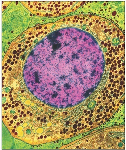
Hormone-producing cell (Somatotrope). Colored transmission electron micrograph of a growth hormone-producing cell from the pituitary gland. The pituitary gland is located at the base of the brain. Here a growth hormone-secreting endocrine cell, known as a somatotroph, is shown (large round). The hormone is in the numerous granules (brown) within the cell cytoplasm (yellow). Visible cell organelles include mitochondria (round, green) and the nucleus (purple, center) with its chromatin (pink). There are large amounts of rough endoplasmic reticulum (thin green) with the protein-synthesizing ribosomes (black dots). Magnification 1,000x.
The endocrine system is self-regulating, as illustrated by the control of the thyroid gland. The hypothalamus releases a messenger molecule called thyrotropin-releasing hormone, which stimulates the release of thyrotropin (also known as thyroid-stimulating hormone) from the pituitary. Thyrotropin, in turn, stimulates the thyroid gland to release thyroid hormones, the most important of which is thyroxin, a hormone that stimulates cell metabolism and growth. The self-regulating feature of this system is the ability of the hypothalamus to monitor the level of thyroxine in the blood. When it gets too high, the hypothalamus signals the pituitary to cut back on the release of thyrotropin, or to stop releasing it altogether.
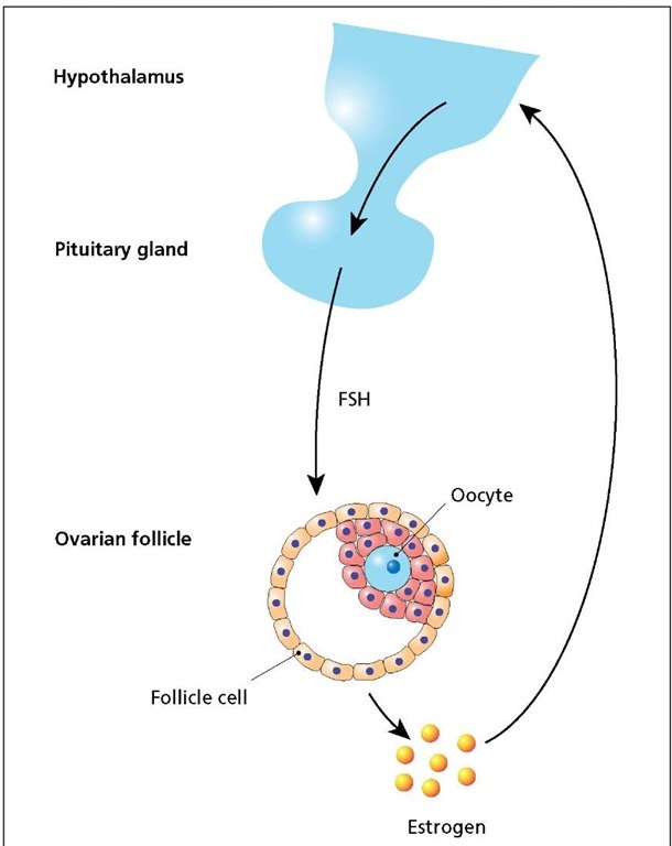
The regulation of the human female reproductive, or ovarian, cycle involves the same general scheme. In this case, the hypothala-mus releases a molecule called gonadotropin-releasing factor, which stimulates the pituitary to release follicle-stimulating hormone (FSH). FSH stimulates growth and development of ovarian follicles, each of which contain an oocyte. As the follicle cells mature, they synthesize and release the female hormone estrogen into the blood. The hypothalamus monitors the level of estrogen in the blood. Low levels of estrogen result in continuous release of FSH from the pituitary gland, but high levels, achieved when the follicle is mature, cause the hypothalamus to block release of FSH from the pituitary and, at the same time, to stimulate release of luteinizing hormone (LH) to trigger ovulation. If the mature oocyte is fertilized and successfully implants in the uterus, cells surrounding the embryo produce large amounts of estrogen to prepare the mother’s body for the pregnancy and to block further release of FSH. If the egg is not fertilized, estrogen levels drop, signaling the hypothalamus to stimulate renewed synthesis and release of FSH to complete the cycle. FSH also promotes development of sperm in the male.
Regulation of the thyroid gland. The hypothalamus instructs the pituitary gland to release thyrotropin, leading to secretion of thyroid hormones, which stimulate the activity of several organs. Thyroid hormone levels are monitored by the hypothalamus. When they get too high, thyrotropin release is reduced or stopped.
Regulation of the ovarian cycle. The hypothalamus instructs the pituitary gland to release follicle-stimulating hormone (FSH), promoting maturation of ovarian follicle cells, which in turn begin synthesizing and releasing estrogen. Low estrogen levels stimulate FSH release. High levels of estrogen inhibit the release of FSH but stimulate the release of a pituitary hormone (not shown) that initiates ovulation.
Hormones of the pituitary gland
|
HORMONE |
DESCRIPTION |
|
Adrenocortico-tropin (ACTH) |
This hormone stimulates release of adrenalin and other steroids from the adrenal cortex. Adrenalin is involved in the "flight-or-fight" response. ACTH is controlled by a hypothalamic messenger called corticotropin-releas-ing hormone. |
|
Antidiuretic hormone (ADH) |
ADH promotes water conservation by the kidneys. It is controlled by sensors that monitor the degree of body dehydration. |
|
Follicle stimulating hormone (FSH) |
This hormone promotes development of sperm in the male and oocyte follicles in the female. Its release is controlled by a hypothalamic messenger called FSH-releasing hormone. |
|
Growth hormone (GH) |
GH stimulates the uptake of glucose and amino acids by all tissues (except neurons). Its release is blocked by a hypothalamic messenger called GH-inhibiting hormone. |
|
Luteinizing hormone (LH) |
LH Stimulates synthesis of testosterone by the testes and ovulation in females. Its release is controlled by a hypothalamic messenger called LH-releasing hormone. |
|
Oxytocin |
Oxytocin stimulates uterine contractions during childbirth and the release of milk from mammary glands. Its release is stimulated by cervical distension and suckling. |
|
Prolactin |
Prolactin stimulates the growth of mammary glands and milk production. Its release is blocked by a hypothalamic messenger called prolactin-inhibiting hormone. |
|
Thyrotropin |
This hormone initiates the release of thyroid hormones from the thyroid gland. Thyroid hormones are growth factors that stimulate cellular activity and growth. The release of thyrotropin is controlled by a hypothalamic messenger called thyrotropin-releasing hormone (TRH). |
Given its breadth of influence, it is no wonder the endocrine system has captured the attention of gerontologists, many of whom believe that aging of the organism as a whole begins with the senescence of the hypothalamus. In this sense, the hypothalamus is like a clock that regulates the rate at which the individual grows older. With the age-related failure of the command center, hormonal levels of the body begin to change, and this in turn produces the physical symptoms of age.
One of the most dramatic age-related changes in humans is the loss of the ovarian cycle in females, generally referred to as the onset of menopause. Menopause usually occurs as women reach 50 years of age and is marked by a cessation in development of ovarian follicles, and as a consequence, a dramatic drop in estrogen levels. Estrogen, aside from its role in reproduction, is important to female physiology for the maintenance of secondary sexual characteristics, skin tone, and bone development. Female mice and rats also go through menopause, although in these animals it is called diestrus, or the cessation of the estrous cycle.
For gerontologists, the onset of menopause in mice and rats provides an experimental system that can be used to test the idea that the hypothalamus is an aging clock; that is, menopause or diestrus occurs because the hypothalamus stops releasing the necessary messenger molecules. When this happens, the reproductive system grinds to a halt. Many experiments were conducted in which pituitary glands or ovaries from old female rats were transplanted into young rats. In general, they showed that old pituitary glands functioned well in young bodies, and that old ovaries regained their estrus cycle. When prepubertal ovaries were transplanted to old female rats the majority of them fail to regain their cycles. Similarly, when young pituitaries are transplanted into old rats, they are usually unable to support a normal estrous cycle.
Additional evidence in support of the role of the hypothalamus in the aging process comes from the observation that the levels of several hormones gradually decrease with age. The overall effect of this change is believed to be the loss of vigor, physical strength, and endurance that is typical in an aging human. Accordingly, many attempts have been made to reverse these effects with hormone therapies that include GH, estrogen, or testosterone supplements. While these therapies have alleviated some of the symptoms of old age, they have not been able to reverse the aging process. With our limited knowledge of the cell and the complexities of human physiology and endocrinology, there are real dangers associated with hormone therapies. Estrogen supplements can minimize bone thinning in menopausal women, but constant exposure to this hormone can lead to breast cancer. Similarly, androgen supplements in men can increase vigor and physical strength, but constant exposure to testosterone is known to be a leading cause of prostate cancer. Growth hormone supplements suffer from similar problems in that they can induce cancers; they can also lead to the development of bone deformations.
Despite its great promise and the fact that it has generated some useful geriatric therapies, the hormonal disregulation or imbalance theory has failed to produce a definitive model of the aging process; nor have any of the hormonal therapies inspired by this theory been able to reverse the effects of age. Instead, the application of this theory, as with the other theories already discussed, merely allows a somewhat healthier old age, an effect that can also be obtained simply by eating well and getting lots of fresh air and exercise.
CONCLUDING REMARKS
With the exception of the age-related role of telomeres, all of the theories just described have been with us for more than 40 years, and during that time scientists have subjected those theories to thousands of experiments. The results have shown clearly that a disregulation of the endocrine system is a central feature of human aging, and that caloric restriction can increase the life span of several mammalian species, including the rhesus monkey. Molecular and genetic models of cellular senescence have been more difficult to pin down, but with the sequence of the human genome now available, the search for longevity genes is well under way, the results of which are expected to revolutionize our understanding of the aging process.


