External Ear
The external ear, also called the pinna or auricle, is the only readily visible part of the ear. It is composed mostly of cartilage and is shaped like a funnel to gather and guide sound waves into its small opening, which extends into a tube called the auditory canal. The opening into the auditory canal is called the external auditory meatus. The lining of the auditory canal is covered with tiny hairs and secretes a waxy substance called cerumen or ear wax. The hairs and wax aid in protecting the ear from foreign objects. The auditory canal is very short, approximately 1 inch (2.54 cm) in an adult, and extends to a thin membrane called the eardrum or tympanic membrane. The tympanic membrane separates the external from the middle ear. The external auditory meatus is angled differently in a child. This has implications for administration of ear drops.
Middle Ear
On the other side of the tympanic membrane is the middle ear, a small air-filled cavity in the temporal bone (see Fig. 21-3). Within this cavity are three tiny bones. The malleus (hammer) has a long process, the manubrium or handle, that is attached to the movable section of the eardrum. The incus (anvil) is the bridge between the malleus and the stapes. The stapes (stirrup) is the smallest bone in the body (0.1-0.3 inches; 2.5-3.3 mm). These three bones, collectively called ossicles, are so small that sound waves can set them in motion. The stapes transmit sound vibrations to the fluid-filled inner ear at the oval window, which separates the middle ear from the inner ear (see Fig. 21-4).
Two small muscles attach to the stapes and the malleus. These muscles reflexively contract at sudden, loud noises. The purpose of this reflex is to stop the vibration of the ossicles, thereby protecting the vital internal organs of the middle and inner ear from injury.
Extending from the middle ear are two openings. One leads into the mastoid cells behind it, and the other leads downward into the eustachian tube, or auditory tube, which communicates with the nasopharynx. To function properly, the pressure in the middle ear must be the same as external atmospheric pressure. The eustachian tube opens during swallowing or yawning. Its function is to equalize the pressure in the middle ear with atmospheric pressure; that is, to equalize the pressure on both sides of the tympanic membrane so that the drum vibrates freely. (If the tympanic membrane cannot vibrate freely, hearing is impaired.) For this reason, it is often suggested that people chew gum (to promote swallowing) or yawn frequently when flying—to equalize the pressures. If the pressures are not equal in flying or in activities such as deep-sea diving, the person will experience pain, which may be severe. A ruptured eardrum may also result. The eustachian tube also helps to drain the middle ear. The middle ear, the eustachian tube, the nasopharynx, and the passage to the mastoid cells are lined with a continuous coating of mucous membrane.
Special Considerations :LIFESPAN
Eustachian Tube
In infants and in children, the eustachian tube is shorter, wider, and positioned at a different angle than in adults. This difference predisposes children to inner ear infections because it is easier for pathogens to migrate into the ear The child’s eustachian tubes can also be blocked more easily than in adults by allergies, enlarged adenoids, or inflammation of the nose and throat.
Polyethylene (PE) Tubes
If the eustachian tubes are chronically blocked, an opening into the eardrum (myringotomy) may be required. A tiny PE tube may be inserted to help drain fluid and to allow air to enter the middle ear and equalize pressure.
Inner Ear
The inner ear is embedded in the temporal bone, the densest bone in the body. Here, the bony labyrinth is filled with a set of tunnels and chambers called the membranous labyrinth (or just labyrinth). Both the bony labyrinth and the membranous labyrinth are filled with a fluid similar to cerebrospinal fluid, called endolymph. The labyrinths are important in the transmission of sound waves and in the determination of balance and positional changes in the body. The three sections of the inner ear are the cochlea, vestibule, and semicircular canals (see Fig. 21-3).
Cochlea. The cochlea is shaped like a hollow snail shell. Inside the bony cochlea lies the organ of Corti, which is very small but very intricate. It is approximately 1.5 inches (3.75 cm) long, and contains approximately 7,500 separate parts. This organ is referred to as the “true organ of hearing” because the transmission of nerve stimuli related to sound begins here. Vestibule. The vestibule, the area between the cochlea and the semicircular canals, contains two membranous sacs called the utricle and the saccule. The utricle and saccule have specialized areas of hair cells that send sensory information to the cerebellum and the midbrain. These areas of the brain relay changes in body position when at rest and during sudden movement (acceleration and deceleration). Semicircular Canals. Another section of the inner ear includes the semicircular canals. Shaped like horseshoes, they lie behind the cochlea. They too contain hair-like nerve endings that are set in motion by fluid within the canals. However, instead of transmitting sound, they transmit information about the body’s position to the brain. Head or body movements cause motion in this fluid. If you look at the semicircular canals in Figure 21-3, you will note that each is on a different plane, or angle, in space. Therefore, the fluid (endolymph) in these semicircular canals flows in various directions and picks up more types of motion than would be the case if all canals were on the same plane. The sensory receptors for equilibrium (balance) while the body is moving are located in these canals. These sensations are interpreted in the brain.
Nerves
Sound is conducted via the pinna and the middle ear to the inner ear’s auditory nerve fibers. The nerve endings that are the receptors for hearing are located within the membranous cochlea in the organ of Corti. These hair-like receptors connect to the cochlear nerve, which becomes a division of the acoustic or auditory nerve (cranial nerve VIII). Sound is conducted to the center for hearing, which is located in the temporal lobe of the brain’s cerebral cortex. The acoustic nerve is actually a combination of two divisions, the cochlear nerve (for transmission of sound) and the vestibular nerve (for transmission of signals relating to balance and position). For this reason, cranial nerve VIII is sometimes called the vestibulocochlear nerve.
System Physiology vision
Vision depends on four factors:
• Size of object
• Brightness (luminance): intensity of light and amount of reflection
• Contrast between object and background
• Speed or time allowed to see object (more difficult to see a fast-moving object)
A person’s two eyes register separate images. The visual areas of the cerebral cortex fuse these separate images into a single image with a three-dimensional effect, called binocular vision. This binocular vision is responsible for depth perception and is made possible because the coordinated muscles of both eyes move the eyeballs in tandem.
Key Concept The vestibulo-ocular reflex (VOR) stabilizes images on the retina when the head moves. Thus, when turning the head, the eyes move in the opposite direction, keeping the image in the center of the field of vision.
A phenomenon called smooth-pursuit movement (SPM) allows the eyes to follow a moving object. This usually requires conscious effort on the part of the person.
Vergence movement in binocular vision causes the eyes to converge (move toward each other) when viewing a close object or diverge for far away objects. This is closely related to accommodation.
Light rays enter the eye through the cornea and pass through the aqueous humor, the pupil opening, the lens, and the vitreous humor before coming into focus on the retina (see Fig. 21-2). Refraction (bending of light rays) occurs within these components.
Key Concept When light rays converge on the retina, the image is upside-down. In the visual cortex of the brain, the image is corrected and perceived as upright.
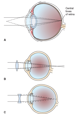
Within the lens, parallel light rays are refracted (bent) so that they focus directly on the retina (Fig. 21-5). This provides a clear image. The ciliary muscles of the eye that cause pupillary constriction or accommodation also play a role in clear image formation. As the eye moves, the exposure is adjusted; this adjustment is performed by the iris and is also the result of a chemical reaction. The formed image is reflected on the retina, the nerve center of the eye. The central fovea (fovea centralis) is a special area on the retina that is responsible for the sharpest vision. The goal of perfect vision is to have the light rays fall precisely on this central fovea. The lens plays the major role in adjusting light rays to facilitate the projection of rays exactly on this retinal area. This adjustment by the lens to make a sharp, clear image is called accommodation; it is controlled by the autonomic nervous system. If the eyeball is too short, the focal point of the light rays is too long and they land behind—instead of directly on—the retina, causing farsightedness (known as hyperopia or hypermetropia). In this instance, the refracted light rays do not come together directly on the retina, but are absorbed by the retina. Therefore, the farsighted person cannot see close objects clearly.
In nearsightedness (myopia), the light rays are focused in front of the retina (the focal length of the rays is too short). Myopia occurs because the muscles of the lens contract too tightly, not allowing enough light to enter the eye, or because the eyeball is too long in relation to the muscles of the lens. The nearsighted person sees close objects clearly, but distant objects are blurry.
FIGURE 21-5 · (A) Accommodation. The solid lines represent rays of light from a distant object, and the dotted lines represent rays from a near object. The lens is flatter for the former, and more convex for the latter;In each case, the rays of light are brought to a focus on the retina.(B) Hyperopia corrected by a biconvex lens, as shown by the dotted lines.(C) Myopia corrected by a biconcave lens, as shown by the dotted lines. In each of these illustrations, the goal is to focus the light rays exactly at the central fovea.
Astigmatism is a condition in which the eye cannot bring horizontal and vertical lines into focus at the same time, causing blurry vision. This condition is a result of irregularities in the curvature of the cornea and lens.
Key Concept Farsightedness is called hyperopia and it is caused when the focal point of light rays falls behind, instead of on, the retina. Nearsightedness is called myopia and it occurs when light rays focus in front of the retina.
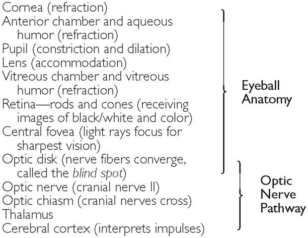
The retina’s receptors send the visual impulses through the nerve fibers to the optic nerve (cranial nerve II). The optic nerve meets the retina at the optic disk (see Fig. 21-2). (This optic disk is called the “blind spot” because it is the region of the eye that is not light sensitive.) The optic nerve then carries the information to the cerebral cortex of the brain’s occipital lobe for interpretation. Box 21-2 summarizes the pathway of light transmission.
BOX 21-2. Light Transmission Through the Eye to the Brain
Key Concept Many vision problems can be corrected with prescription eyeglass lenses, contact lenses, or surgery including laser eye surgery (LASIK).
HEARING
Sound is a form of energy that moves in waves of pressure; these sound waves are perceived by the brain via firing of nerve cells in the auditory system. The perception of sound is called audition; hearing is measured by an audiometer. Sound waves enter through the ear’s external auditory canal and strike the tympanic membrane (eardrum). The tympanic membrane vibrates at various speeds in response to various pitches of sounds. The ossicles within the middle ear are in contact with each other and act as a movable bridge to transmit these vibrations to the oval window.
Because the oval window is so much smaller than the tympanic membrane, sound is concentrated onto a smaller space, thereby amplifying the sound waves (the hydraulic principle). The base of the stapes fits into the oval window. When stimulated, the stapes vibrates against this membrane, setting the fluid of the cochlea of the inner ear in motion. The wave-like action of this fluid passes the vibrations onto the tiny hair-like nerve endings (receptors) in the organ of Corti. This organ contains microscopic protein filaments (“hair cells”) which are mechanoreceptors. They release a chemical neurotransmitter when stimulated. (The sound waves push the filaments over, causing them to fire.) The stimuli from the nerve endings in the organ of Corti are sent to the vestibulocochlear nerve (cranial nerve VIII) and then to the temporal lobe in the cerebral cortex, where the sounds are interpreted.
Sound Amplification
Figure 21-4 shows the pathway of sound transmission.
Sound waves are amplified in three ways:
• The ear canal is open, and the resonance there approximately doubles the sound waves.
• The ossicles (hammer, anvil, and stirrups) act as levers. This mechanical advantage amplifies the sound approximately threefold. (This, when multiplied by the amplification in the ear canal, now equals a total of a sixfold amplification.)
• The relative sizes between the eardrum and the oval window amplify or increase the sound waves approximately 30 times more, as depicted here:
Hearing can be damaged by extremely loud sounds or music (noise trauma), by certain drugs (ototoxicity), blast injuries, foreign objects, or specific illnesses. The ensuing hearing loss can be temporary or permanent.
Key Concept A process of localizing the direction from which a sound originates is called the "Echo Positioning System” (EPS). The central nervous system analyzes the sound from each ear to locate the source of the sound.
BALANCE AND EQUILIBRIUM
The sense of static balance (when the person is at rest) is centered in structures called the utricle and saccule of the inner ear. Balance with movement is associated with the semicircular canals. Cranial nerve VIII (vestibulocochlear nerve) has an important role in both balance and hearing. This cranial nerve transmits impulses to other cranial nerves that are responsible for the control of head and neck movement. This nerve also sends and receives information (about the body’s position at rest and during movement) from structures in the inner ear, including the semicircular canals, in order for the body to maintain positional balance and equilibrium.
Balance is a complex process. In addition to the receptors in the ears, proprioceptors exist in the muscles. Visual input and tactile skin receptors also contribute to our sense of position in space. Sometimes, the motion of fluid in the ears without accompanying visual reference can confuse the sensory system. For example, think of the sensation you feel on a carnival ride if you close your eyes. This dizzy—sometimes ill— feeling or sense of being rotated is called vertigo. Vertigo does not have the same meaning as dizziness. True vertigo is a sensation that either you or the room is spinning. Dizziness is more a sensation of light-headedness. Diseases of the inner ear or defects in the conductive pathways and central nervous system cause true vertigo. Nausea often accompanies vertigo, as does tinnitus, a high-pitched buzzing sound or “ringing in the ears.”
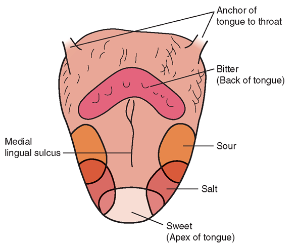
FIGURE 21-6 · The dorsal surface of the tongue, viewed from above, showing the location of specific taste buds.


