Sympathetic Division
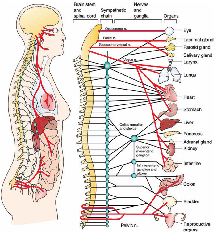
The sympathetic division of the ANS produces a response that prepares individuals for an emergency, extreme stress, or danger. This “fight or flight” response readies people to defend themselves or to flee from danger. During an emergency, the heart beats faster and the breathing rate increases. The skin becomes pale, secondary to diversion of blood flow away from the skin. Blood flow also decreases to structures such as the external genitalia and abdominal organs. Thus, body processes such as digestion slow or stop, allowing more blood to flow to more vital organs: the brain, lungs, and large muscles that move the body during an emergency. Involuntary defecation or urination can occur. Obviously, the body can sustain an emergency awareness for only a limited time. The homeostatic mechanism that balances the sympathetic nervous system (SNS) is the parasympathetic nervous system.
FIGURE 19-10 · Anatomy ofthe autonomic nervous system. The red lines represent the parasympathetic nervous system (craniosacral division). The black lines represent the sympathetic nervous system (thoracolumbar division). (The oculomotor, facial, glossopharyngeal, and vagus nerves are cranial nerves, shown here to illustrate their autonomic functions.)
TABLE 19-3. Effects of the Sympathetic and Parasympathetic Systems on Selected Organs
|
EFFECTOR |
SYMPATHETIC SYSTEM |
PARASYMPATHETIC SYSTEM |
|
Pupils of eye |
Dilation |
Constriction |
|
Sweat glands |
Stimulation |
None |
|
Digestive glands |
Inhibition |
Stimulation |
|
Heart |
Increased rate and strength of beat |
Decreased rate and strength of beat |
|
Bronchi of lungs |
Dilation |
Constriction |
|
Muscles of digestive system |
Decreased contraction |
Increased contraction (peristalsis) |
|
Kidneys |
Decreased activity |
None |
|
Urinary bladder |
Relaxation |
Contraction and voluntary emptying |
|
Liver |
Increased release of glucose |
None |
|
Penis |
Ejaculation or relaxation |
Erection |
|
Adrenal medulla |
Stimulation |
None |
|
Blood vessels to |
||
|
Skeletal muscles |
Dilation |
Constriction |
|
Skin |
Constriction |
None |
|
Respiratory system |
Dilation |
Constriction |
|
Digestive organs |
Constriction |
Dilation |
Parasympathetic Division
The parasympathetic division of the ANS is involved in relaxation. The parasympathetic division generally produces responses that are normal functions of the body while at rest or not under unusual or extreme stress. The effects are usually opposite to the effects of the sympathetic division. Unlike the sympathetic division, however, the parasympathetic system does not normally activate in a way that affects the body overall. For example, it can decrease the body’s heart rate without affecting other organs. To return to homeostasis after a “fight or flight” episode, the parasympathetic nerves return the heart rate to normal, resume digestive processes, and restore blood flow to the skin, abdominal organs, and genitalia. Previously normal patterns of defecation and urination return.
Key Concept The ANS functions independently without conscious effort, to innervate cardiac muscle, and smooth visceral muscle and glands. It has two divisions: the sympathetic and parasympathetic. The sympathetic division prepares the body for "fight or flight.” It acts "in sympathy” with the body in an emergency The parasympathetic division maintains normal body functions and returns a person’s body back to normal after a stressful situation. These opposing reactions are called "antagonism” and they operate to maintain homeostasis.
System Physiology
TRANSMISSION OF NERVE IMPULSES
Messages from one part of the body to another can take several possible nerve pathways. The body is thrifty in its use of its resources and in patterns of automatic activities. As a rule, it uses the quickest route to send a message. The body builds patterns (reflexes) and habits by using the same nerve pathways repeatedly. The same kind of message tends to follow the same path every time. Repeated motions become more or less automatic.
Resting Potential and Action Potential
Electrical and chemical influences make it possible for nerve impulses to occur. An unequal distribution of ions (electrically charged particles) exists between the inside and outside of the neuron’s cellular membrane. Outside the membrane are sodium (Na+) and some potassium (K+) ions. Inside the nerve cell are a number of negatively charged ions. This charge difference is called the resting potential.
A stimulus, or nerve impulse, causes an organized, rapid shift of sodium and potassium ions across the cell membrane (called “firing,” as in firing a gun), instantly reversing the polarity (ionization or electrical charge) of the cell. (Sodium crosses first, then potassium.) The impulse spreads like an electric current along the membrane, beginning at one spot and spreading along the length of the nerve cell. This electrical charge can actually be measured in millivolts.The changed polarity of the neuron’s plasma membrane and the electrical impulse generated by ion movement is called the action potential. An action potential takes only milliseconds (tiny fractions of a second). As a result, many neurons can transmit impulses at over several meters per second.
After this, there is a refractory period, during which the membrane cannot be stimulated. (This prevents nerve impulses from going backward.) The potassium channels open to allow the potassium to again pass to the outside of the membrane. This reversal of the cell’s polarity, back to its original state, occurs instantly (within 0.1-1.0 millisecond [a millisecond is 1/1000 second]). Therefore, as quickly as polarity occurs, it reverses itself to bring the cell back to its resting state. This alternating depolarization and repolarization moves as a wave along the length of the neuron and becomes a transmitted message.
Key Concept Nerve cell at rest =resting potential (plasma membrane more positive on outside).
Nerve stimulus—Sodium moves inside membrane, followed by potassium = action potential (membrane now more positive on inside) S nerve impulse.
Potassium ions flow out of nerve cell, followed by sodium = refractory period (resting potential restored).
Neurotransmitters
For nerve impulses to cross the synaptic cleft, neurotransmitters are required. There are more than 30 known neurotransmitters. These chemicals are stored in small synaptic or neurotransmitter vesicles, clustered at the tip of the axon (see Fig. 19-3). When an action potential arrives at a neuron, some of the vesicles move to the end of the axon and discharge their neurotransmitters into the synaptic cleft. These substances diffuse across the cleft and bind to receptors on the next cell’s membrane, causing ion channels to open and enabling the desired action. This allows nerve impulses to cross the synapse and transmit an impulse, which either excites or inhibits the target cell. After the action, neurotransmitters are immediately destroyed by enzymes in the synaptic cleft, diffuse out, or are reabsorbed by the cell, preventing a continuous impulse.
An example of a neurotransmitter is acetylcholine, which is inactivated by an enzyme called cholinesterase. Dopamine, norepinephrine, and serotonin, as well as specific hormones, are also neurotransmitters. These names may be familiar to you because many of their properties have been developed into useful pharmacological products.
Key Concept Parkinson’s disease is caused by a deficiency of the neurotransmitter; dopamine.
Many drugs, such as cocaine, LSD, caffeine, and insecticides, are known to interfere with conduction of impulses across synapses.
The All-or-None Law
Either a nerve impulse is transmitted across a particular synapse or it is not—with no exceptions. Because an impulse cannot be partially transmitted, this law is known as the all-or-none response of nerve tissue.
Electroencephalogram
An electroencephalogram (EEG) is a visual record of the electrical wave activity of the millions of neurons in the brain. Brain wave activity helps diagnose neurologic problems. In many states, cessation of brain wave activity has become one important legal consideration in the confirmation of biological death.
Actions of Three Types of Neurons
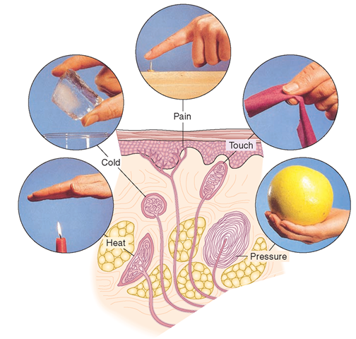
Sensory Neurons. The sensory neurons (neurons of sensation) are also known as afferent neurons because they carry impulses to the brain or spinal cord from the periphery of the body by means of receptors, end organs that initially receive stimuli from outside or within the body (Fig. 19-11).
Receptors are classified in three ways. Exteroceptors (related to the external environment) are involved in touch, cutaneous (skin) pain, heat, cold, smell, vision, and hearing. Proprioceptors carry sensations of position and balance, or placement of the body in space. Interoceptors (related to the body’s internal environment) respond to changes in the internal organs (viscera), such as visceral pain, hunger, or thirst.
After the receptors receive an impulse, fibers of sensory neurons carry the sensation to the CNS. Here, interneurons analyze and distribute the impulses. Some interneurons act as integrators, and the impulse is carried directly to a motor neuron.
Motor Neurons. If an action is required after receiving a stimulus, the CNS sends an impulse via motor neurons to a muscle or gland to cause the proper response. Motor neurons are also called efferent neurons because they carry impulses away from the CNS. The structures that carry out activity are called effectors. Effector neurons are classified as somatic-voluntary or visceral-involuntary.
To understand the difference between the types of effector neurons, consider an insect bite. A sensory (afferent) neuron makes the initial sting known to the brain. The brain interprets the sensation and sends a message via somatic-voluntary (efferent) neurons to appropriate muscles. A person manifests a response by slapping the insect. This action is voluntary, in that it can be controlled. An example of a visceral-involuntary response is the peristaltic movement of food through the digestive system. Such movement is not under a person’s voluntary control; rather, it depends on the nervous system’s ability to process information and relay appropriate messages.
Interneurons. Neurons that integrate signals between neurons of various parts of the CNS are called interneurons. Interneurons are found only in the CNS (see Fig. 19-7B).
FIGURE 19-11 · The receptors (exterocep-tors) in the skin receive stimuli related to temperature, pain, touch, and pressure. These neurons are sensory (afferent) neurons, which carry stimuli via the sensory pathways to the central nervous system for interpretation.
They carry out or integrate sensory or motor impulses. They assist with thinking, learning, and memory. They can be thought of as a link between sensory and motor neurons.


