Accessory Structures
The three major accessory structures of the central nervous system are the meninges, cerebrospinal fluid, and ventricles.
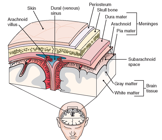
Meninges. The brain and spinal cord (the CNS) are covered with three protective membranes called meninges (Fig.19-8). The dura mater, the outer layer, is a tough, fibrous covering that adheres to the bones of the skull. The middle layer is a delicate web of tissue called the arachnoid membrane. The pia mater, the inner layer, lies closely over the brain and spinal cord. It is thin and vascular, containing many blood vessels that bring oxygen and nutrients to the nervous tissue. The space between the arachnoid membrane and the pia mater is the subarachnoid space. The subarachnoid space contains cerebrospinal fluid, the tissue fluid of the CNS.
Key Concept Closed head trauma (CHT)can result in intracranial (within the skull) bleeding, with blood accumulating under specific meninges: under the dura mater (subdural) or under the arachnoid (subarachnoid). This causes increased and potentially dangerous intracranial pressure. Disorders can also result when the brain is smashed against the bones of the skull from a blow to the head. Some disorders are caused by an imbalance of neurotransmitters (e.g., Parkinson’s disease, a dopamine deficiency). Alzheimer’s dementia indicates an accumulation of protein plaques in the brain. Various drugs, such as cocaine, heroin, marijuana, and alcohol, block or enhance specific neurotransmitters, causing related dysfunction or disorders.
FIGURE 19-8 · Frontal (coronal) section of the top of the head, showing meninges and related structures.
BOX 19-2.
Functions of Cerebrospinal Fluid
♦ Acts as shock absorber for the brain and spinal cord
♦ Carries nutrients to the brain
♦ Carries wastes away from the brain
♦ Keeps the brain and spinal cord moist, thus preventing friction
♦ Can be tested to determine the presence of some disorders
♦ Can be used to transmit medications
Cerebrospinal Fluid. Cerebrospinal fluid (CSF) is a lymph-like fluid that forms a protective cushion around and within the CNS (Box 19-2). CSF allows the brain to “float” within the cranial vault, which changes the effective weight of the brain from about 1,500 g (grams) to about 50 g. The CSF removes cellular waste from nerve tissue and lessens damage caused on impact by spreading out the force of trauma. It is produced in specialized capillary networks (the choroid plexuses) in the ventricles of the brain. About 800 mL of CSF are produced each day, although only 150 to 200 mL circulate at any one time. Arachnoid villi reabsorb some CSF into the blood, where it becomes blood plasma again.
Nursing Alert If a client sustains head trauma and fluid is noted leaking from the ears or nose, this fluid should be tested for glucose, which is present in CSF.
If the glucose test is positive, notify a healthcare provider immediately because leaking CSF usually indicates a life-threatening situation.
A physician may withdraw a small amount of CSF through the space between two vertebrae. This procedure is called a lumbar puncture (LP) or spinal tap. Laboratory studies of CSF can reveal bleeding into the CNS and infections of the brain or its meninges. A lumbar puncture can also introduce medications such as antibiotics or anesthetics into the CSF and can measure CSF pressure, which must be maintained within very close tolerances.
Nursing Alert The pressure of the CSF reflects the pressure of the fluid in and around the brain (intracranial pressure [ICP]). Increased ICP can be a sign of a serious disorder, such as bleeding within the brain, brain tumors, swelling of the brain as a result of trauma, hydrocephalus, or an infection within the CNS.
Ventricles. Deep within the brain are four ventricles, or cavities. These are lined with ependymal cells. They also contain many blood vessels from the pia mater, which make up the choroid plexuses. The choroid plexuses produce CSF, which fills the ventricles.
Nursing Alert The skull is a rigid container that contains brain tissue, CSF, and blood. The volume of these components determines ICR A large increase in any of these factors can increase ICR This can cause brain hypoxia (oxygen deprivation), herniation of the brain (brain contents being pushed through an opening), necrosis (death) of brain tissue in a specific area, or death of the individual.
Peripheral Nervous System
The peripheral nervous system (PNS) consists of bundles of neurons that connect the brain and spinal cord (the CNS) to the rest of the body. The PNS is made up of two nerve groups: cranial nerves and spinal nerves. The terms cranial and spinal indicate that the nerves begin either in the brain or in the spinal cord. The nerves of the PNS are sensory, motor, or mixed.
The afferent (sensory) division of the PNS conveys information to the brain, primarily from sensory organs, such as the skin. Muscle spindles convey information regarding posture and joint position. The sense of proprioception conveys awareness of where parts of the body are, in relation to space. For example, if you close your eyes and wave your hand, you still know where your hand is located. Several areas of the brain (the cerebellum and red nucleus) coordinate movements and positioning, using this proprioceptive feedback. Deeper,muscles, such as those controlling posture, are controlled by nuclei in the brainstem and basal ganglia. (Much of the sensory input of the PNS remains below conscious awareness. However, if you think about it, some of the sensory input can be brought into consciousness.)
The efferent (motor) division of the PNS sends voluntary and involuntary commands from the CNS to muscles and stimulates glands to secrete hormones. Signals to muscles nearly always originate in the primary motor cortex. This is located just in front of the central sulcus, dividing the frontal and parietal lobes of the brain.
Mixed nerves allow signals to pass both to and from the CNS.
Reflexes, both afferent and efferent, are directly managed at the spinal cord level.
The autonomic nervous system and its subdivisions (discussed later in this section) are also classified as part of the PNS.
Cranial Nerves
The 12 pairs of cranial nerves attach directly to the brain. Many cranial nerves are mixed; that is, they carry impulses both to and from the brain and various structures around the head. One pair (cranial nerve X), however, acts on the organs of the thorax and the abdomen. Cranial nerves are given Roman numerals and numbered in the order in which they originate in the brain (from front to back). Table 19-2 lists cranial nerves and their functions.
TABLE 19-2. The Cranial Nerves and Their Functions
|
NUMBER |
NAME |
MAIN FUNCTION |
DISTRIBUTION |
|
I |
Olfactory |
Smell (Sensory) |
Nasal mucous membrane |
|
Optic |
Vision (Sensory) |
Retina |
|
|
III |
Oculomotor |
Eye movements (Motor) |
Most ocular muscles |
|
IV |
Trochlear (smallest cranial nerves) |
Voluntary eye movements (Motor) |
Superior oblique muscle of eye |
|
V |
Trigeminal (largest cranial nerves) |
Sensations of head and face; movement of mandible (Both) |
Skin of face; tongue; teeth; muscles of mastication (chewing) |
|
Ophthalmic branch |
Sensations from front of head and face, eye sockets, and upper nose (Sensory) |
||
|
Maxillary branch |
Sensations from nose, mouth, upper jaw, cheek, and upper lip (Sensory) |
||
|
Mandibular branch |
Sensations of tongue, lower teeth, chin, clenched teeth (Both) |
||
|
VI |
Abducent (Abducens) |
Eye movements (Motor) |
Lateral rectus muscle of eye |
|
VII |
Facial |
Taste; facial expressions (Both) |
Muscles of expression; taste buds |
|
VIII |
Vestibulocochlear (Acoustic) Cochlear division |
Hearing and balance Conduct impulses related to hearing (Sensory) |
Internal auditory meatus |
|
Vestibular division |
Conduct impulses related to equilibrium (balance) (Sensory) |
Inner ear |
|
|
IX |
Glossopharyngeal |
Controls swallowing; gives information on pressure and oxygen tension of blood (Both) |
Rharynx, posterior third of tongue, parotid |
|
X |
Vagus ("wanderer”) (the only cranial nerve not restricted to head and neck) |
Somatic motor function; parasympathetic functions; speech (Both) |
Rharynx, larynx, heart, lungs, esophagus, stomach, abdominal viscera |
|
XI |
Accessory (Spinal accessory) |
Rotation of head; raising of shoulder (Motor) |
Arising from medulla and spinal cord |
|
XII |
Hypoglossal |
Movement of tongue (Motor) |
Intrinsic muscle of tongue |
BOX 19-3.
Mnemonic for Names of Cranial Nerves
|
On |
I Olfactory |
|
Old |
II Optic |
|
Olympus |
III Oculomotor |
|
Towering |
IV Trochlear |
|
Top |
V Trigeminal |
|
A |
VI Abducens |
|
Finn |
VII Facial |
|
And |
VIII (Acoustic) Vestibulocochlear |
|
German |
IX Glossopharyngeal |
|
View |
X Vagus |
|
Some |
XI (Spinal) Accessory |
|
Hops |
XII Hypoglossal |
A common mnemonic used to remember the 12 cranial nerves is: On Old Olympus’ Towering Top A Finn And German View Some Hops (Box 19-3).
To remember the classification of functions of these nerves, use the mnemonic: Some Say Marry Money But My Brother Says Bad Business Marry Money (Box 19-4). The S represents sensory nerves, M stands for motor nerves, and B means that the corresponding nerve has both sensory and motor functions (also known as a mixed nerve). The oculomotor, trochlear, abducens, accessory, and hypoglossal are actually mixed nerves; however, they are primarily motor. Therefore, this mnemonic reflects these nerves’ primary function (motor).
Although all the cranial nerves are important, one deserves special attention. The vagus nerve (cranial nerve X) serves a much larger portion of the body than the others. It affects many body functions that are beyond conscious control. Branches of the vagus nerve innervate muscles of the pharynx, larynx, respiratory tract, heart, esophagus, and parts of the abdominal viscera. Therefore, the vagus nerve has reflex control of heart rate, sneezing, hunger, secretions from glands in the stomach, and constrictions within the respiratory tract. It is also involved in sympathetic and parasympathetic responses. For this reason, it is called “the wanderer.”
The gross functioning of most of the cranial nerves can be assessed with simple actions, such as asking the client to move or clench the jaw (cranial nerve V: mandibular branch of the trigeminal nerve).
BOX 19-4.
Mnemonic for Functions of Cranial Nerves
|
Some |
Sensory |
|
Olfactory |
|
Say |
Sensory |
II |
Optic |
|
Marry |
Motor |
III |
Oculomotor |
|
Money |
Motor |
IV |
Trochlear |
|
But |
Both |
V |
Trigeminal |
|
My |
Motor |
VI |
Abducens |
|
Brother |
Both |
VII |
Facial |
|
Says |
Sensory |
VIII |
(Acoustic) Vestibulocochlear |
|
Bad |
Both |
IX |
Glossopharyngeal |
|
Business |
Both |
X |
Vagus |
|
Marry |
Motor |
XI |
(Spinal) Accessory |
|
Money |
Motor |
XII |
Hypoglossal |
Spinal Nerves
The 31 pairs of spinal nerves attach to the spinal cord. Each group of spinal nerves is named for its corresponding attachment site on the spinal cord: cervical (8 pairs), thoracic (12 pairs), lumbar (5 pairs), sacral (5 pairs), and coccygeal (1 pair). Each spinal nerve contains a dorsal (posterior) root that receives sensory information, and a ventral (anterior) root that carries motor impulses to muscles and glands. On the dorsal root of each spinal nerve is a collection of nerve cell bodies called the dorsal root ganglion (plural: ganglia). (There are many ganglia in the body, nearly all of which are located outside the CNS.)
A group of spinal nerves forms a plexus (plural: plexus or plexuses). Examples include the cervical plexus, where the phrenic nerve (which controls the diaphragm) arises; the brachial plexus, where the nerves to the upper arms (e.g., the radial and ulnar nerves) arise; the lumbosacral plexus, from which the sciatic nerve arises; and the pudendal plexus, from which nerves to the perineum arise. An injury to any of these could cause nerve damage resulting in weakness, numbness, or diminished movement. The cervical plexus and phrenic nerve have an important role in respiration. If the cervical plexus is damaged above the area of the phrenic nerve, respiratory arrest will occur.
The spinal nerves carry impulses such as temperature, touch, pain, muscle tone, and balance. They also transport motor impulses to skeletal muscles. In some situations, physicians prescribe medication to block these nerves to reduce pain or discomfort.
Key Concept
Spinal Nerves
Cervical: 8 pairs: (cervical plexus—phrenic nerves—diaphragm) Thoracic: 12 pairs
Lumbar: 5 pairs: (lumbosacral plexus—sciatic nerve)
Sacral: 5 pairs Coccygeal: 1 pair
Spinal Cord Injuries. Injury to the spinal cord causes swelling, which can result in temporary paralysis. If the spinal cord is cut through completely (transection), paralysis below the level of injury is permanent. Paralysis occurs when nerve impulses are interrupted and can no longer reach the spinal nerves and brain. Damage is permanent because the spinal cord cannot regenerate itself. If the injury is close to the junction of the brain and spinal cord, damage to the respiratory and other vital centers can result in death.
Autonomic Nervous System
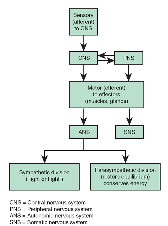
The autonomic nervous system (ANS) is composed of portions of the CNS and PNS (Fig. 19-9). It is generally classified under the PNS; however, the ANS is highly specialized. The ANS functions independently and without conscious effort. It innervates organs that are not usually under voluntary control, particularly cardiac muscle, glands, and smooth (visceral) muscles, such as those in the intestine, bladder, and uterus. The ANS contains visceral motor neurons to these areas. Figure 19-10 illustrates the ANS and the organs it influences. The body’s ability to maintain homeostasis is largely due to the ANS.
FIGURE 19-9 • Organization of the nervous system.
The ANS has two divisions: sympathetic and parasympathetic. As shown in Figure 19-10, stimuli to the illustrated structures originate in both divisions, but they often function in opposition to each other (antagonistic reactions). For instance, sympathetic nerves increase heart rate and dilate the pupil of the eye; parasympathetic nerves slow heart rate and constrict the pupil. Table 19-3 summarizes the effects of both divisions of the ANS on selected organs.
Many chemicals are produced by the ANS that also function in a similar manner as do the autonomic nerves. Simply stated, some chemical substances “turn on” or mimic either the sympathetic or parasympathetic nervous system. Other drugs “turn off” or “lyse” the sympathetic or parasympathetic subdivisons of the ANS.


