DIVISIONS OF THE NERVOUS SYSTEM
The nervous system is divided into two major parts: the central nervous system and the peripheral nervous system. The central nervous system, the largest part of the nervous system, consists of the brain and the spinal cord. The peripheral nervous system consists of the cranial nerves, spinal nerves, and the autonomic (automatic) nervous system.
Central Nervous System
The central nervous system (CNS) is made up of the brain, spinal cord, and accessory structures. The spinal cord functions as a major pathway to carry information between the body and the brain. The brain is responsible for interpreting this information and for directing body responses.
The Brain
The human brain alone consists of approximately 100 billion neurons, as well as innumerable synapses. It weighs approximately 1.36 kg (kilograms) (3 lb), which is about 2% of the body’s weight. However, the brain uses approximately 20% of the body’s circulating blood flow (about 750 mL/min). The human brain, spread out, would be about the size of a pillowcase. (An infant’s brain triples in size during the first year of life.) The brain is also referred to as the encephalon; the word element pertaining to the brain is encephal(o).
The brain has an extensive and specialized vascular (blood vessel) supply. It has a higher metabolic rate than the rest of the body, and this rate remains constant during physical or mental exercise. The brain must have a constant flow of oxygen and nutrients, particularly glucose. A failure of blood flow for as little as 10 seconds will result in unconsciousness. The brain is also very sensitive to toxins and drugs. Although the brain has many parts, it functions as an integrated whole. Its major divisions are the cerebrum, cerebellum, and medulla oblongata. Table 19-1 summarizes the components and functions of the brain.
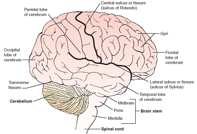
Cerebrum. Of the brain’s volume, 80% is the cerebrum (Fig. 19-4), which fills the upper part of the cranium (skull cavity). The cerebrum is sometimes called “the seat of consciousness.” It coordinates sensory data and motor functioning and governs intelligence, reasoning, learning, memory, and other complex behaviors. The cerebrum is divided into two layers and two halves (hemispheres). The longitudinal fissure, or cleft, runs from the front of the brain to the back, thus forming the two halves. Each portion of the cerebrum has individualized functions. All the portions, however, are integrated, and many areas overlap.
Cerebral Cortex. The adjective cerebral pertains to the unique human abilities of learning, intelligent reasoning, and judgment. The cerebral cortex is the thin (1-4 mm) outside layer of the brain. It is made of soft gray matter, mostly nerve cell bodies. The cortex is wrinkled and folded back on itself many times. These folds are called convolutions or gyri (singular: gyrus). The crevices between the folds are called fissures or sulci (singular: sulcus). Because of the folds, the cortex has a large surface area and therefore is able to contain its millions of neurons.
The cerebral cortex is divided into four lobes. These lobes have the same names as the overlying cranial bones: frontal, parietal, temporal, and occipital. These special centers in the cerebrum enable humans to associate impressions and information, which becomes knowledge.
• The frontal lobe is larger in humans than in all other animals. It controls the areas for written and motor speech. It is largely responsible for the ability of humans to achieve higher levels of mental functioning, including conception, judgment,speech, and communication. The frontal lobe is also involved with motor functions that direct body movements.
TABLE 19-1. Components of the Brain and Their Functions
|
COMPONENT |
DESCRIPTION |
FUNCTION |
|
Cerebrum (forebrain) |
Largest portion of brain |
Center of conscious thought and higher mental functioning (intelligence, learning, memory) |
|
Cerebral cortex |
Outer coating of cerebrum; gray matter (nerve cell bodies) |
Contains convolutions (grooves) and elevations (gyri) that increase brain’s surface area |
|
Inner portion of brain |
White matter |
Location of billions of connections, due to presence of dendrites and myelinated axons |
|
Lobes Frontal |
Located at front of skull, forehead |
Location of higher mental processes (intelligence, motivation, mood, aggression, planning); site for verbal communication and voluntary control of skeletal muscles |
|
Parietal |
Between frontal and occipital lobes |
Location of skin, taste, and muscle sensations; speech center; enables formation of words to express thoughts and emotions; interprets textures and shapes |
|
Temporal |
Located at sides of skull |
Location of sense of smell and auditory interpretation; stores auditory and visual experiences; forms thoughts that precede speech |
|
Occipital |
Located at back of skull |
Location of eye movements; integrates visual experiences |
|
Hemispheres Corpus callosum |
Longitudinal fissure divides brain into right and left halves Connects hemispheres internally |
|
|
Diencephalon (Interbrain) Thalamus |
Located between cerebral hemispheres and brainstem Located within cerebral hemispheres |
Central relay point for incoming nerve messages ("switching center”)—consolidates sensory input (especially extremes and pain); influences mood and body movements; associated with strong emotions |
|
Hypothalamus |
Located below thalamus, at base of cerebrum |
Regulates homeostasis—center for body temperature regulation, hunger, peristalsis, thirst and water balance, sexual response, and sleep-wake cycle; controls heart rate and blood vessel diameter; influences pituitary gland secretions; controls muscles of swallowing, shivering, and urine release; links nervous and endocrine systems |
|
Limbic system |
Consists of hippocampus and reticular formation |
Responsible for learning, long-term memory, wakefulness, and sleep |
|
Cerebellum ("little brain”) |
Second largest part of the brain (part of hindbrain); attached to back of brainstem, below curve of cerebrum. Connected, via midbrain, to spinal cord and motor area of cortex |
Location of involuntary movement, coordination, muscle tone, balance, and equilibrium (semicircular canals); coordinates some voluntary muscles |
|
Brainstem Midbrain (mesencephalon) Pons (bridge) |
Smallest and most primitive part of brain Located at top of brainstem, below thalamus |
Connects cerebral hemispheres with spinal cord Acts as visual and auditory reflex center; righting reflex located here |
|
Medulla (oblongata) |
Between cerebrum and medulla Located at floor of skull below midbrain; connects brain to spinal cord |
Carries messages between cerebrum and medulla; acts as respiratory center to produce normal breathing patterns Vital for life; descending nerve tracts from brain cross here to opposite side; contains centers for many body functions (cardiac, vasomotor; and respiratory center; swallowing, coughing, and sneezing reflexes) |
• The sensory area is located in the parietal lobe. This area interprets sensations such as touch, taste, pressure, temperature, and pain, which are received from the skin. Spatial ability (the ability to recognize shapes and sizes) is also located in this area.
• The temporal lobe receives and interprets auditory signals and processes language. It controls the sensations of hearing, auditory interpretation, and smell.
• Visual transmissions and interpretation occur in the visual areas of the occipital lobe. These areas direct a person’s visual experiences.
White Matter. The interior of the brain consists of white matter. It lies under the cerebral cortex. Billions of synapses between axons and dendrites are located here. This area is white because of the myelinated axons that connect the lobes of the cerebrum to each other and to all other parts of the brain.
FIGURE 19-4 · Lateral view (right side) of the external surface of the brain, indicating lobes of the cerebrum, as well as the cerebellum and brain stem. Major sulci are also shown.
Cerebral Hemispheres. The right hemisphere (right half) of the cerebrum controls the muscles of the left side of the body and receives sensory information from it as well. The left hemisphere controls the muscles of, and receives sensory information from, the right side. This phenomenon is a result of decussation (crossing) of the nerve tracts within the brain’s medulla.
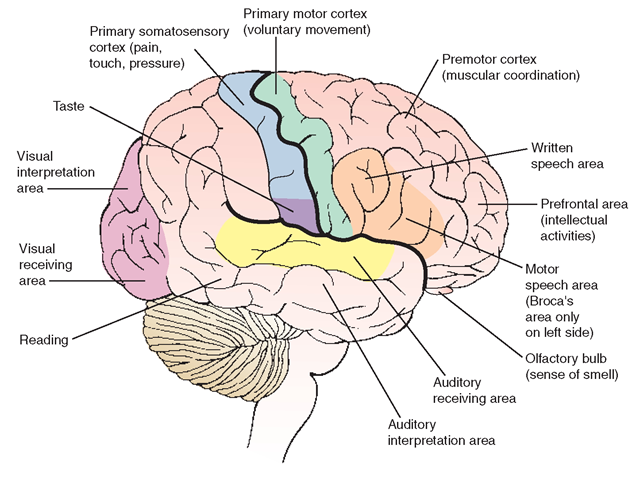
Figure 19-5 shows some functional areas of the cerebrum. The two hemispheres of the brain process information differently. The right side is associated with spatial perception, pictures, art, and musical ability. The left hemisphere is connected with analytic and verbal skills (e.g., reading and writing, words, symbols, mathematics, and speech) and walking.
Key Concept Individuals who have had a cerebrovascular accident (e.g., stroke) affecting one brain hemisphere show external symptoms on the opposite side of the body Studies have shown that most people process I anguage and speech on the left side of the brain. For this reason, a left-sided stroke often causes speech and language impairment, although the physical symptoms (e.g., paralysis) will be on the right side of the body
♦ Language comprehension (Wernicke’s area)—damage impairs ability to comprehend written and spoken language, but the person can still speak
♦ Speaking ability (Broca’s area)—damage causes speech impairment, but does not affect language comprehension
FIGURE 19-5 · Lateral view of the brain, indicating major functional areas.
The corpus callosum looks like a band of matted white fibers and contains approximately 200 million neurons. It lies between and interconnects the right and left hemispheres of the cerebrum, deep within the longitudinal fissure. The corpus callosum allows one cerebral hemisphere to share information with the other. If this structure is severed, the right hand will literally not know what the left hand is doing.
Thalamus. The thalamus is located in the diencephalon portion of the brain, between the hemispheres and brainstem (Fig. 19-6). This is in the posterior area of the forebrain. The thalamus lies just superior and posterior to the hypothalamus. It is a relay station between cutaneous (skin) receptors and the cerebral cortex for all sensory impulses, except smell. The thalamus integrates sensations so a person perceives a whole experience and not just individual impulses. For example, touching a snowball produces sensations of cold, pressure, texture, and shape. The thalamus integrates all of these sensations to present the whole picture. The thalamus also has some crude awareness of pain. It may suppress unimportant sensations to permit the cerebrum to concentrate on important aspects of daily activities.
Hypothalamus. Although small, the hypothalamus is vital to human functioning because it regulates homeostasis. It is located below the thalamus and is also the part of the diencephalon portion of the brain (see Fig. 19-6). Most functions of the hypothalamus relate directly or indirectly to the regulation of visceral activities. The hypothalamus has a role in increasing or decreasing body functions, and it regulates the release of hormones from the pituitary gland which, in turn, regulate many of the body’s other hormones.These functions include regulation of body temperature, water balance (thirst), sleep, hunger, sexual urges, and emotions. The hypothalamus is a major link between the nervous and endocrine systems.
FIGURE 19-6 · Brain, midline cross section, showing the thalamus, hypothalamus, and pituitary
Key Concept The diencephalon portion of the forebrain consists of the thalamus and hypothalamus, as well as structures called the metathalamus and epithalamus. Another area, called the subthalamus, is usually considered to be a separate division of the forebrain.
Limbic System. The limbic system is located between the cerebrum and the inner brain. (The term “limbic” literally means “pertaining to a margin or edge.”) It is largely responsible for maintaining a person’s level of awareness. The limbic system includes the hippocampus and the reticular formation. The hippocampus functions in learning and longterm memory. The reticular formation plays a role in allowing sensory input to enter the cerebral cortex. It also governs wakefulness and sleep, through a portion of the reticular formation called the “reticular activating system” (RAS). This system is affected by environmental stimuli via the eyes (light) and ears, which activate the RAS to assist a person in awakening and maintaining alertness.
Cerebellum. The cerebellum is the second largest part of the brain (see Fig. 19-4). Its functions are concerned with movement: muscle tone, coordination, posture, and equilibrium. It coordinates the action of voluntary muscles and adjusts to impulses from proprioceptors within muscles, joints, and sense organs. (Proprioceptors relay information about balance and body position.) Adjustments made in the cerebellum help a person to maintain balance when walking, and to make graceful movements, throw a ball, or participate in activities such as inline skating.
Brainstem. The brainstem is continuous with the spinal cord and connects the cerebral hemispheres with the spinal cord. It is the smallest part of the brain. It includes the midbrain, pons, and medulla (see Figs. 19-4 and 19-6).
The midbrain (mesencephalon) is located at the very top of the brainstem and functions as an important reflex center. Visual and auditory reflexes are integrated here. When you turn your head to locate a sound, you are using the midbrain. The righting reflex, or the ability to keep the head upright and maintain balance, is also located here. The red nucleus (ruber) is a pinkish colored oval mass of gray matter in the area of the midbrain which is involved in motor function.
The word “pons” means “bridge.” The term indicates that the pons contains nerve tracts that carry messages between the cerebrum and medulla. The pons has respiratory centers that work with the medulla to produce a normal breathing pattern.
The medulla (also known as medulla oblongata) lies just below the pons and rests on the floor of the skull. It is continuous with, but not part of, the spinal cord. Nerve tracts that descend from the brain cross to the opposite side here (decussation). The medulla contains centers for many vital body functions, including the cardiac center (regulates heart rate), vasomotor center (regulates the diameter of blood vessels, thereby regulating blood pressure), and respiratory center (regulates breathing). Other activities of the medulla are concerned with reflexes, such as swallowing, coughing, sneezing, hiccupping, and vomiting. Because the medulla is responsible for regulating so many vital body processes, any injury to the occipital bone at the base of the skull can be instantly fatal because of its proximity to the medulla.
The Spinal Cord
The spinal cord is a long mass of nerve cells and fibers extending through a central canal on the dorsal side (back) of the body. It contains gray matter, mostly composed of cell bodies and dendrites. This is surrounded by white matter made up of interneuronal axons (tracts), arranged in bundles. Some nerve tracts are ascending (to the brain) and are sensory tracts. Some are descending (from the brain) and are motor tracts. The spinal cord extends from the medulla to the approximate level of the first or second lumbar vertebra
Its position within the vertebral column helps protect the spinal cord from shocks and injuries. This protection is essential because the nerve fibers of the spinal cord cannot regenerate after an injury.
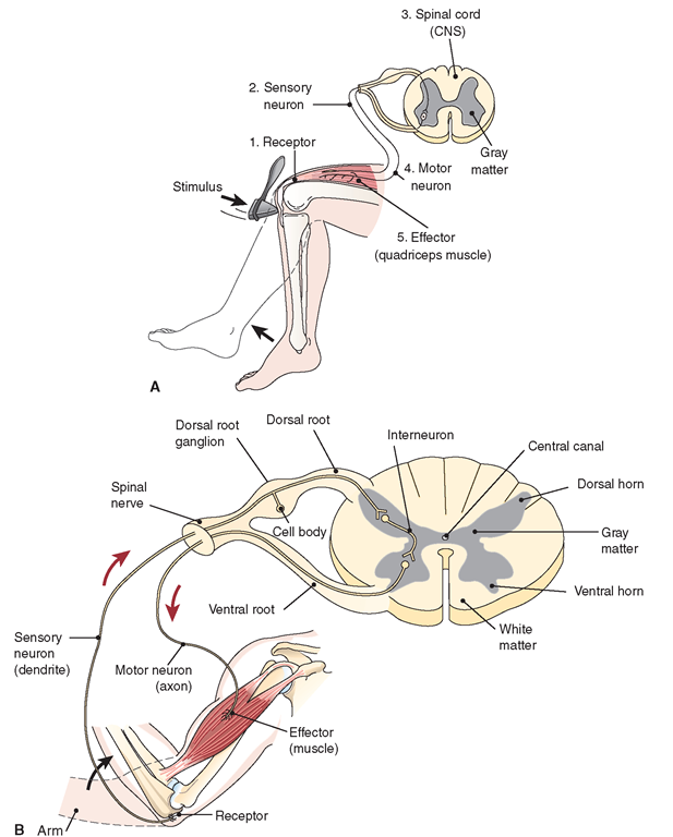
The spinal cord has two main functions: to conduct impulses to and from the brain and to act as a reflex center. The reflex center in the cord receives and sends messages through the nerve fibers without entering the brain. This “circle” is known as a reflex arc (Fig. 19-7). The reflex center acts as a substation for messages and relieves the brain of routine work.
FIGURE 19-7 · Reflex arcs, showing pathways of impulses in response to a stimulus. (A) The cross-section of the spinal cord shows the simplest reflex, the patellar (knee-jerk) reflex, which involves only sensory and motor neurons. (B) The response to a painful stimulus, such as a flame, also involves a central neuron. This is a three-neuron reflex arc.
Key Concept The nerve fibers of the spinal cord cannot regenerate after an injury The central nervous system is protected by the skull and the vertebrae of the spinal column


