Learning Objectives
1. List the four classifications of bones according to shape.
2. Locate and name the major bones of the body and describe their functions.
3. Explain the function of red bone marrow.
4. Name three types of joints and give an example of each.
5. Differentiate between the axial.
6. List the five divisions of the vertebral column and the number of vertebrae in each division.
7. Differentiate between an adult and an infant skull; identify the anterior and posterior fontanels on a newborn, explaining their functions.
8. Compare and contrast skeletal, smooth, and cardiac muscles and their functions.
9. Identify the major muscle groups in the body, including the functions of each group.
10. Discuss factors that influence bone growth.
11. Explain the process by which muscles produce heat.
12. Explain types of exercise, give examples, and state the purpose of each.
13. Differentiate between tendons and ligaments.
|
IMPORTANT TERMINOLOGY |
||||
|
acetabulum |
elasticity |
irritability |
osteoblast |
sacrumscapula |
|
articulation |
epiphysis |
isometric |
osteoclast |
sinus |
|
atrophy |
extensibility |
isotonic |
osteocyte |
sternum |
|
bursa |
extension |
joint |
patella |
symphysis pubis |
|
calcaneus |
femur |
ligament |
pelvis |
tendons |
|
carpal |
fibula |
malleolus |
periosteum |
thorax |
|
cartilage |
flexion |
mandible |
phalanx |
tibia |
|
clavicle |
gait |
marrow |
pubic arch |
ulna |
|
coccyx |
hematopoiesis |
maxilla |
radius |
vertebral column |
|
contractility |
humerus |
muscle tone |
range of motion |
|
|
diaphysis |
ilium |
ossification |
reabsorption |
|
Most people take for granted the fact that their bodies can move. By working in harmony, the components of the musculoskeletal system provide movement and independence. The musculoskeletal system includes the skeleton, joints, bursae, ligaments, muscles, and tendons. The skeleton provides the bony framework for the human body and the muscles assist the body to move. The musculoskeletal system works in coordination with other systems of the body, such as the nervous system.
Structure and Function
A systematic way to study the structure and function of the musculoskeletal system is to study the skeleton, the muscles, and how they work together. Although each component is distinct and unique, the system overall cannot function without cooperative action between the separate parts. An examination of the structure and function of the musculoskeletal system must consider both the skeleton and the muscles.
THE SKELETON
The functions of the skeleton are support (structure), protection, movement, hematopoiesis (blood formation), and storage (Box 18-1). Movement is possible because many of the bones of the skeleton are used as levers. Not only is the skeleton the framework of the body, it is also a living structure. Although bone itself is hard and filled with calcium deposits, the cells of the bone (osteocytes) are living organisms. Each bone is made up of several types of tissue; therefore, bones are considered organs.
Key Concept Without muscles, the bones would be unable to move.
Bones
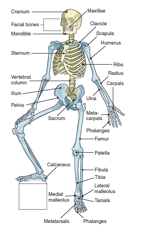
The adult human body has 206 bones (Fig. 18-1). More than half of bones in the body are found in the hands, wrists, feet, and ankles. Bones, the marrow within certain bones, and the minerals of which bones are made (calcium and phosphorus) contribute to the homeostatic functioning of the body.
Special Considerations: LIFESPAN
A baby has approximately 300 bones at birth. Fusion of some of them takes place and the process is complete by about age 25.
Classification
Bones are classified according to shape: long, short, flat, or irregular. Long bones have an extended shape and provide the body with support and strength. Short bones are approximately cube shaped. Flat bones are shaped exactly as the name suggests, and provide broad surfaces for muscle attachments.
BOX 18-1.
Functions of the Skeleton
|
Support |
|
♦ Supports the body ♦ Provides framework for the body ♦ Gives shape to the body |
|
Protection |
|
♦ Protects vital organs ♦ Protects soft tissues |
|
Movement |
|
♦ Provides locomotion (walking, movement) by attachment of muscles, tendons, and ligaments |
|
Hematopoiesis |
|
♦ Produces red blood cells ♦ Produces white blood cells ♦ Produces platelets |
|
Storage |
|
♦ Provides calcium ♦ Provides phosphorus |
FIGURE 18-1 · The skeleton.
Irregular bones are similar to short bones, but are irregular in shape. The irregular bone classification includes small, rounded bones called sesamoid bones. Sesamoid bones develop within joints and tendons. The patella (kneecap) is the largest sesamoid bone. Other special irregular bones are those in the middle ear. The smallest bone in the body is the stapes (stirrup) in the middle ear. Table 18-1 lists the classifications of bones, along with their functions and examples.
Structure
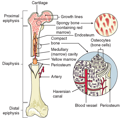
Bone tissue comes in two types. Compact bone is hard and dense. It comprises the shaft of long bones and the outer layer of other bones. Spongy bone (cancellous bone) is composed of small bony plates. It contains more spaces than compact bone and resembles a sponge.
The hollow inner part of most bones, the medullary cavity, is lined with endosteum and contains a soft substance called marrow. There are two types of bone marrow. Yellow marrow (as seen in soup bones) is in the medullary cavity of long bones and is mostly fat. Red marrow is found in the ends of long bones, in the bodies of vertebrae, and in flat bones. Red bone marrow is responsible for manufacturing red blood cells, white blood cells, and platelets. This process is called hematopoiesis.
The periosteum is a thin, hard, fibrous, dense connective tissue membrane that covers the outside of most bones. It often merges with tendons and ligaments. The periosteum contains blood vessels that supply oxygen and nutrients to bone cells (osteocytes), keeping them alive. The circulation also supplies minerals and other bone-building substances that fill intercellular spaces.
TABLE 18-1. Classification of Bones
|
CLASSIFICATION |
FUNCTIONS |
LOCATIONS |
EXAMPLES |
|
L°ng |
Act as levers; support frame |
Arms, legs |
Femur, tibia, radius |
|
Short |
Facilitate movement; transfer forces |
Wrists, ankles, feet |
Metatarsals, phalanges |
|
Flat |
Serve as muscle attachment and for protection |
Head, chest, shoulders, hips |
Cranial, ribs, scapula, pelvis, ilium |
|
Irregular |
For attachment of other structures or articulations, or for special functions |
Facial, vertebrae, patella Bones of middle ear (malleus, incus, stapes) |
Two types of osseous (bony) tissue are involved in construction of the long bones of the extremities. The diaphysis, or shaft of the long bone, is hard and compact. The end of the long bone, the epiphysis, is sponge-like and is covered by a shell of harder bone known as compact bone (Fig. 18-2). The diaphysis and epiphysis do not fuse until full adult growth is achieved. The place where the diaphysis and epiphysis meet is called the epiphyseal growth plate.
Key Concept The layers of a bone are, from the outside-in: Periosteum, compact bone (on the epiphysis), cancellous bone (spongy bone), endosteum, and bone marrow within the medullary cavity (thick, jelly-like; makes blood cells).
There are several recognizable contours with structural significance on bones. A facet (fah-set’) is a small plane or smooth area. The vertebrae of the spinal column contain facets, which are the locations for articulation (a joint) with the heads of the ribs.
FIGURE 18-2 · The structure of a long bone; the composition of compact bone.
Special Considerations :LIFESPAN
Damage to the Epiphyseal Growth Plate
Damage to the epiphyseal growth plate by trauma usually causes cessation of growth in long bones and results in shortening of the limb involved. The younger the child is when an injury occurs and the greater the severity of the injury, the greater will be the final deficit in length between the injured limb and the uninjured limb.
A condyle (e.g., the head of the femur) is a large, rounded projection, usually for articulation with another bone. A tuberosity is a large, elevated, knob-like projection, usually for muscle attachment. A tubercle is a small, rounded knob or nodule, usually for attachment of a tendon or ligament. The tibia has condyles and tuberosities on both ends, where it articulates with the thigh and ankle bones and connects with muscles and tendons (see Fig. 18-1).
A flat projection or area is called a plate. The dental plate (dorsal or roof plate) makes up the roof of the mouth. The foot plate is the flat portion of the stapes bone in the middle ear.
Any prominence or projection of bone is called a bony process. A spine (spina) is a sharp process; a ridge or crest is a thin or narrow process. (A distinct border or ridge, usually on the superior aspect of a bone, is most commonly called a crest.) The great (greater) trochanter of the femur is a large bony process. A familiar crest in the body is the iliac crest, which lies at the top of the ileum, or iliac bone of the pelvis. Each bone of the vertebral column also contains several processes, including the spinous process and the transverse process. The pedicle is another process that forms part of the vertebral arch of the vertebral column.
Key Concept The giraffe and the human have the same number of vertebra; the difference is in the size of each.
Bones also have recognizable structural openings (holes or open areas). A hole through which blood vessels, ligaments, and/or nerves pass is called a foramen (plural: foramina). A long, tube-like hole is sometimes called a canal. Within the bones of the spinal column are found the transverse foramen, through which blood vessels and nerves pass, and vertebral foramina, through which the spinal cord passes. Other important foramina and canals in the body provide passages for blood vessels and nerves. These include the apical foramen, an opening in the root of each tooth; the sciatic foramen in the hip bone; Alcock’s canal in the perineal area; the carotid canal, through which the carotid blood vessels pass into the cranium (head); and the infraorbital canal in the eye socket.
A sinus is a sponge-like air space within a bone, such as the paranasal sinuses within the skull bones.A dent, trench, or depression usually is called a fossa (plural: fossae). The cranial or cerebral fossae are depressions in which the brain rests. The olfactory bulb (for the sense of smell) lies in the ethmoid fossa, and the mandible (lower jaw bone) lies in the mandibular or glenoid fossa.
Joints
The points at which bones attach to each other are called joints or articulations (Table 18-2 ). Joints determine which motions are possible because of the way the bones are attached. Joints are classified according to the degree of movement they permit:
• Synarthroses (synarthrodial, fibrous, or fixed joints; also called sutural ligaments) are immovable. The most familiar synarthroses are the joints between the bones of the skull. These joints are not firmly fixed in infants, but become fused by adulthood because of interlocking projections and fibrous connective tissue growth. They are also called sutures because they are so tightly bound, as if they are sewn together. A gomphosis is a fibrous joint in which a conical process is inserted into a socket, as is a tooth in its bony socket.
• Amphiarthroses (amphiarthrodial or cartilaginous joints) are only slightly movable. Examples are the symphysis pubis or the articulations between the ribs and spinal column. Here, cartilage lies between articulating bones. A synchrondosis is a joint in which cartilage is converted to bone by adulthood, as in the coccyx.
• Diarthroses (synovial joints) are freely movable, allowing movement in various directions, as at the ends of long bones. These joints also contain ligaments and cartilage (discussed later). A thin, smooth layer of articular cartilage covers and pads synovial joints, and an articular capsule encloses the ends of the bones. The capsules are lined with synovial membrane, which secretes synovial fluid, a lubricating material.
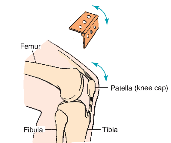
FIGURE 18-3 · Hinge joint (knee).
Some synovial joints, notably knees, shoulders, elbows, and hips, also contain bursae, fluid-filled sacs that cushion the movements of muscles and tendons.
Synovial joints are further classified according to their structure and range of movement.
• Hinge (ginglymus) joints (Fig. 18-3) allow movement in only one plane, similar to the hinge of a door—as in the elbow, knee, and the finger and toe joints between phalanges. (The jaw, knee, and ankle joints are hinge joints, but can also move slightly from side to side.
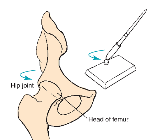
• Ball-and-socket (spheroidal) joints (Fig. 18-4) consist of a rounded end of one bone that moves within a cup-shaped depression (the acetabulum) in the other bone, allowing movement in every direction. The hip and the shoulder are examples of ball-and-socket joints.
• Pivot joints (Fig. 18-5) consist of a bone pivoting or turning within a bony or cartilaginous ring. An example is found in the atlas (first cervical vertebra) and the head rotating on the axis (second cervical vertebra).
• Gliding (arthrodial, plane) joints are where the bones slide against each other, as in the intervertebral joints and parts of the wrist and ankle joints.
TABLE 18-2. Classification of Joints
|
TYPE OF JOINT |
ACTIONS |
EXAMPLES |
|
Synarthroses: immovable, fibrous |
No motion |
Bones of the skull fitted together with interlocking notches (in adults) (see Fig. 18-6) |
|
Amphiarthroses: slightly movable, cartilaginous |
Slight degree of motion or flexibility |
Vertebral column (see Figs. 18-8 and 18-9) Symphysis pubis |
|
Diarthroses: freely movable (synovial) Hinge joints Ball-and-socket joints Pivot joints Gliding joints Condyloid joints Saddle joints |
Motion like a door on hinges Rotation Motion like that of turning a doorknob Gliding motion Allow motions in two planes at right angles Opposing surfaces are concavoconvex (fit together like two saddles with riding surfaces together); allow a wide range of movements |
Finger; elbow, and knee joints (see Fig. 18-3) Shoulders and hips (see Fig. 18-4) Elbow (turn forearm) (see Fig. 18-5) Wrist Wrist, foot, hand Thumb, vertebrae, ankle |
FIGURE 18-4 · Ball-and-socket joint (hip).
• Condyloid joints involve the oval-shaped head of one bone moving within the elliptical cavity in another, permitting all movements except axial rotation. Examples are the wrist (between the radius and carpal bones) and at the base of the index finger.
• Saddle joints allow movements that can be shifted in several directions, as in the ankle and the base of the thumb.


