Learning Objectives
1. Describe lymphocytes, their functions, and where they are produced.
2. Differentiate between B cells and T cells (lymphocytes).
3. Describe nursing implications related to a lack of or a decrease in antibody production.
4. Differentiate between non-specific and specific immunity. Compare and contrast naturally acquired and artificially acquired active and passive immunities.
5. Describe the process of antibody-mediated immunity. Explain how the “lock-and-key” concept applies to the antigen-antibody complex.
6. Explain the mechanisms antibodies use to destroy antigens.
7. Describe the effects of aging on the immune system.
|
IMPORTANT TERMINOLOGY |
||
|
acquired immunity |
cytokine |
naturally acquired |
|
antibody-mediated |
gamma globulin |
immunity |
|
immunity |
humoral immunity |
non-specific immunity |
|
artificially acquired |
immunity |
specific immunity |
|
immunity |
immunization |
T cells/T lymphocytes |
|
B cells/B lymphocytes |
inborn immunity |
thymus |
|
cell-mediated immunity |
macrophage |
vaccine |
|
complement fixation |
||
|
Acronyms |
|
|
Ab |
IgD |
|
Ag |
IgE |
|
!g |
IgG |
|
IgA |
IgM |
The human body must always protect itself against foreign invasion. It does so using a “layered defense” system, which includes the skin and chemical barriers, as well as the innate and adaptive immune systems. This topic is primarily concerned with innate and adaptive immune responses (immunity).
Immunity is the body’s ability to recognize and destroy specific pathogens, such as bacteria and viruses, as well as parasites, and to prevent infectious diseases. The immune system also recognizes some tumor cells. When the immune system is compromised, immunodeficiency diseases may occur. When the immune system is overreactive, disorders such as allergies and autoimmune disorders may result.
Key Concept The immune system must distinguish between "self" (normal components of the body) and "non-self” (foreign tissues or substances). In some cases, this mechanism is faulty and the body destroys its own cells.
Structure and Function
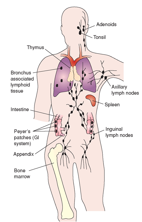
The body’s immune system includes the bone marrow, lymphoid organs, and the mononuclear phagocyte system (also called the reticuloendothelial system). Primary functions of the immune system include defense, homeostasis, and surveillance. Box 24-1 describes these functions. Figure 24-1 shows many of the specific organs and tissues involved in the immune system.
BONE MARROW AND LYMPHOCYTE PRODUCTION
Cells in the bone marrow are capable of developing into any of three types of blood cells: erythrocytes (red blood cells [RBCs]), leukocytes (white blood cells [WBCs]), or thrombocytes (platelets). WBCs defend the body against disease organisms, toxins, and irritants. The two types of WBCs are granular (neutrophils, basophils, and eosinophils) and agranular (monocytes and lymphocytes). The lymphocytes are found in blood, lymph, and lymphoid tissues, such as the lymph nodes and tonsils. They form the immune cells and their precursors. This topic focuses on lymphocytes.
BOX 24-1.
Functions of the Immune System
Defense
♦ Resists invasion by foreign microorganisms, including viruses and intracellular parasites
♦ Attacks some pathogens directly
♦ Attacks foreign antigens—usually proteins (including transplanted organs)
♦ Helps body to fight cancer cells
♦ Produces antibodies and immunoglobulins
♦ Produces inflammatory response
♦ Produces memory cells
Homeostasis
♦ Digests and removes damaged cellular substances
♦ Kills diseased cells (especially those infected with viruses)
Surveillance
♦ Recognizes and destroys cellular mutations
♦ Recognizes and destroys foreign cells
♦ Monitors for presence of antigens
FIGURE 24-1 · Central and peripheral lymphoid organs and tissues involved in the immune system.
• Lymphocytes are the “cornerstone” of the immune system; they alone have the ability to recognize foreign substances in the body.
• Differentiation of lymphocytes into special lymphocytes called B cells (B lymphocytes) and T cells (T lymphocytes) must occur before detection of foreign invaders begins. T lymphocytes help protect against viral infections and can detect and destroy some cancer cells. B lymphocytes develop into cells that produce antibodies (plasma cells).
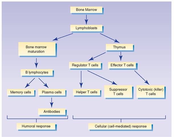
Figure 24-2 illustrates the development of immune system cells.
Key Concept Lymphocytes formed in the bone marrow and lymphatic tissues are able to transform into specialized cells called B cells and T cells. B cells provide humoral immunity by reacting to the presence of antigens to produce antibodies. (The term, antigen, is an abbreviation for "antibody generator") Antibodies then target antigens for destruction.
T cells, which proliferate at the direction of thymic hormones, attack infected cells and provide cell-mediated (cellular) immunity
B Lymphocytes
Stem cells in the bone marrow are responsible both for production of B lymphocytes and for their maturation. After they mature, B cells can produce antibodies. Exposure to an antigen in the bloodstream activates B cells to enlarge and multiply rapidly to produce colonies of clones, although B cells do not respond to all pathogens. Most of the clones become plasma cells, which produce specific antibodies to circulate in the blood. These antibodies provide the form of immunity called humoral immunity (humoral means body fluid). In the process of humoral immunity, macrophages (large cells) engulf and destroy antigens after antibodies have identified them for destruction.
Those clones that do not become plasma cells remain in the body as memory cells. On repeated exposure to an antigen, the memory cells are ready to produce antibodies immediately. This “immunologic memory,” in many cases, makes a person immune to reinfection after having had a disease (Cohen & Wood, 2005), but this is not true for all diseases.
Key Concept The second exposure to an antigen can cause a quicker and more dramatic response than the first because of "immunologic memory" The first exposure causes a more delayed reaction because it takes time to form antibodies to the antigen. Antibodies are ready for the second exposure and act quickly This is an important concept when related to allergic reactions.
FIGURE 24-2 · Development of the cells of the immune system.
B lymphocytes are found predominantly in organized lymphoid tissues, such as the spleen. They constitute only about 10%-20% of circulating lymphocytes in the blood. Even fewer B cells are found in lymph.
Key Concept The B cell recognizes whole pathogens without antigen processing. Each specific B cell recognizes a specific antibody
Antigens and Antibodies
An antigen (Ag) is any foreign substance or molecule entering the body that stimulates an immune response (the activity of B or T lymphocytes). Most antigens are large protein molecules found on the surface of foreign organisms, RBCs, or tissue cells; on pollen; and in toxins and foods. Some carbohydrates and lipids also act as antigens (Cohen & Wood, 2005).
An antibody (Ab) is a protein substance that the body produces in response to an antigen. B lymphocytes are responsible for antibody production. All antibodies are contained in a portion of the blood plasma called the gamma globulin fraction. Therefore, antibodies are commonly called gamma globulins or immunoglobulins (Ig) (Cohen & Wood, 2005). The five basic groups of immunoglobulins are:
• IgM: Stimulates complement activity. This is the antibody produced on initial exposure to an antigen (e.g., after a first tetanus immunization). IgM is abundant in the blood,but is not usually present in organs and tissues and is not transferred across the placenta.
• IgG: Protects the fetus before birth against antitoxins, viruses, and bacteria. (It is the only antibody transferred from mother to fetus across the placenta.) IgG is the most common antibody and it is produced on second and future exposures to an antigen (e.g., after a tetanus booster). It is present in the blood (intravascular) and in the tissues (extravascular). IgG (often called gamma globulin) is the main component of commercial immunoglobulin.
• IgA: Protects mucosal surfaces. The major component of secretions such as saliva, tears, and bronchial fluids, IgA is transported across mucous membranes. It is important in the defense against invasion of microbes via the nose, eyes, lungs, and intestines. IgA is found in blood, as well as in gastrointestinal (GI) and mucosal secretions. It is also found in breast milk.
• IgE: Responsible for immediate-type allergic reactions, including latex allergies. Although this antibody causes problems in developed countries, it is helpful in the developing world in fighting against parasitic infections, such as river blindness.
• IgD: Believed to function as an antigen receptor. It is present in the blood in very small amounts.
Each antigen (foreign invader) stimulates the production of its own specific antibody. The body can make about 1 million individual antibodies. Antibodies do not destroy antigens themselves, but label antigens for destruction by other substances.
Key Concept IgG is the most abundant immunoglobulin found in the blood. Maternal IgG crosses to the fetus via the placenta, thereby transferring immunity to the fetus for the first few months of an infant’s life.
Nursing Alert Latex allergies may be caused by an IgE-mediated reaction. Latex allergies can occur owing to contact with the skin or mucous membrane, or inhalation. Persons exposed may have signs of urticaria (hives), dermatitis (usually of the hands), asthma, or severe anaphylaxis (exaggerated allergic shock response).
Special Considerations :LIFESPAN
A child with spina bifida (a congenital defect in the spinal column) is at increased risk for latex allergies because the mucous membranes of the bladder and rectum are exposed to latex during frequent examinations and procedures, such as urinary catheterization. It is suggested that non-latex gloves and other materials be used as much as is possible for all children, particularly those with this disorder (This would help prevent development of latex allergy in healthcare workers as well.)
T Lymphocytes
Some immature stem cells produced in the bone marrow migrate to the thymus gland to become T cells (thymus-derived lymphocytes). T cells make up the remaining 80%-90% of lymphocytes found in circulating blood. While in the thymus, T cells proliferate and become sensitized (capable of combining with specific foreign antigens). T lymphocytes produce an immunity called cell-mediated immunity.
T lymphocytes are generally responsible for fighting cancer cells, viruses, and intracellular parasites. They are particularly important in dealing with viruses because they kill the host cell and prevent replication. T cells enable the body to differentiate between “self’ and “non-self.” Usually this is helpful in fighting off foreign pathogens. However, this function can also become a problem, for example when T cells cause tissue or organ rejection after transplantation. This is because T cells recognize these tissues as “non-self” and work to eliminate them. (Specific anti-rejection medications must be given to neutralize this rejection response in recipients of most transplants.)
Several types of T lymphocytes exist, each of which has its own function. For a T cell to react with a specific antigen, the antigen must first be presented to the T cell on the surface of a macrophage. Macrophages, when combined with T cells, release substances called interleukins, which stimulate T-cell growth (Cohen & Wood, 2005). Table 24-1 identifies the types and associated functions of the B lymphocytes and T lymphocytes.
One type of T cell is called the helper T cell. There are several subtypes of helper T cells. These cells often have no cytotoxic (cell-killing) function per se. They help regulate innate and adaptive immune responses. They control immune responses by instructing other cells to kill infected cells or pathogens. Many receptors on the helper T cell must be bound by specific antigens to activate the helper cell. They require a longer time of exposure. They also release cytokines, which help macrophages and also increase the killer T cells to do their work. They also help to activate antibody-producing B cells.
Killer T cells (cytotoxic T cells) are another subgroup of T cells. These cells kill cells infected with pathogens, or which are otherwise damaged or defective. Killer T cells search for cells where the receptors have a specific antigen. Then, these cells release a specific cytotoxin, such as perforin. (Perforin allows pores [holes] to form in the plasma membrane of the target cells. This allows the cellular contents to flow out and substances to enter the targeted cells [osmotic lysis] and kill them.) Unlike the helper T cells, killer T cells can be activated when bound to a single antigen molecule.
TABLE 24-1. Lymphocytes Involved in Immune Responses
|
CELL TYPE |
FUNCTION |
TYPE OF IMMUNE RESPONSE |
|
B cell |
Produces antibodies or immunoglobulins (IgA, IgD, IgE, IgG, IgM) |
Humoral |
|
T cell |
Cellular (cell-mediated) |
|
|
Helper T4 |
Attacks foreign invaders (antigens) directly |
|
|
Initiates and augments inflammatory response |
||
|
Helper T1 |
Increases activated cytotoxic T cells |
|
|
Helper T2 |
Increases B-cell antibody production |
|
|
Suppressor T |
Suppresses the immune response |
|
|
Memory T |
Remembers contact with an antigen and on subsequent exposures mounts an immune response |
|
|
Cytotoxic T (killer T) |
Lyses cells infected with virus; plays a role in graft rejection |
|
|
Non-T or B lymphocytes |
Nonspecific |
|
|
Null cells |
Destroys antigens already coated with antibody |
|
|
Natural killer (NK) (granular lymphocyte) |
Defends against microorganisms and some types of malignant cells; produces cytokines |
Key Concept T cells recognize a "non-self” target only after antigens have been processed and presented, combined with a "self-receptor” called a major histocompatabil-ity (MHC) molecule.
B cells are involved in humoral immune response. T cells provide the cell-mediated (cellular) immune response. Both B cells and T cells have receptor molecules that recognize specific target cells.


