Significance of Changes in On-Center and Off-Center Bipolar and Ganglion Cell Activities
As described earlier, the changes in membrane potential of on-center and off-center bipolar cells and firing of corresponding ganglion cells to light stimulus to the receptive field center and receptive field surround are opposite. This mechanism renders the bipolar and ganglion cells sensitive to contrast in illumination that falls in the receptive field center and receptive field surround. The sensitivity of the bipolar and ganglion cells to the contrast properties, rather than to an absolute level of illumination, renders brightness or darkness of objects constant over a wide range of lighting conditions. For example, on a printed page, the darkness of the print or white background of the page appears the same whether we look at the page inside a room or in sunlight. In experimental conditions, when on-center ganglion cells are damaged, the animals can still see objects that are darker than the background. However, they cannot see objects brighter than the background. These observations indicate that information about brightness and darkness of objects in a visual field is transmitted to the brain by on-center and off-center ganglion cells, respectively.
There are at least two types of retinal ganglion cells: M and P cells. P cells are more numerous than the M cells. M-type retinal ganglion cells project to magnocellular layers of the lateral geniculate nucleus located in the thala-mus, whereas P cells project to the parvocellular layers of the same nucleus (see the section titled "Visual Pathways"). M cells have larger cell bodies, dendritic fields, and axons compared to P cells. The responses of M ganglion cells to visual stimuli are transient, whereas those of P ganglion cells are sustained. M cells cannot transmit information about color, whereas P cells can. The P cells are capable of transmitting color information because their centers and surrounds contain different types of cones. For example, the center of P retinal ganglion cells may contain cones sensitive to long wavelength light (red color), while their surrounds may contain cones sensitive to medium wavelength light (green color). These P retinal ganglion cells will be sensitive to differences in wavelengths of light falling on their center and surround regions.
Color Vision
There are three types of cone receptors, each of which contains a different photopigment that is sensitive to one of the primary colors (red, blue, and green). The relative frequency of impulses from each cone determines the sensation of any particular color. Besides cones, other cells in the retina that are involved in the processing of color vision include the horizontal cells (which are either hyperpolar-ized or depolarized by monochromatic colors) and ganglion cells (which are either turned "on" or "off" by monochromatic colors). Information following stimulation of a particular cone preferentially by a monochromatic color (e.g., green) is processed by the visual cortex and interpreted as a particular color (green in this case). If two different types of cones are stimulated equally by two different monochromatic colors (e.g., red and green), the visual cortex interprets them as a yellow color. The visual cortex contains cells that can differentiate between brightness and contrast and cells that respond to a particular monochromatic color. Processing of color vision in the visual cortex involves integration of the responses of the cones, horizontal cells, ganglion cells, and lateral geniculate body cells.
Blood Supply of The Retina
The retina receives blood supply from branches of the ophthalmic artery. One branch (central retinal artery) enters the eye at the optic nerve disc and supplies the inner portion of the neural retina. The other branch ( ciliary artery) penetrates the sclera near the exit of the optic nerve and supplies the choriocapillaris, a part of the choroid. The outer portions of retina, including the rods and cones, are metabolically dependent on the blood supply to the choroidal layer because they receive nutrients from the choriocapillaris.
Visual and Retinal Fields
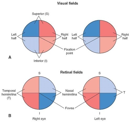
To understand visual deficits, it is helpful first to make a distinction between the visual and retinal fields. The visual field of each eye is the region of space that the eye can see looking straight ahead without movement of the head. The fovea of each retina is aligned with a point, called the fixation point, in the visual field. A vertical line can divide the visual field of each eye into two halves: the left-half field and right-half field. A horizontal line can divide each visual hemifield into superior and inferior halves. Each half can be further divided into quadrants. The vertical and horizontal lines dividing the visual field of each eye intersect at the fixation point (Fig. 16-7A). Similarly, the surface of the retina may be divided into two halves by a vertical line drawn through the center of the fovea: a nasal hemiretina that lies medial to the fovea and a temporal hemiretina that is located lateral to the fovea. A horizontal line drawn through the center of the fovea can divide the retina into superior and inferior halves. The vertical and horizontal lines dividing the retina intersect at the center of the fovea (Fig. 16-7B). Each hemiretina is further subdivided into quadrants.
FIGURE 16-7 Visual and retinal fields. (A) Vertical lines divide the visual field of each eye in space into right and left halves. Horizontal lines divide the visual field of each eye into superior and inferior halves. These lines intersect at the fixation point. (B) Vertical lines divide the retina of each eye into temporal and nasal hemiretinae. Horizontal lines divide the retina of each eye into superior and inferior halves. These lines intersect at the fovea.
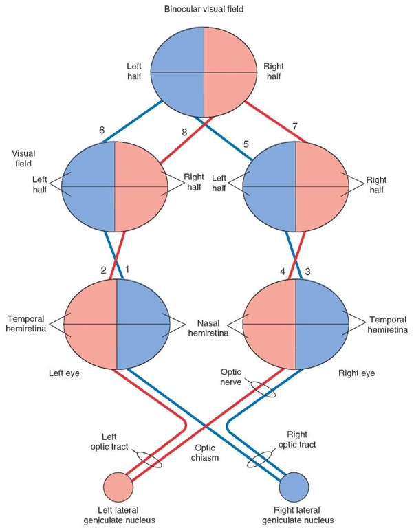
The images of objects in the visual field are right-left reversed and inverted on the retina. Accordingly, images present in the left half of the visual field of the left eye fall on the nasal hemiretina of the left eye, and images present in the right half of the visual field of the left eye fall on the temporal hemiretina of the left eye (Fig. 16-8, lines 1 and 2). Similarly, images present in the left half of the visual field of the right eye fall on the temporal hemiret-ina of the right eye, and images present in the right half of the visual field of the right eye fall on the nasal hemiretina of the right eye (Fig. 16-8, lines 3 and 4). A similar relationship exists between the superior and inferior halves of the visual fields of the superior and inferior hemiretinae of each eye (not shown in Fig. 16-8).
FIGURE 16-8 Relationship between the visual fields and retinae. 1 and 2: The nasal half of the left eye sees objects in the left half of the visual field of the left eye (shown in blue) and the temporal half of the left eye sees objects in the right half of the visual field of the left eye (shown in red). 3 and 4: Relationship between the visual fields and hemiretinae of the right eye is similar to that of the left eye. 5 and 6: When the visual fields of the two eyes are superimposed, the left halves of the two eyes coincide to form the left half of the binocular visual field (shown in blue). 7 and 8: When the visual fields of the two eyes are superimposed, the right halves of the two eyes coincide to form the right half of the binocular visual field (shown in red). Each optic nerve contains axons from the nasal and temporal hemiretinae. At the optic chiasm, the axons from the nasal hemiretinae cross to the contralateral side, whereas the axons from the temporal retinae remain uncrossed. The crossed and uncrossed axons on each side form the optic tracts.
The central portion of the visual field of each eye can be seen by both retinae. This portion of full visual field is called a binocular visual field. In a simplified diagram of the binocular visual field (Fig. 16-8), the visual fields of the two eyes are superimposed; the left half of the binocular visual field (shown in blue) represents the left half of the visual field of each eye (Fig. 16-8, blue lines 5 and 6), and the right half of the binocular visual field (shown in red) represents the right half of the visual field of each eye (Fig. 16-8, red lines 7 and 8).
Visual Pathways
The axons of ganglion cells travel towards the posterior pole of the eye where the optic disc is located. At this point, the axons become myelinated and exit the eye as the optic nerve. Extensions of meninges covering the brain also ensheath the optic nerves. The optic nerves and tracts and their relationship with visual fields of the eyes are shown in Figure 16-8. When the optic nerves of the two eyes reach the brain, they join to form the optic chiasm. At this site, the fibers representing the nasal half of the retina of each eye cross to the contralateral side, while the fibers representing the temporal half of the retina of each eye remain uncrossed. After leaving the optic chiasm, the crossed and uncrossed fibers on each side join to form the optic tracts. The left optic tract contains axons from the temporal hemiretina of the left eye and the nasal hemi-retina of the right eye. As mentioned earlier, the temporal hemiretina of the left eye sees objects in the right half of the visual field of the left eye, and the nasal hemiretina of the right eye sees objects in the right half of the visual field of the right eye (Fig. 16-8, red lines 2 and 4). This means that the left optic tract contains fibers that convey visual information from the right half of the visual field of each eye or the right half of the binocular visual field (Fig. 16-8, red lines 7 and 8).
The right optic tract contains axons from the temporal hemiretina of the right eye and the nasal hemiretina of the left eye. Recall that the temporal hemiretina of the right eye sees objects in the left half of the visual field of the same eye, and the nasal hemiretina of the left eye sees objects in the left half of the visual field of the left eye (Fig. 16-8, blue lines 3 and 1). Thus, the right optic tract contains fibers that convey visual information from the left half of the visual field of each eye or the left half of the binocular visual field (Fig. 16-8, blue lines 5 and 6). The optic tracts on each side project to the corresponding lateral geniculate nucleus of the thalamus (see next section).
The Lateral Geniculate Nucleus of Thalamus
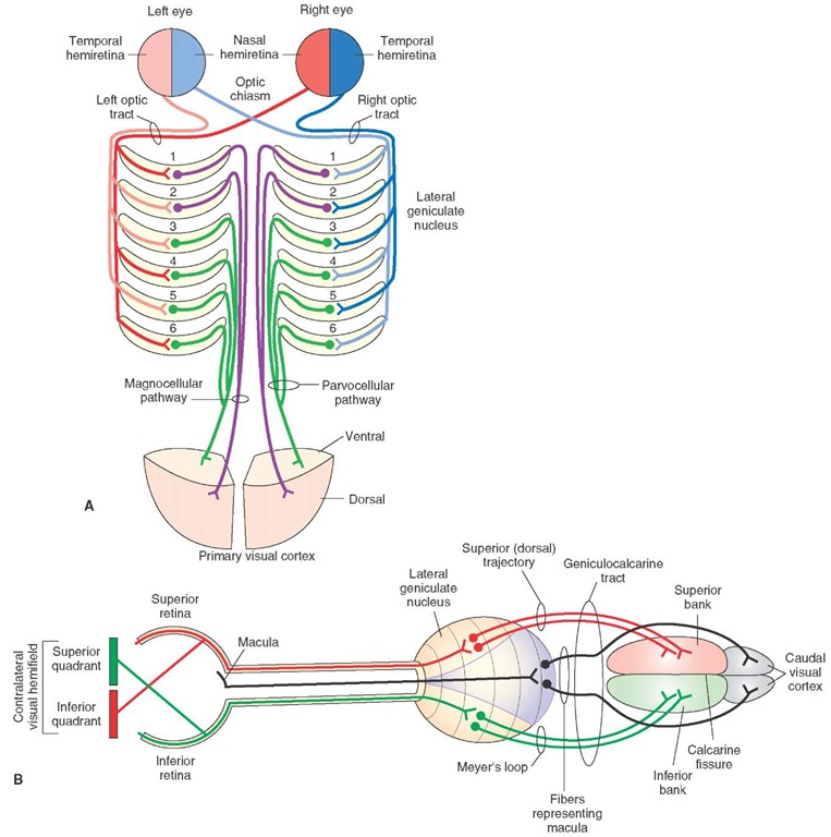
The projections of the optic tracts to the lateral geniculate nuclei are shown in Fig. 16-9A. This nucleus consists of 6 layers. The ventral layers (layers 1 and 2) are called magnocellular layers because they contain large cells, while the dorsal layers (layers 3, 4, 5, and 6) are called parvocellular layers because they contain cells of smaller size. Injury to magnocellular layers reduces the ability to detect fast-moving visual stimuli, but there is little or no effect on visual acuity or color perception. On the other hand, damage to the parvocellular layers eliminates color vision and impairs visual acuity without affecting perception of fast-moving visual stimuli. Thus, the parvocellular system seems to be concerned with color and detailed form, while the magnocellular system is concerned with location and movement. In general, the parvocellular system projects to more ventral portions (deeper regions) of the primary visual cortex, and the magnocellular system projects to more dorsal portions (superficial regions) of the primary visual cortex. Axons from the contralateral nasal hemiretina project to layers 1, 4, and 6, whereas axons from the ipsilateral temporal hemiretina project to layers 2, 3, and 5. The macular or central areas in the retina are represented to a greater extent in the lateral geniculate nucleus than are the peripheral areas of the retina.
The Geniculocalcarine Tract
Axons of the neurons in the parvocellular and magnocel-lular layers of the lateral geniculate nucleus project through the geniculocalcarine tract (also known as optic radiations) to the primary visual cortex located on the medial aspect of the occipital lobe of the cortex. The course of different fiber bundles in this tract is shown in Figure 16-9B. The fibers in the geniculocalcarine tract serving the inferior quadrant of the contralateral visual hemifield (via superior retina) arise from the dorsomedial region of the lateral geniculate nucleus and use a superior (dorsal) trajectory to synapse on the superior bank of the calcarine fissure of the visual cortex. The axons conveying information from the superior quadrant of the contralat-eral visual hemifield (via inferior retina) arise from the ventrolateral region of the lateral geniculate nucleus and use a relatively more inferior trajectory traveling toward the tip of the temporal horn of the lateral ventricle, looping caudally (called Meyer’s loop) in the inferior part of the temporal lobe and terminating in the inferior bank of the calcarine fissure of the visual cortex. The axons conveying information from the macula (including fovea) arise from the central portion of the lateral geniculate nucleus and synapse on the caudal pole of the occipital cortex.
Visual Cortex
Different areas in the primary visual cortex are shown in Figure 16-10A. The primary visual cortex ([V1] Brod-mann’s area 17) is located on the superior and inferior banks of the calcarine sulcus on the medial side of the occipital lobe and receives projections from the lateral geniculate nucleus of the thalamus. The secondary visual cortex ([V2] Brodmann’s area 18) and tertiary visual cortex ([V3 and V5] Brodmann’s area 19) are located adjacent to the primary visual cortex.
FIGURE 16-9 Course of axons from the retinal halves. (A) On each side, the axons from ipsilateral temporal hemiretinae in the optic tract synapse in layers 2, 3, and 5 of the lateral geniculate nucleus, and the axons from the contralateral nasal hemiretinae synapse in layers 1, 4, and 6. The postsynaptic axons emerging from the lateral geniculate nucleus on each side project to the primary visual cortex. (B) The axons from the superior retinae and inferior retinae project to the lateral geniculate nuclei. The postsynaptic neurons in the lateral geniculate nuclei receiving inputs from the inferior retinae form Meyer’s loop and project to the inferior bank of the calcarine fissure. The postsynaptic neurons in the lateral geniculate nuclei receiving inputs from the superior retinae form the superior trajectory and synapse on neurons in the superior bank of the calcarine fissure. The fibers conveying visual information from the macula synapse in the central regions of the lateral geniculate ganglion, and axons from the postsynaptic neurons in the lateral geniculate nucleus project to the caudal pole of the occipital cortex.
The secondary and tertiary visual areas are also known as association, extras-triate, or prestriate areas. Visual area V4 is located in the inferior occipitotemporal area (not visible in Fig. 16-10A). V3 is associated with form, V4 is associated with color, and V5 is associated with motion. The portion of area V5 that is concerned with motion of an object lies in the middle temporal gyrus. The primary visual cortex sends projections to the secondary visual cortex; from here, this information is relayed to the tertiary visual cortex. Thus, information from the nasal retina of the left eye and temporal retina of the right eye (representing the left visual field of both eyes) is directed to the right visual cortex (Fig. 16-10B). Likewise, information from the nasal retina of the right eye and temporal retina of the left eye (representing the right visual field of both eyes) is directed to the left visual cortex. The overall representation of the retina in the primary visual cortex is as follows: the macular part of the retina is represented in the posterior part of the visual cortex, the peripheral part of the retina is represented in the anterior part of the visual cortex, the superior half of the retina relating to the inferior visual fields is represented in the superior visual cortex, and the inferior half of the retina relating to the superior visual fields is represented in the inferior part of the visual cortex.
FIGURE 16-10 The visual cortex. (A) Note the location of the primary visual cortex ([V1] Brodmann’s area 17), the secondary visual cortex ([V2] Brodmann’s area 18), and the visual areas V3 and V5 (Brodmann’s area 19). (B) Information from the nasal retina of the left eye and temporal retina of the right eye (representing the left visual field of both eyes) is directed to the right visual cortex. Likewise, information from the nasal retina of the right eye and temporal retina of the left eye (representing the right visual field of both eyes) is directed to the left visual cortex (not shown).
The Superior Colliculus
The superior colliculus controls saccadic (high velocity) eye movements. The superficial layers of the superior col-liculus receive converging inputs related to visual processes from the retina (via the optic tracts) and from the visual cortex. The superior colliculus receives additional inputs from somatic sensory and auditory systems. Based on information received from these three sensory systems, the deeper layers of the superior colliculus control motor mechanisms responsible for saccadic movements and orientation of the eyes towards the stimulus. The superior colliculus achieves this function by virtue of its projections to cranial nerve nuclei of III and VI, which regulate horizontal movements of the eye. In addition, descending fibers of the tectospinal tract (from the superior colliculus) reach the upper spinal cord, activate neurons controlling neck muscles, and, thus, produce reflex neck movements in response to visual input. Other tectal fibers innervate the cerebellum indirectly through synaptic connections in the ventral pontine and paramedian reticular nuclei. These connections enable the cerebellum to coordinate eye and head movements.


![The visual cortex. (A) Note the location of the primary visual cortex ([V1] Brodmann's area 17), the secondary visual cortex ([V2] Brodmann's area 18), and the visual areas V3 and V5 (Brodmann's area 19). (B) Information from the nasal retina of the left eye and temporal retina of the right eye (representing the left visual field of both eyes) is directed to the right visual cortex. Likewise, information from the nasal retina of the right eye and temporal retina of the left eye (representing the right visual field of both eyes) is directed to the left visual cortex (not shown). The visual cortex. (A) Note the location of the primary visual cortex ([V1] Brodmann's area 17), the secondary visual cortex ([V2] Brodmann's area 18), and the visual areas V3 and V5 (Brodmann's area 19). (B) Information from the nasal retina of the left eye and temporal retina of the right eye (representing the left visual field of both eyes) is directed to the right visual cortex. Likewise, information from the nasal retina of the right eye and temporal retina of the left eye (representing the right visual field of both eyes) is directed to the left visual cortex (not shown).](http://what-when-how.com/wp-content/uploads/2012/04/tmp15F64_thumb.jpg)
