Corticospinal Tract
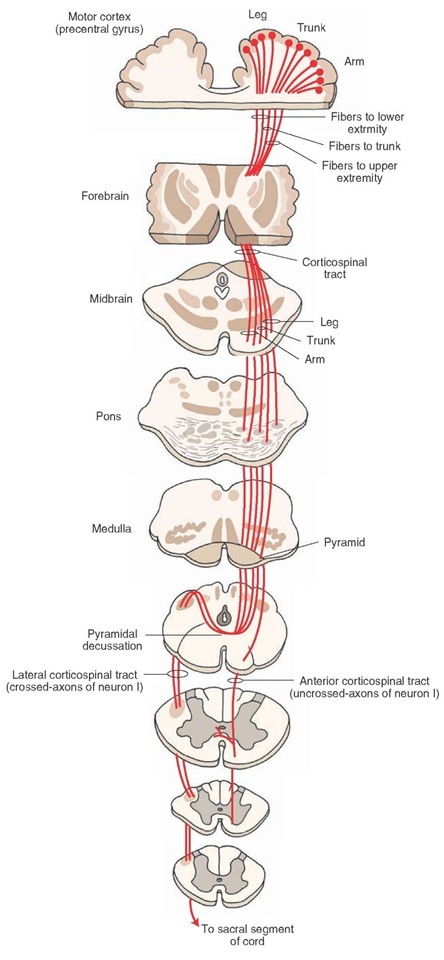
This tract is involved in the control of fine movements. It is the largest and perhaps the most important descending tract in the human CNS. A schematic representation of the course of this tract is shown in Figure 9-12. It arises from the cerebral cortex, passes through the medullary pyramids, and terminates in the spinal cord.In brief, the cells of origin functionally associated with the arm are located in the lateral convexity of the cortex, whereas the cells of origin functionally associated with the leg are located along the medial wall of the hemisphere. This somatotopic organization is called a cortical homunculus. Many of these axons control the fine movements of the distal parts of the extremities. The axons arising from the cortex converge in the corona radiata and then descend through the internal capsule, crus cerebri in the midbrain, pons, and medulla. A majority (about 90%) of the fibers cross to the contralateral side at the juncture of the medulla and spinal cord, forming the lateral corticospinal tract, which descends to all levels of the spinal cord and terminates in the spinal gray matter of both the dorsal and ventral horns. The remaining fibers do not cross at the juncture of the medulla and spinal cord; these uncrossed fibers constitute the anterior corticospinal tract. The fibers in this tract descend through the spinal cord and, ultimately, cross over at different segmental levels to synapse with anterior horn cells on the contralateral side.
FIGURE 9-12 The corticospinal tract. This tract arises from the motor cortex (precentral gyrus), passes through the medullary pyramids, and terminates in the spinal cord. Note that a majority of corticospinal fibers cross to the contralateral side in the caudal medulla (pyramidal decussation) and descend as the lateral corticospinal tract and the remaining fibers descend ipsilaterally as anterior corticospinal tract. neuron I = first-order neuron.
There is a continuation of the anterior median fissure of the spinal cord on the ventral (anterior) surface of the medulla. The medullary pyramids are located on either side of this fissure. The pyramids carry descending corticos-pinal and corticobulbar fibers. As mentioned earlier, the corticospinal fibers in the pyramids cross to the contralat-eral side at the juncture of the medulla and spinal cord. This decussation, known as the pyramidal decussation, forms the anatomical basis for the voluntary motor control of one half of the body by the contralateral cerebral hemisphere. The corticobulbar fibers leave the pyramids as they descend in the medulla and project to the cranial nerve and other brainstem nuclei.
The corticospinal tract controls voluntary movements of both the contralateral upper and lower limbs. Depending on the extent of the lesion, these functions are lost when the corticospinal tract is damaged. After a stroke, first the affected muscles lose their tone. After several days or weeks, the muscles become spastic, (i.e., they resist passive movement in one direction) and hyperreflexia occurs (i.e., the force and amplitude of the myotatic reflexes is increased, particularly in the legs). The superficial reflexes (abdominal, cremasteric, and normal plantar) are either lost or diminished. A Babinski sign, which usually indicates damage of the corticospinal tract, is also present. This sign is characterized by an abnormal plantar response (extension of great toe while the other toes fan out) when the sole of the foot is stroked by a blunt instrument.The normal plantar response consists of a brisk flexion of all toes when the sole of the foot is stroked by a blunt instrument.
At this time, it is useful to define the terms "upper" and "lower motor neurons" and their corresponding forms of paralyses. A lower motor neuron is a neuron whose cell body lies in the CNS but whose axon innervates muscles. An upper motor neuron is a neuron that descends from the cerebral cortex to the brainstem or spinal cord or a neuron that descends from the brainstem to spinal cord and synapses with a lower motor neuron. In general, a lower motor neuron is usually thought of as spinal cord motor neuron or cranial nerve motor neuron, whereas upper motor neurons are thought of as corticospinal or corticobulbar (neurons projecting from cerebral cortex to the brainstem nuclei) neurons.
The symptoms of damage to the corticospinal tract (i.e., loss of voluntary movement, spasticity, increased deep tendon reflexes, loss of superficial reflexes, and Babinski sign) comprise an upper motor neuron paralysis. The symptoms of lower motor neuron paralysis include: loss of muscle tone, atrophy of muscles, and loss of all reflex and voluntary movement. When both the arm and leg on one side of the body are paralyzed, the disorder is referred to as hemiplegia. When one limb on a given side is paralyzed, it is called monoplegia. A paralysis involving both arms is called diplegia. Paralysis of both legs is called paraplegia. Quadriplegia refers to paralysis of all limbs.
Rubrospinal Tract
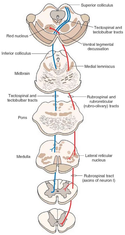
The course of this tract is shown in Figure 9-13. The rubrospinal tract arises from neurons in the red nucleus.This nucleus is located in the rostral half of the midbrain tegmentum. The axons of these neurons cross the midline in the ventral midbrain (called the ventral tegmental decussation) and descend to the contralateral spinal cord. Fibers in the rubrospinal tract are somatotopi-cally arranged. The cervical spinal segments receive fibers from the dorsal part of the red nucleus, which, in turn, receive inputs from the upper limb region of the sensori-motor cortex; the lumbosacral spinal segments receive fibers from the ventral half of the red nucleus, which in turn, receive inputs from the lower limb region of the sensorimotor cortex. The fibers of the rubrospinal tract end on interneurons that, in turn, project to the dorsal aspect of ventral (motor) horn cells. In humans most of the efferent fibers emerging from the red nucleus terminate in the inferior olive. The neurons in the red nucleus are activated monosynaptically by projections from the cortex. The function of this tract is to facilitate flexor motor neurons and inhibit extensor motor neurons. Although the effects of lesions restricted to the red nucleus are not known in humans, lesions of the midbrain tegmentum including the red nucleus are known to elicit contralateral motor disturbances (e.g., tremor, ataxia, and choreiform activity).
Tectospinal Tract
The neurons from which this tract arises are located in the superior colliculus (Fig. 9-13). The axons of these neurons terminate in upper cervical segments. This tract is believed to aid in directing head movements in response to visual and auditory stimuli.
Lateral Vestibulospinal Tract
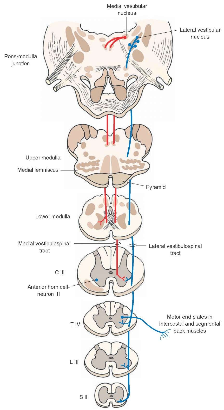
A schematic representation of the course of this tract is shown in Figure 9-14. This tract arises from neurons of the lateral vestibular nucleus, which is located at the border of the pons and medulla. The fibers in this tract are uncrossed and descend the entire length of the spinal cord. The descending fibers terminate predominantly on interneu-rons that activate motor neurons innervating extensor muscles of the trunk and ipsilateral limb. The lateral ves-tibular nucleus receives inhibitory inputs from the cerebellum and excitatory inputs from the vestibular apparatus. Impulses transmitted to the spinal cord by the lateral ves-tibular nucleus powerfully facilitate ipsilateral extensor motor neurons and their associated gamma motor neurons, thereby increasing extensor motor tone. Thus, the main functions of this tract are to control the muscles that maintain upright posture and balance.
FIGURE 9-13 Rubrospinal and tectospinal tracts. The rubrospinal tract (shown in red) arises from neurons in the red nucleus (located in the midbrain). The axons of these neurons cross the midline in the ventral midbrain (called the ventral tegmental decussation) and descend to the contralateral spinal cord. Fibers of the rubrospinal tract end on interneurons that, in turn, project to the dorsal aspect of ventral (motor) horn cells. The neurons giving rise to the tectospinal tract (shown in blue) are located in the superior colliculus. The axons of these neurons descend around the periaqueductal gray, cross the midline (called the dorsal tegmental decussation), join the medial longitudinal fasciculus in the medulla, and descend in the anterior funiculus of the spinal cord. They terminate in upper cervical segments. neuron I = first-order neuron.
Medial Vestibulospinal Tract
This tract arises from ipsilateral and contralateral medial vestibular nuclei, descends in the ventral funiculus of the cervical spinal cord, and terminates in the ipsilateral ventral horn (Fig. 9-14). The main function of this tract is to adjust the position of the head in response to changes in posture, such as keeping the head stable while walking.
Reticulospinal Tracts
The reticular formation gives rise to three functionally different fiber systems. One component of this system mediates motor functions, a second component mediates autonomic functions, and a third component modulates pain impulses.
Fibers arising from the medulla emerge from a group of large cells located medially called the nucleus reticularis gigantocellularis. These cells project bilaterally to all levels of the spinal cord, and this pathway is referred to as the medullary (lateral) reticulospinal tract (Fig. 9-15). A key function of this pathway is that it powerfully suppresses extensor spinal reflex activity. In contrast, a separate retic-ulospinal pathway arises from two distinct nuclear groups in the medial aspect of the pontine reticular formation; these two nuclear groups are called the nucleus reticularis pontis caudalis and nucleus reticularis pontis oralis. These neurons project ipsilaterally to the entire extent of the spinal cord, and their principal function is to facilitate extensor spinal reflexes. This fiber bundle is called the pontine (medial) reticulospinal tract (Fig. 9-15).
A second group of descending reticulospinal fibers mediates autonomic functions. These fibers arise largely from the ventrolateral medulla and project to the IML of the thoracolumbar cord. This fiber system excites sympathetic preganglionic neurons in the IML, and these neurons provide sympathetic innervation to visceral organs.
A third group of descending fibers is involved in pain modulation. The first limb of this pathway consists of enkephalinergic neurons located in the midbrain PAG that project to serotonergic neurons located in the nucleus raphe magnus of the medulla. The second limb of this pathway consists of projections of these serotonergic neurons to the dorsal horn of the spinal cord, making synaptic contacts with a second group of enkephalinergic interneu-rons, which, in turn, synapse upon primary afferent pain fibers. Therefore, a key function of this descending fiber system is to modulate the activity of pain impulses that ascend in the spinothalamic system.
Medial Longitudinal Fasciculus
The medial longitudinal fasciculus (MLF) is a bundle of fibers comprising mainly ascending fibers. However, it also contains a descending component. Descending fibers of the MLF arise from the medial vestibular nucleus, the reticular formation, and the superior colliculus (tectospinal fibers). The fibers in the MLF project primarily to the ipsilateral upper cervical spinal cord segments and mono-synaptically inhibit motor neurons located in these segments. By virtue of its connections with motor neurons in the cervical cord, the MLF controls the position of the head in response to excitation by the labyrinth of the ves-tibular apparatus.
Fasciculi Proprii
The fasciculi proprii include fibers (ascending and descending, crossed and uncrossed) that arise and end in the spinal cord. They connect different segments of the spinal cord. These fibers mediate intrinsic reflex mechanisms of the spinal cord, such as the coordination of upper and lower limb movements. Signals entering the spinal cord at any segment are thus conveyed to upper or lower segments and finally transmitted to ventral horn cells either directly or through interneurons.
FIGURE 9-14 Lateral and medial vestibulospinal tracts. The lateral vestibulospinal tract (shown in blue) is uncrossed and arises from neurons of the lateral vestibular nucleus (located at the border of the pons and medulla) and descends the entire length of the spinal cord. The descending fibers terminate on interneurons that activate motor neurons innervating extensor muscles in the trunk and ipsilateral limb. The medial vestibulospinal tract (shown in red) arises from ipsilateral and contralateral (not shown) medial vestibular nuclei, descends in the ventral funiculus of the cervical spinal cord, and terminates in the ipsilateral ventral horn. neuron III = third-order neuron; C = cervical; T = thoracic; L = lumbar; S = sacral.


