Many of the structures discussed thus far appear to relate to unitary functions of the nervous system. For example, structures associated with either visual, auditory, vestibular, taste, or somatosensory information are dedicated to the transmission of a single sensory process. Likewise, other regions of the brain that we have just considered provide mechanisms related to motor processes but not to other functions. Additional structures and pathways considered in previous topics are dedicated to autonomic functions.
In this topic, we will examine the anatomical and functional organization of a region forming the core of the brainstem that is referred to as the reticular formation. The reticular formation of the brainstem differs from other regions of the brain in that it participates in a variety of processes. These functions include sensory, motor, and autonomic functions; sleep and wakefulness cycles; consciousness; and the regulation of emotional behavior. The questions that this topic addresses are: (1) Which structures contribute to the processing of sensory information, and what are their contributions? (2) What are the regions of the reticular formation that contribute to motor functions, and what are their contributions? (3) How does the reticular formation contribute to autonomic functions? (4) How does the reticular formation help to regulate sleep and wakefulness cycles and states of consciousness? And, finally, (5) what is the role of the reticular formation in the regulation of emotional behavior? The remainder of this topic attempts to answer these questions by analyzing the input-output relationships of the various regions of the reticular formation and their respective roles in mediating the different processes.
Anatomical Organization of the Reticular Formation
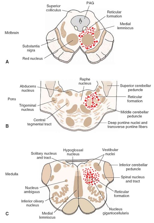
The reticular formation represents a phylogenetically older region of the brain because it is found in the core of the brainstem of lower forms of many species. Its appearance in lower forms is also similar to that found in humans. The reticular formation extends from the caudal medulla rostrally up to and including the midbrain (Fig. 23-1). At the upper end of the midbrain, the reticular formation becomes continuous with several nuclei of the thalamus, with which it is both anatomically and functionally related. These nuclei include the midline and intralaminar nuclei and are described later in this topic. The reticular formation is surrounded laterally, dorsally, and ventrally by cranial nerve nuclei, which are the ascending and descending fiber pathways of the brainstem. The reticular formation, itself, consists of many cell groups situated among large numbers of fiber bundles. The name, "reticular formation," was given to this region on the basis of its histo-logical appearance, which can be described as a reticular network of fibers and cells.
FIGURE 23-1 Position of the reticular formation in the brainstem. Diagrams illustrate the approximate positions of the reticular formation within the (A) midbrain, (B) pons, and (C) medulla as indicated by the dotted areas. Larger-sized dots represent magnocellular (large-celled regions), and smaller-sized dots represent parvocellular (small-celled regions). PAG = periaqueductal gray matter.
General Characteristics
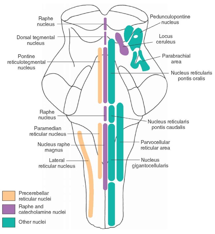
As a general rule, the nuclei of the reticular formation are arranged in the following manner. The lateral third of the reticular formation contains small-sized cells (called par-vocellular regions) whose function is to receive afferent fibers from both neighboring regions of the brainstem as well as from distant structures. The medial two thirds of the reticular formation contains different groups of large-sized cells (called magnocellular regions), which, in most instances, serve as the effector regions (i.e., the cell groups that give rise to the efferent projections of the reticular formation [Fig. 23-2]). At least two distinct magnocellular regions in the pons and one large region in the medulla have been identified. Additional groups of cells, which lie along or adjacent to the midline of the upper medulla, pons, and midbrain, are also included in the reticular formation. These cell groups comprise the raphe nuclei. These cells are important because they produce serotonin that is distributed to wide regions of the brain and spinal cord. As will be discussed later in this topic, the other types of monoamine neurons that supply the brain and spinal cord, namely, norepinephrine and dopamine, are also located at different levels within the reticular formation.
Afferent Connections
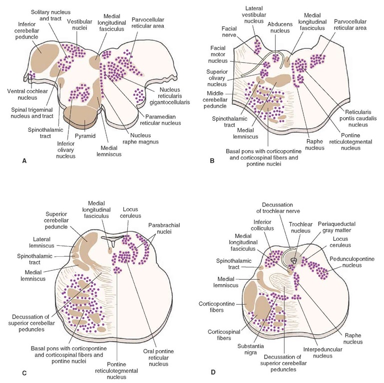
Cranial nerve nuclei and secondary sensory fibers form an outer shell that surrounds the reticular formation (and, particularly, the parvocellular region) on its dorsal, lateral, and ventral sides (Figs. 23-2 and 23-3). Because of this arrangement, the reticular formation is strategically positioned so that the various sensory systems can easily provide inputs to it. In this manner, the reticular formation is endowed with a capacity to modulate a wide variety of sensory processes.
The reticular formation receives inputs from brainstem and forebrain regions associated with sensory modalities as well as from structures associated with motor functions, which include the cerebral cortex and cerebellum. Moreover, structures associated with autonomic and higher order visceral processes, such as the hypothalamus and limbic system, also contribute inputs to the reticular formation.
FIGURE 23-2 The positions of the magnocellular and parvocellular regions of the reticular formation.
FIGURE 23-3 Cross sections of the brainstem. Note the principal nuclei of the reticular formation as well as other (nonreticular) nuclei situated adjacent to them. Many of these (other) nuclei make synaptic connections with nuclei of the reticular formation. (A) Medulla at level of rostral aspect of inferior olivary nucleus. (B) Level of caudal pons at the level of the abducens and facial nerves. (C) Rostral pons at level of the nucleus locus ceruleus and superior cerebellar peduncle. (D) Oblique section in which the dorsal aspect is at the level of the inferior colliculus of the caudal midbrain and the ventral aspect is at the level of the rostral aspect of the basilar pons.
The nature of these afferent sources to the reticular formation is considered in the following sections.
Sensory Systems
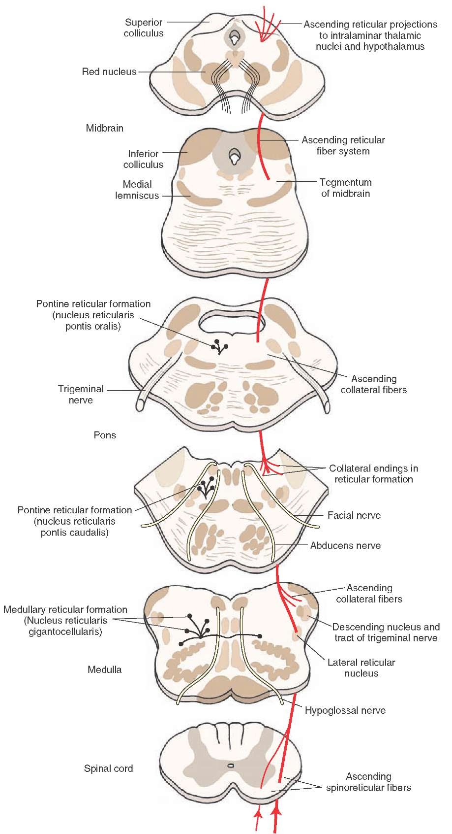
It is now well established that fibers arising from parts of the spinal cord gray matter (lamina VII) give off collaterals that terminate in specific parvocellular regions of the reticular formation, mainly adjacent to the magnocellular nuclei of the medulla (nucleus gigantocellularis) and pons (nucleus reticularis pontis caudalis and nucleus reticularis pontis oralis [Figs. 23-3 and 23-4]). In turn, these sensory signals are then transmitted from the parvocel-lular to the magnocellular nuclei. Neuronal groups with the reticular formation synapse with spinoreticular fibers and give rise to long ascending fibers. Accordingly, the fibers projecting from spinal cord to the brainstem may be thought of as the first neurons in a chain of fibers that innervate the intralaminar nuclei of the thalamus, and these nuclei ultimately supply the cerebral cortex. It is believed that the information transmitted to the thalamus from the reticular formation mediates affective components of pain.Nociceptive inputs into the PAG represent part of a circuit whose descending component serves to inhibit ascending pain signals to the cerebral cortex.
FIGURE 23-4 The connections of the lateral spinothalamic tract, including those made with the reticular formation.
The results of various studies have shown that other types of sensory signals can also produce cortical excitation by virtue of their connections with the brainstem reticular formation. On the basis of these studies dating back to the period of the 1940s and 1950s, the term "reticular activating system" was proposed as a mechanism by which sensory and other inputs to the reticular formation can modulate cortical neurons, cortical excitability, and states of consciousness. The importance of the reticular activating system is considered later in this topic. Other Secondary Sensory Systems. All sensory systems contribute fibers that ultimately make their way into the reticular formation. Trigeminal (for somatosensory information), solitary (for taste information), auditory, and vestibular nuclei lie adjacent to the reticular formation. Information is thus transmitted to the reticular formation from these structures, either as a result of the dendritic arborizations of neurons whose cell bodies lie in the l ateral aspect of the reticular formation but which extend into these regions or by short axons that arise from these sensory regions that pass into the lateral aspects of the reticular formation.
In addition to these sensory inputs, impulses mediated from regions associated with olfactory and visual systems also contribute inputs to the reticular formation. Secondary olfactory signals are received by a number of limbic structures (amygdala, hippocampal formation, and septal area) that have known projections to the reticular formation.Visual signals can reach the superior colliculus via optic tract fibers. From the superior colliculus, fibers project directly to the reticular formation of the midbrain and pons.
Thus, it is evident that the reticular formation receives multiple sensory inputs from all sensory systems. The retic-ular formation cannot maintain the specificity of the information that it receives from each of these sensory systems. Therefore, information of a nonspecific nature is transmitted via ascending fibers to the thalamus; this is in contrast to the specific transmission lines characteristic of other ascending sensory systems such as the dorsal column-medial lemniscus pathway. As indicated earlier, an important function of these signals that enter the reticular formation is to allow the reticular activating system to alter excitability levels of target neurons in the thalamus.


