The forebrain constitutes all parts of the brain that lie rostral to the midbrain and are derived from the prosencephalon. It includes the diencephalon (i.e., thalamus, subthalamus, epithalamus, and hypothalamus), basal ganglia, limbic system, internal capsule, anterior commissure, and cerebral cortex. These structures mediate a wide variety of functions, which include sensory, motor, autonomic, and complex behavioral and visceral processes. These functions will be discussed separately in detail in subsequent topics.
Diencephalon
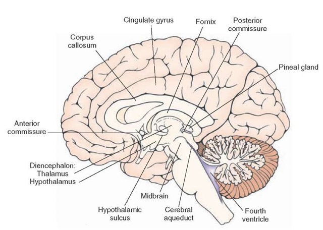
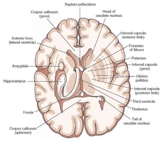
The diencephalon consists of two major regions called the thalamus and hypothalamus and two additional but smaller areas called the epithalamus and subthalamus. The boundaries of the diencephalon include the following: (1) the rostral boundary is the anterior commissure and the lamina terminalis (i.e., the rostral limit or wall of the third ventricle); (2) the caudal boundary is the posterior commissure, which divides the diencephalon from the midbrain; (3) the dorsal boundary is the roof of the diencephalon; (4) the ventral boundary is the base of the hypothalamus; (5) the lateral boundary is the internal capsule that divides it from the lenticular nuclei (i.e., globus pallidus and putamen); and (6) the third ventricle constitutes its medial limits (Figs. 13-1 through 13-3).
FIGURE 13-1 Mid-sagittal section of the brain depicting the medial aspect of the forebrain. Note that the diencephalon is bounded on its rostral end by the anterior commissure and at its caudal end by the posterior commissure. Within the diencephalon, the thalamus is separated from the hypothalamus by the hypothalamic sulcus.
FIGURE 13-2 Horizontal section of the brain depicting:
(1) on one side, the relationships of the internal capsule to the lateral ventricles, thalamus, and basal ganglia; and
(2) on the other side, the positions occupied by the hip-pocampal formation, fornix, and amygdala.
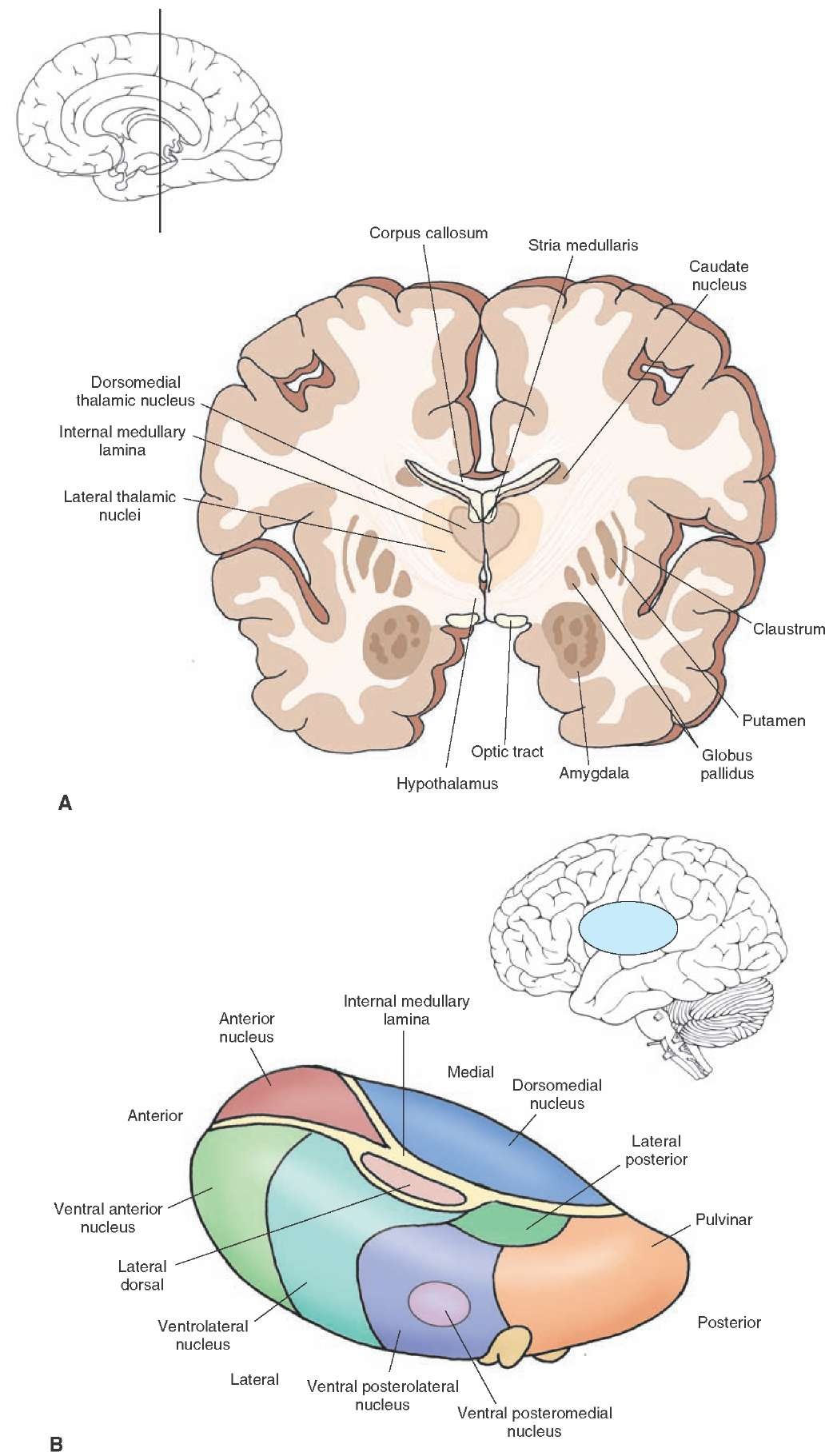
FIGURE 13-3 Arrangement of thalamic nuclei. (A) Cross section of the brain taken through the level of the anterior third of the diencephalon. The cross section indicates the relationship of the diencephalon to the internal capsule and basal ganglia as well as the position occupied by the amygdala in the temporal lobe. The inset indicates the approximate cut of the section. (B) The relative positions of the thalamic nuclei to one another and the position of the thalamus within the brain.
Thalamus
The thalamus constitutes the largest component of the diencephalon. It appears as an oval-shaped region and is bounded rostrally by the anterior commissure and caudally by the posterior commissure. Its dorsal limit is the roof of the thalamus (subarachnoid space in the form of the transverse cerebral fissure), and it extends ventrally to the level of the hypothalamic sulcus (of the third ventricle) (Fig. 13-1). It is composed of a variety of nuclei that serve diverse functions. In general, these nuclei serve as relays by which information is transmitted from different regions of the central nervous system (CNS) to the cerebral cortex.
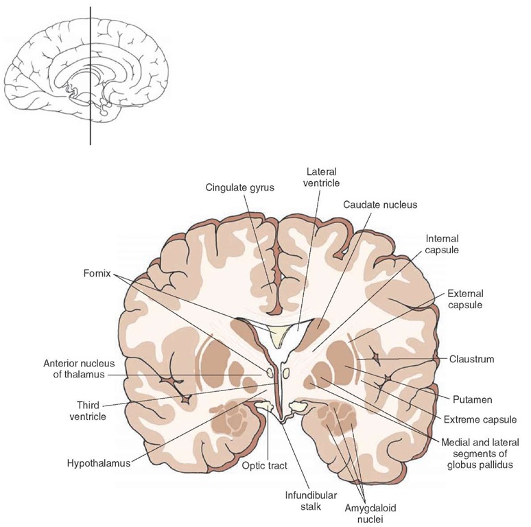
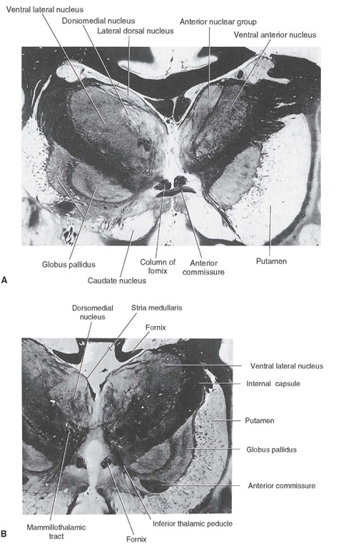
The major nuclear divisions of the thalamus, the thalamic nuclei, include anterior, medial, and lateral nuclei (Fig. 13-3B). The anterior nucleus of the thalamus forms a rostral swelling of the thalamus (Figs. 13-4 and 13-5A).
It has anatomical connections with the hippocampal formation, cingulate gyrus, and mammillary bodies. Because the hippocampal formation and cingulate gyrus play a role in selective aspects of memory function, it is likely that the anterior thalamic nucleus is involved with some aspects of memory function as well. Previously, it was believed this nucleus was also associated with the regulation of emotional behavior.
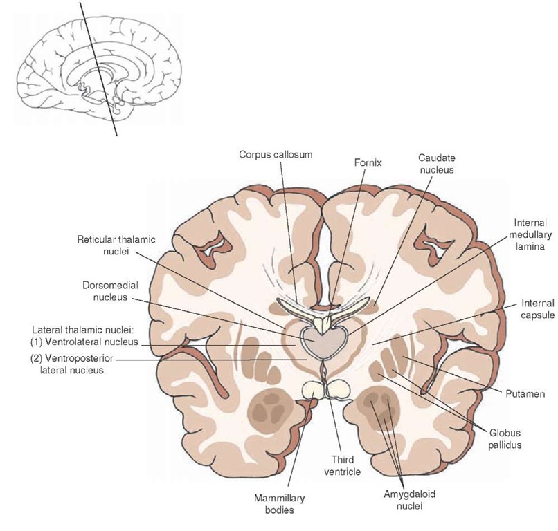
Posterior to the anterior nucleus lies a large structure called the dorsomedial nucleus (Figs. 13-5B and 13-6), which extends from its border with the anterior nucleus posteriorly to the level of the posterior thalamus. The dorsomedial nucleus is separated from the lateral thala-mus by a thin plate of myelinated fibers called the internal medullary lamina (Figs. 13-3 and 13-6). It is associated with mediation of affective processes and emotional behavior.
Within the lateral thalamus, the following structures can be identified beginning from the most anterior level of thalamus.
FIGURE 13-4 Cross section of the brain depicting the position occupied by the anterior thalamic nucleus in the anterior aspect of the thalamus. The inset indicates the approximate cut of the section.
FIGURE 13-5 Two levels of the thalamus. (A) Photograph of an asymmetrical section taken through the level of the rostral aspect of the thalamus (on the right side) in which the anterior nucleus and ventral anterior nucleus are clearly shown. (B) Photograph of a cross section taken through the middle third of the thalamus in which the dorsome-dial nucleus is clearly shown. Note also the positions occupied by the ventrolateral thalamic nucleus and stria medullaris.
The ventral anterior nucleus (VA) is situated at approximately the same anterior-posterior (rostro-caudal) plane as the anterior nucleus (Fig. 13-3B). Just posterior to the ventral anterior nucleus lies the ventral lateral nucleus (VL). Both the VA and VL are intimately involved in the control of motor functions. At the level of VL, a second structure can be identified. It lies just off the dorsolateral aspect of the VL and is called the lateral dorsal nucleus (LD). Functions of the LD are not well understood. Posterior to the VL is the ventral basal complex. The ventral basal complex consists of two nuclei called the ventral posterior lateral (VPL) and ventral posterior medial (VPM) nuclei (Fig. 13-6). Both the VPL and VPM serve as relay nuclei for the transmission of somatosensory information from the body and head, respectively, to different regions of the postcentral gyrus. Adjoining the dorsolateral surface of the ventrobasal complex is the lateral posterior nucleus (LP). LP may be associated with integration of different modalities of sensory inputs and cognitive functions associated with them.
FIGURE 13-6 Cross section of the brain depicting the position occupied by the dorsomedial thalamic nucleus situated in the middle third of the thalamus. Note that the medial and lateral nuclei of thalamus are separated by an internal medullary lamina. The inset indicates the approximate cut of the section.
FIGURE 13-7 Cross section of the brain depicting the positions occupied by the pulvinar, medial geniculate, and lateral geniculate nuclei in the caudal aspect of the thalamus and proximal to the mid-brain. The inset indicates the approximate cut of the section.
Within the posterior aspect of the thalamus lie three important (lateral) thalamic nuclei adjacent to the rostral midbrain; they can usually be detected when sections of the rostral midbrain are taken (Fig. 13-7). The largest of the structures is the pulvinar nucleus. It is relatively massive in size and appears to be associated with cognitive functions involving auditory and visual stimuli. The other two structures include the medial geniculate nucleus, which serves as an important relay for the auditory system, and the lateral geniculate nucleus, which lies ventrola-teral to the medial geniculate nucleus and which serves as a key relay of visual impulses to the visual cortex from both retinas.
In addition to the major nuclei of the medial and lateral thalamus, which have significant inputs into the cerebral cortex, there is another class of neuronal groups that lie either along or near the midline of the thalamus (i.e., mid-line thalamic nuclei) or along the lateral margin of the thalamus (i.e., reticular nucleus [Fig. 13-6]) or are enclosed by the internal medullary lamina (i.e., intralaminar nuclei, the most important of which is the centromedian nucleus [Fig. 13-3B, see also Fig. 13-8]). These nuclei modulate the activity of the major projection nuclei of the thalamus and have wide-ranging effects upon cortical neurons throughout the cortex.
