We further indicated that highly precise, complex, and purposeful voluntary movements result from the transmission of signals passing through the corticospinal tract to the spinal cord where they act on lower motor neurons of the spinal gray matter, which produce movements by virtue of their actions on different muscle groups. If the production of movement and sequences of movements are due to the actions of the cerebral cortex and its descending axons of the corticospinal tract, then it is possible to consider that the basal ganglia provides a postural platform off of which purposeful movement can be made.
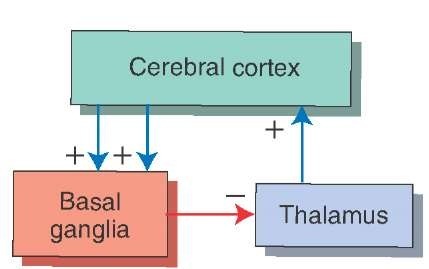
The primary function of the basal ganglia is to provide a feedback mechanism to the cerebral cortex for the initiation and control of motor responses (Fig. 20-1). Much of the output of the basal ganglia, which is mediated through the thalamus, is to reduce or dampen the excitatory input to the cerebral cortex. When there is a disruption of this mechanism, disturbances in motor function ensue. In general, it may be stated that when the discharge patterns of the basal ganglia become excessive, the effect on motor systems is to produce abnormal slowing of movements. At other times, however, lesions of the basal ganglia produce a reduced output, the result of which is the presence of abnormal, involuntary movements that occur during periods of rest. This form of disorder is called dyskinesia. To understand how the basal ganglia functions as a feedback mechanism, it is essential to know the following:
(1) the nature of the afferent supply to the basal ganglia;(2) the internal circuitry; (3) the efferent projections of the basal ganglia; and (4) the functional role of the neuro-transmitters in the basal ganglia.
Composition of the Basal Ganglia
As a brief review, the basal ganglia consist of the neostria-tum (caudate nucleus and putamen), paleostriatum (globus pallidus), and two additional nuclei, the subthalamic nucleus and substantia nigra, which are included with the basal ganglia because of their anatomical connections (made with different nuclei of the basal ganglia [Fig. 20-2]). The primary regions of the basal ganglia that serve as afferents (receiving areas) are the caudate nucleus and putamen. The major outputs of the basal ganglia arise from neurons located in the medial pallidal segment. These neurons give rise to two fiber bundles, the ansa lenticularis and lenticular fasciculus, which supply thalamic nuclei (see Fig. 13-12).
FIGURE 20-1 The feedback circuit between the cerebral cortex and basal ganglia. Cortical inputs into the basal ganglia are excitatory (+), and the outputs of the basal ganglia back to the cortex are mediated through the thalamus. Because the outputs of the thalamus to the cortex are generally excitatory (+), the inhibitory inputs into the thalamus from the basal ganglia (-) serve to dampen thalamic excitation of cortical neurons.
Afferent Sources of the Basal Ganglia
The largest afferent source of the basal ganglia arises from the cerebral cortex. In fact, most regions of the cortex contribute projections to the basal ganglia. These include inputs from motor, sensory, association, and even limbic areas of the cortex. While the caudate nucleus and puta-men serve as the primary target regions of afferent projections from the cortex, the source of cortical inputs to these regions of the basal ganglia differ. The principal inputs from the primary motor, secondary motor, and primary somatosensory regions of cortex are directed to the puta-men. These inputs to the putamen are somatotopically organized, which means that different regions of the puta-men receive sensory and motor inputs that are associated with different parts of the body. The caudate nucleus, on the other hand, receives inputs from cortical association regions, frontal eye fields, and limbic regions of cortex (Fig. 20-3). Thus, the putamen appears to be concerned primarily with motor functions, while the caudate nucleus, which appears to receive more varied and integrated cortical inputs, is likely involved with cognitive aspects of movement, eye movements, and emotional correlates of movement (relegated to the ventral aspect of the neos-triatum).
The neostriatum also receives an indirect source of cortical input. The source of this input is the centromedian nucleus of the thalamus. This nucleus receives afferent fibers primarily from the motor cortex and projects its axons topographically to the putamen as thalamostriate fibers. An additional but highly important source of dopaminergic inputs to the neostriatum is the substantia nigra. The function of this afferent source to the neostria-tum is considered later in this topic.
The modular organization of the neostriatum is as follows. The projections from the neocortex and thalamus are not distributed uniformly to the neostriatum. Instead, the projections end in compartments within different parts of the neostriatum. The smaller of these compartments is referred to as a patch or striosome and is surrounded by a larger compartment referred to as a matrix. These two compartments are distinguished from each other because they have different neurochemical properties, contain different receptors, and receive inputs from different cortical regions. The matrix is acetylcholinesterase rich. The strio-somes, however, are acetylcholinesterase poor but contain peptides, such as somatostatin, substance P, and enkepha-lin. The striosomes receive their inputs mainly from limbic regions of cortex, and neurons from this region preferentially project to the substantia nigra. In contrast, the neurons of the matrix receive inputs from sensory and motor regions of cortex, and many of them project to the globus pallidus. The overwhelming majority of projection neurons from both of these compartments of the neostriatum are g-aminobutyric acid (GABA)-ergic. Their significance is considered in the following paragraph.
FIGURE 20-2 The anatomical structures that comprise the basal ganglia. (A) The principal components of the basal ganglia situated within the forebrain: the caudate nucleus, putamen, globus pallidus, and the subthalamic nucleus, which is linked to the basal ganglia by its connections with the primary structures comprising the basal ganglia. (B) The position of the substantia nigra within the midbrain, which is also linked to the basal ganglia because of its connections with the neos-triatum.
FIGURE 20-3 Relationship of the cerebral cortex to the putamen and caudate nucleus. The diagram illustrates that the putamen receives fibers from motor regions of the cerebral cortex, whereas the caudate nucleus receives inputs from association regions (temporal and parietal lobes) as well as inputs from other regions of the frontal lobe, including the prefrontal cortex.
The presumed role of the neostriatum in motor functions can be illustrated by the following example. During active movement of a joint of a finger, the cells within a certain striosomal or matrix compartment become active, whereas neurons embedded within other compartments become active only after there is passive movement of the same joint. This would suggest that the neurons embedded within given compartments function in a similar manner to those present in functional homogenous regions of the cerebral cortex with respect to movement (i.e., specific regions of motor cortex can be subdivided into distinct functional columns in which the neurons in one column respond to one feature of movement of a limb, whereas those neurons situated in an adjoining column respond to a different feature of movement of that limb.
Internal Connections of the Basal Ganglia
The anatomical relationships between different components of the basal ganglia are extensive. The most salient of the connections include the following: (1) the projections from the neostriatum to the globus pallidus, (2) the reciprocal relationships between the neostriatum and substantia nigra, and (3) the reciprocal relationships between the globus pallidus and the subthalamic nucleus. In examining these relationships, the overall role of the basal ganglia in motor functions should be kept in mind. Namely, as signals are transmitted through the basal ganglia in response to cortical inputs, they ultimately result in a distinct response transmitted back to the motor areas of the cerebral cortex. Moreover, the circuits within the basal ganglia by which signals are transmitted back to the cerebral cortex may be direct or indirect. The differences between the direct and indirect routes are discussed in the following section.
Connections of the Neostriatum With the Globus Pallidus
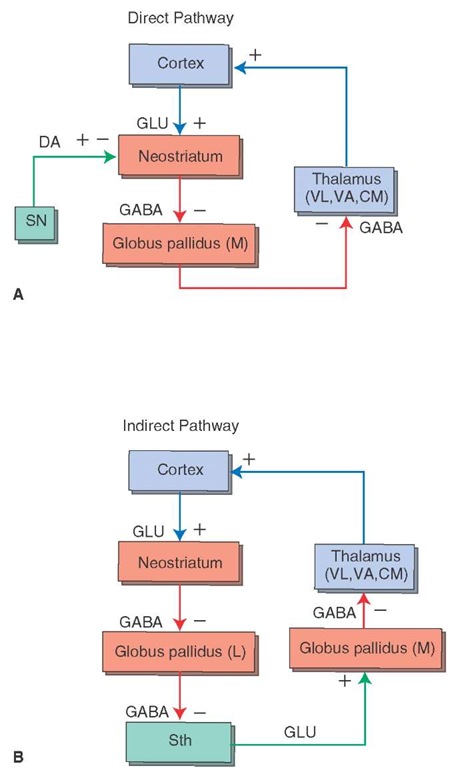
There are two basic projection targets of the neostriatum: the globus pallidus and the substantia nigra. The neostria-tum projects to two different regions of the globus pallidus: the medial (internal) pallidal segment and the lateral (external) pallidal segment (Fig. 20-4). GABA mediates the pathway from the neostriatum to the medial pallidal segment; likewise, the pathway from the neostriatum to the lateral pallidal segment also primarily uses GABA as a likely neurotransmitter.
Each of these projections forms the initial links of two different circuits within the basal ganglia. Because the primary output of the basal ganglia is from the internal palli-dal segment, the projection (neostriatum — globus pallidus [internal] — thalamus — neocortex) is called the direct pathway (Fig. 20-4). In contrast, the external segment of the globus pallidus shares reciprocal projections with the subthalamic nucleus. Thus, this circuit of the basal ganglia can be outlined as follows: neostriatum — globus pallidus (external) — subthalamic nucleus — globus pallidus (internal) – thalamus – neocortex. Because this circuit involves a loop through the subthalamic nucleus, it is called the indirect pathway (Fig. 20-4). The neurotrans-mitters involved in the reciprocal pathway between the globus pallidus and subthalamic nucleus and the overall functional significance of these pathways are discussed later in this topic.
FIGURE 20-4 The detailed anatomical and functional nature of the input, internal, and output circuits of the basal ganglia. Note the distinction between the direct and indirect pathways. (A) The direct pathway involves projections from the neostriatum to the medial (internal) pallidal segment, which in turn, projects to the thalamus and then to the cerebral cortex. (B) The indirect pathway involves projections from the neostriatum to the lateral pallidal segment (L), which in turn, projects to the subthalamic nucleus. The subthalamic nucleus then projects to the medial pallidal segment (M), and the remaining components of the circuit to the cerebral cortex are similar to that described for the direct pathways. Red arrows depict inhibitory pathways, and blue arrows indicate excitatory pathways. The green pathways represent dopaminergic projections to the neostriatum, which have opposing effects on D, and D2 receptors (see Fig. 20-5), and the excitatory projection from the subthalamic nucleus (Sth) to the medial pallidal segment. When known, the neurotransmitter for each of the pathways is indicated. DA = dopamine; GLU = glutamate; CM = centromedian nucleus; VA = ventral anterior nucleus; VL = ventrolateral nucleus; (+) = excitation; (-) = inhibition.
FIGURE 20-5 Key relationships of the substantia nigra. The pars reticulata of the substantia nigra receives an inhibitory (red line) gamma aminobutyric acid (GABA)-ergic input from the neostriatum. In turn, there are two important outputs of the substantia nigra. The first is a dopaminergic (DA) projection to the neostriatum (which is excitatory when acting through D, receptors and inhibitory when acting through D2 receptors). The second is an inhibitory GABAergic projection from the pars reticulata to the ventral anterior (VA) and ventrolateral (VL) thalamic nuclei as well as to the superior colliculus. GPL = lateral segment of the globus pallidus; GPM = medial segment of the globus pallidus; (+) = excitation; (-) = inhibition.


