Types of Neurons
Based on morphological characteristics, the neurons have been classified into the following groups: multipolar, bipolar, pseudo-unipolar, and unipolar.
Multipolar Neurons
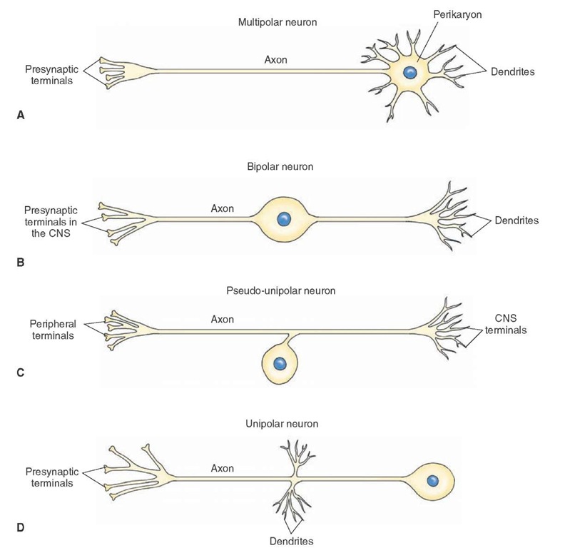
Multipolar neurons are most common in the brain and spinal cord. They possess three or more dendrites and one long axon issuing from the cell body (Fig. 5-3A). A large motor neuron of the anterior horn of the spinal cord is one example of such a neuron.
Bipolar Neurons
In bipolar neurons, two processes, one on each end, arise from an elongated cell body (Fig. 5-3B). One process ends in dendrites, and the other process, an axon, ends in terminals in the CNS. These neurons have sensory functions and transmit information received by the dendrites on one end to the CNS via the axon terminals on the other end. Retinal bipolar cells, sensory cells of the cochlea, and ves-tibular ganglia are included in this category.
Pseudo-Unipolar Neurons
In pseudo-unipolar neurons, a single process arises from the cell body and divides into two branches. One of these branches projects to the periphery, and the other projects to the CNS (Fig. 5-3C). Each branch has the structural and functional characteristics of an axon.
Information collected from the terminals of the peripheral branch is transmitted to the CNS via the terminals of the other branch. Examples of this type of cell are sensory cells in the dorsal root ganglion and barore-ceptor-sensitive cells in the nodose ganglion. The baroreceptor-sensitive cells sense changes in the systemic blood pressure and transmit this information to neurons in the dorsal medulla.
Unipolar Neurons
Unipolar neurons are relatively rare in vertebrates. In these neurons, dendrites arise from one end of the neuron, and an axon arises from the site where the dendrites are located (Fig. 5-3D).
Other Types of Neurons
Neurons can also be divided into two groups:
1. Principal or projecting neurons are also known as type I or Golgi type I neurons. Principal neurons (e.g., motor neurons in the ventral horn of the spinal cord) possess very long axons and form long fiber tracts in the brain and the spinal cord. 2. Intrinsic neurons are also known as type II or Golgi type II neurons. Intrinsic neurons have very short axons. These neurons are interneurons and are considered to have inhibitory function. They are abundant in the cerebral and cerebellar cortex.
FIGURE 5-3 Different types of neurons. (A) Multipolar neuron. (B) Bipolar neuron. (C) Pseudo-unipolar neuron. (D) Unipolar neuron. CNS = central nervous system.
Neuralgia
The supporting cells located in the CNS are called neuro-glia or simply glial cells. They are more numerous (5-10 times) than neurons. Glial cells do not propagate action potentials. They are involved in maintaining the appropriate environment for normal neuronal function and providing structural support for the neurons. Neuroglia have been classified into the following groups: astrocytes, oli-godendrocytes, microglia, and ependymal cells.
Astrocytes
Among the glial cells, astrocytes are the largest and have a stellate (star-shaped) appearance because their processes extend in all directions. Their nuclei are ovoid and centrally located. The astrocytes provide support for the neurons, a barrier against the spread of transmitters from synapses, and insulation to prevent electrical activity of one neuron from affecting the activity of a neighboring neuron. Some transmitters (for example, glutamate and Y-aminobutyric acid [GABA]), when released from nerve terminals in the CNS, are taken up by astrocytes, thus terminating their action. The neurotransmitters taken up by astrocytes are processed for recycling.The intermediate filaments in astrocytes contain a distinctive protein called glial fibrillary acidic protein(GFAP). Astrocytes are capable of proliferation in the mature brain. This may be the reason for the majority of CNS tumors to be of astrocytic origin.
When extracellular K+ increases in the brain due to local neural activity, astrocytes take up K+ via membrane channels and help to dissipate it over a large area. Excess K+ is distributed to several adjacent astrocytes via gap junctions connecting them and is finally distributed to perivascular spaces. Astrocytes are further subdivided into the following subgroups: protoplasmic astrocytes, fibrous astrocytes, and Muller cells.
Protoplasmic Astrocytes
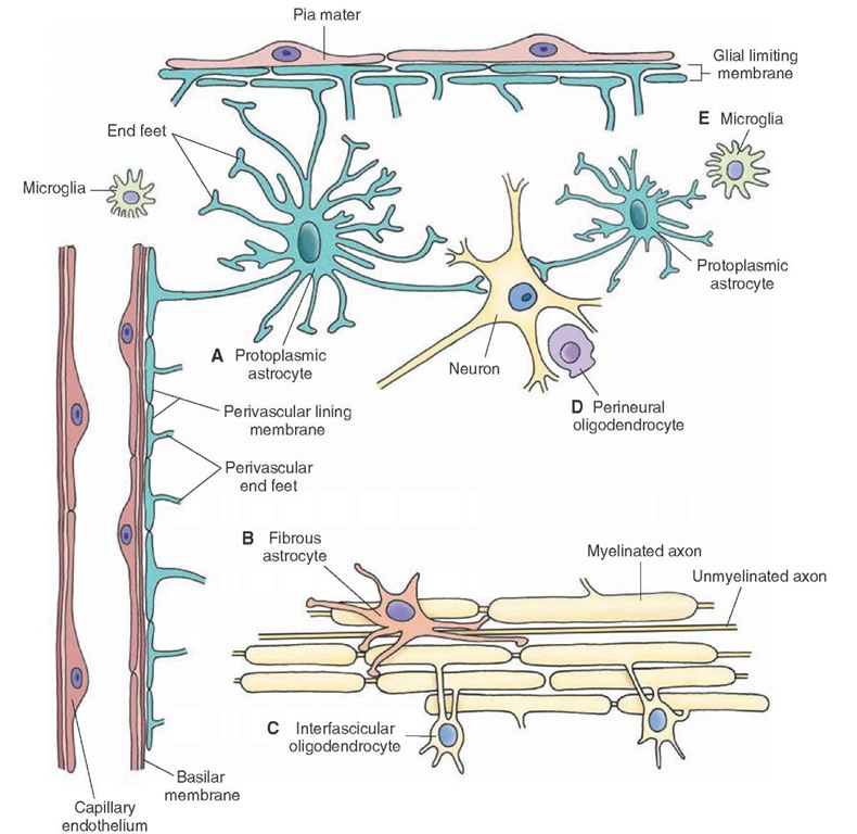
Protoplasmic astrocytes are present in the gray matter in close association with neurons. Because of their close association with the neurons, they are considered satellite cells and serve as metabolic intermediaries for neurons. They give out thicker and shorter processes, which branch profusely. Several of their processes terminate in expansions called end-feet (Fig. 5-4A). The neuronal cell bodies, dendrites, and some axons are covered with end-feet of the astrocytes. The end-feet join together to form a limiting membrane on the inner surface of the pia mater (glial limiting membrane) and outer surface of blood vessels (called perivascular lining membrane) (Fig. 5-4A). The perivascular end-feet may serve as passageways for the transfer of nutrients from the blood vessels to the neurons. Abutting of processes of protoplasmic astrocytes on the capillaries as perivascular end-feet is one of the anatomical features of the blood-brain barrier.
Fibrous Astrocytes
These glial cells are found primarily in the white matter between nerve fibers (Fig. 5-4B). Several thin, long, and smooth processes arise from the cell body; these processes show little branching. Fibrous astrocytes function to repair damaged tissue, and this process may result in scar formation.
Muller Cells
Modified astrocytes, Muller cells are present in the retina.
Oligodendrocytes
These cells are smaller than astrocytes and have fewer and shorter branches. Their cytoplasm contains the usual organelles (e.g., ribosomes, mitochondria, and microtu-bules), but they do not contain neurofilaments. In the white matter, oligodendrocytes are located in rows along myelinated fibers and are known as interfascicular oli-godendrocytes (Fig. 5-4C). These oligodendrocytes are involved in the myelination process (described later). The oligodendrocytes present in the gray matter are called perineural oligodendrocytes (Fig. 5-4D).
Microglia
These are the smallest of the glial cells (Fig. 5-4E). Micro-glia usually have a few short branching processes with thorn-like endings. These processes arising from the cell body give off numerous spine-like projections. They are scattered throughout the nervous system. When the CNS is injured, the microglia become enlarged, mobile, and phagocytic. In the CNS, microglia may be activated in response to trauma or stroke and release cytokines (small proteins that mediate and regulate immunity and inflammation) such as tumor necrosis factor-alpha (TNF-a). Some of these released cytokines are neurotoxic. Thus, activation of microglia may be responsible for secondary neuronal damage in such situations.
Ependymal Cells
Ependymal cells consist of three types of cells: ependymo-cytes, tanycytes, and choroidal epithelial cells.
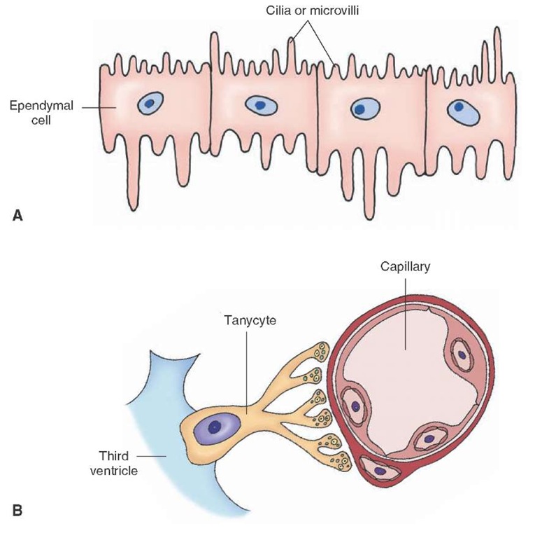
Ependymocytes are cuboidal or columnar cells that form a single layer of lining in the brain ventricles and the central canal of the spinal cord. They possess microvilli and cilia (Fig. 5-5A). The presence of microvilli indicates that these cells may have some absorptive function. The movement of their cilia facilitates the flow of the cerebro-spinal fluid (CSF).
Tanycytes are specialized ependymal cells that are found in the floor of the third ventricle, and their processes extend into the brain tissue where they are juxtaposed to blood vessels and neurons (Fig. 5-5B). Tanycytes have been implicated in the transport of hormones from the CSF to capillaries of portal system and from hypothalamic neurons to the CSF.
FIGURE 5-4 Different types of neuroglia. (A) Protoplasmic astrocyte. (B) Fibrous astrocyte. (C) Interfascicular oligodendrocyte. (D) Perineural oligodendrocyte. (E) Microglia.
Choroidal epithelial cells are modified ependymal cells. They are present in the choroid plexus and are involved in the production and secretion of CSF. They have tight junctions that prevent the CSF from spreading to the adjacent tissue.
Myelinated Axons
Myelinated axons are present in the peripheral nervous system (PNS) and the CNS.
Peripheral Nervous System
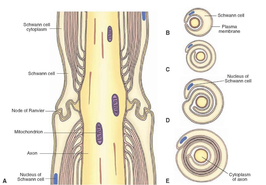
In the PNS, Schwann cells (Fig. 5-6A) provide myelin sheaths around axons. The myelin sheaths are interrupted along the length of the axons at regular intervals at the nodes of Ranvier (Fig. 5-6A). Thus, the nodes of Ranvier are uninsulated and have a lower resistance. These nodes of Ranvier are rich in Na+ channels, and the action potential becomes regenerated at these regions. Therefore, the action potential traveling along the length of the axon jumps from one node of Ranvier to another. This type of propagation enables the action potential to conduct rapidly and is known as saltatory conduction. During the myelina-tion, the axon comes in contact with the Schwann cell, which then rotates around the axon in clockwise or counterclockwise fashion. As the Schwann cell wraps around the axon, the cytoplasm becomes progressively reduced, and the inner layers of the plasma membrane come in contact and fuse together (Fig. 5-6, B-E).
Central Nervous System
Within the brain and the spinal cord, oligodendrocytes form the myelin sheaths around axons of neurons. Several glial processes arise from one oligodendrocyte and wrap around a portion of the axon (Fig. 5-7). The intervals between adjacent oligodendrocytes are devoid of myelin sheaths and are called the nodes of Ranvier (Fig. 5-7). Unlike in peripheral axons, the process of an oligodendro-cyte does not rotate spirally on the axon. Instead, it may wrap around the length of the axon. The cytoplasm is reduced progressively, and the sheath consists of concentric layers of plasma membrane. Unlike in peripheral nerves, one oligodendrocyte forms myelin sheaths around numerous (as many as 60) axons of diverse origins.
FIGURE 5-5 (A) Ependymocytes are cuboidal or columnar in shape and line the brain ventricles. (B) Tanycytes are specialized ependymal cells that are found in the floor of the third ventricle.
FIGURE 5-6 Myelination of peripheral nerves. (A) A longitudinal section showing a myelinated nerve fiber. The myelin sheaths are discontinuous along the length of the axons, and the intervals between these sheaths are called nodes of Ranvier. (B-E) Cross sections showing different stages of formation of a myelin sheath.


