The Neuron
Signals from one nerve cell to another are transferred across special zones of contact between the neurons that are known as synapses. The mechanism by which neurons communicate with each other is called synaptic transmission. Chemical neurotransmission is the most prevalent mechanism of communication between neurons. It involves release of chemical substances (neurotransmit-ters) from the presynaptic terminals of neurons, which excite or inhibit one or more postsynaptic neurons.
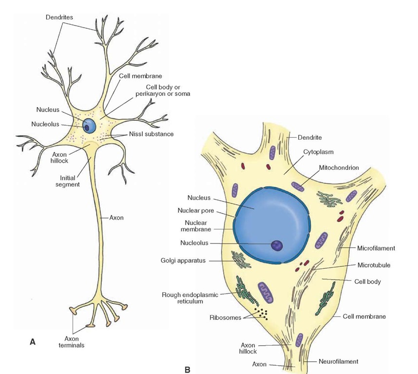
The human brain consists of about 1011 nerve cells. Each nerve cell (neuron) consists of a cell body (peri-karyon or soma) from which numerous processes (neur-ites) arise. The neurites that receive information and transmit it to the cell body are called dendrites (Fig. 5-1, A and B). A long neurite conducts information from the cell body to different targets and is known as an axon (Fig. 5-1, A and B). The cytoskeleton of a neuron consists of fibrillar elements (e.g., neurofilaments and microfilaments) and their associated proteins (Fig. 5-1B). Descriptions of the different components of the neuron are provided in the following sections.
The Cell Membrane
The cell (plasma) membrane (Fig. 5-1, A and B) forms the external boundary of the neuronal cell body and its processes. It is about 6 to 8 nanometers (nm) thick and consists of a double layer of lipids in which proteins, including ion channels, are embedded. Inorganic ions enter and leave the neuron through the ion channels.
The Nerve Cell Body
The neuronal cell body, also called perikaryon or soma (Fig. 5-1, A and B), consists of a mass of cytoplasm bounded by an external membrane. The presence of neurites increases the surface area of the cell body for receiving signals from axons of other neurons. The total volume of cytoplasm present in the neurites is much greater than in the cell body proper. The cell body contains the nucleus and various organelles and is the metabolic and trophic (relating to nutrition) center of the neuron. Accordingly, the synthesis of most proteins, phospholipids, and other macromolecules occurs in the soma.
Figure 5-1 A schematic representation of a neuron. (A) Note the orientation of the dendrites, axon, and nucleus. The f irst few microns of the axon as it emerges from the axon hillock represent the initial segment of the axon. (B) Components of the neuron: the cell membrane, nucleus, nuclear membrane, nucleolus, and the organelles.
The Nucleus
Histologically, the term nucleus refers to the round structure that is usually located in the center of the cell body (Fig. 5-1). It should be noted that, in neuroanatomy, the term "nucleus" also refers to a collection of neurons in the brain or spinal cord with similar morphological characteristics (e.g., nucleus ambiguus). The contents of the nucleus are enclosed within a nuclear membrane. The nuclear membrane is double-layered and contains fine pores through which substances can diffuse in and out of the nucleus (Fig. 5-1B). The genetic material of the nucleus, consisting of deoxyribonucleic acid (DNA), is called chromatin. When the nucleoplasm is homogenous and does not stain with basic dyes, the DNA is said to be widely dispersed and in euchromatin form. The nucleus contains a prominent (relatively large) nucleolus that is concerned with the synthesis of ribonucleic acid (RNA) and stains deeply. In the female, the Barr body represents one of the two X chromosomes and is located at the inner surface of the nuclear membrane.
The Cytoplasm
The following organelles and inclusions are present in the cytoplasm.
Nissl Substance or Bodies
This granular material is present in the entire cell body and proximal portions of the dendrites. However, it is not present in the axon hillock (portion of the soma from which the axon arises) and the axon (Fig. 5-1A). The Nissl substance consists of RNA granules called ribosomes (Fig. 5-1A). In all neurons, a net-like meshwork consisting of a highly convoluted single membrane, called the endo-plasmic reticulum, extends throughout the cytoplasm. Many ribosomes are attached to the membrane of the endo-plasmic reticulum and create regions known as rough endoplasmic reticulum (Fig. 5-1B). Many ribosomes lie free in the cytoplasm. The Nissl substance is basophilic and stains well with basic dyes (e.g., toluidine blue or basic aniline dyes). It is responsible for the synthesis of proteins that are carried into the dendrites and the axon.
Mitochondria
These spherical or rod-shaped structures consist of a double membrane (Fig. 5-1B). The inner membrane consists of folds projecting into the interior of the mitochondria, and many enzymes involved in the tricarboxylic cycle and cytochrome chains of respiration are located on this membrane. The mitochondria are present in the soma, dendrites, and the axon of the neuron and are involved in the generation of energy for the neuron.
Golgi Apparatus
The Golgi apparatus (Fig. 5-1B) consists of aggregations of flat vesicles of various sizes made up of smooth endoplasmic reticulum. Protein-containing vesicles that bud off from the rough endoplasmic reticulum are transported to the Golgi apparatus where the proteins are modified (with processes such as glycosylation or phosphorylation), packaged into vesicles, and transported to other intracellular locations (such as nerve terminals).
Lysosomes
Lysosomes are small (300-500 nm) membrane-bound vesicles formed from the Golgi apparatus that contain hydro-lytic enzymes. They serve as scavengers in the neurons.
Cytoskeleton
This cytoplasmic component, the cytoskeleton, is the main determinant of the shape of a neuron. It consists of the following three filamentous elements: microtubules, neurofilaments, and microfilaments.
1. Microtubules (Fig. 5-1B) consist of helical cylinders made up of 13 protofilaments, which, in turn, are linearly arranged pairs of alpha and beta subunits of tubulin. They are 25 to 28 nm in diameter and are required in the development and maintenance of the neuron’s processes.
2. Neurofilaments (Fig. 5-1B) are composed of fibers that twist around each other to form coils. Two thin proto-filaments form a protofibril. Three protofibrils form a neurofilament that is about 10 nm in diameter. Neuro-filaments are most abundant in the axon. In Alzheimer’s disease, neurofilaments become modified and form a neurofibrillary tangle.
3. Microfilaments (Fig. 5-1B) are usually 3 to 7 nm in diameter and consist of two strands of polymerized globular actin monomers arranged in a helix. They play an important role in the motility of growth cones during development and in the formation of presynaptic and postsynaptic specializations.
Dendrites
Dendrites are short processes arise that from the cell body. Their diameter tapers distally, and they branch extensively (Fig. 5-1, A and B). Small projections, called dendritic spines, extend from dendritic branches of some neurons. The primary function of dendrites is to increase the surface area for receiving signals from axonal projections of other neurons. The presence of dendritic spines further enhances the synaptic surface area of the neuron. The dendritic spines are usually the sites of synaptic contacts. The cytoplasmic composition in the dendrites is similar to that of the neuronal cell body. Nissl granules (ribosomes), smooth endoplasmic reticulum, neurofila-ments, microfilaments, microtubules, and mitochondria are found in the dendrites.
Axon
A single, long, cylindrical and slender process arising usually from the soma of a neuron is called an axon. The axon usually arises from a small conical elevation on the soma of a neuron that does not contain Nissl substance and is called an axon hillock (Fig. 5-1A). The plasma membrane of the axon is called the axolemma, and the cytoplasm contained in it is called axoplasm. The axoplasm does not contain the Nissl substance or Golgi apparatus, but it does contain mitochondria, microtubules, and neurofilaments. The first 50 to100 |lm of the axon, after it emerges from the axon hillock, is known as the initial segment (Fig. 5-1A). The action potential originates at the axon hillock. An action potential is a brief fluctuation in the membrane potential, which moves like a wave along the axon in order to transfer information from one neuron to another. Membrane potential is the voltage difference across the cell membrane brought about by differences in extracellular and intracel-lular ionic distributions.Axons are either myelinated or unmyelinated; the mechanism of myelina-tion is described later in this topic. Usually, the axons do not give off branches near the cell body. However, in some neurons, collateral branches arise from the axon near their cell body; these branches are called recurrent collaterals. At their distal ends, the axons branch extensively (Fig. 5-1A); their terminal ends, which are mostly enlarged, are called synaptic terminals (synaptic boutons).
Axonal Transport
Various secretory products produced in the cell body are carried to the axon terminals by special transport mechanisms. Likewise, various constituents are carried from the axon terminals to the cell body. Three main types of axonal transport are fast anterograde transport, slow anterograde transport, and fast retrograde transport.
Fast Anterograde Transport
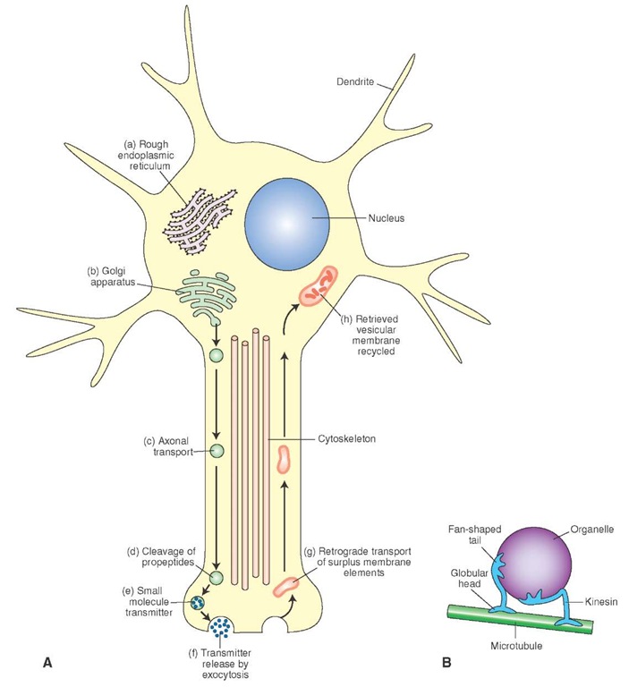
Fast anterograde transport (orthograde or forward flowing) is involved in the transport of materials that have a functional role at the nerve terminals (e.g., precursors of peptide neurotransmitters, enzymes needed for the synthesis of small molecule neurotransmitters, and glycoproteins needed for reconstitution of the plasma membrane) from the cell body to the terminals. Polypeptides much larger than final peptide neurotransmitters (pre-propeptides) and enzymes needed for the synthesis of small molecule neuro-transmitters are synthesized in the rough endoplasmic reticulum (Fig. 5-2A, a). Vesicles containing these propep-tides and enzymes bud off from the rough endoplasmic reticulum and are transported to the Golgi apparatus (Fig. 5-2A, b) where they are modified and packaged into vesicles. The vesicles formed in the Golgi apparatus then become attached to the microtubules and are transported by fast axonal transport (at a rate of 100-400 mm/d) into the nerve terminal (Fig. 5-2A, c). The propeptides are then cleaved to smaller peptides (Fig. 5-2A, d). Small molecule neurotransmitters are synthesized in the neuronal terminal. The neurotransmitters (either small peptides or small molecule neurotransmitters) are packaged in vesicles (Fig. 5-2A, e). The neurotransmitters are then released into the synaptic cleft by exocytosis. During the process of exocytosis, the membranes of the vesicles and terminal membrane fuse together, an opening develops, and the contents of the vesicle are released in the synaptic cleft (Fig. 5-2A, f).
The rapid axonal transport depends on the microtu-bules. The microtubule provides a stationary track and a microtubule-associated ATPase (kinesin) forms a cross-bridge between the organelle to be moved and the micro-tubule. On one end, kinesin contains two globular heads that bind to the microtubule and, on the other end, it has a fan-shaped tail that binds to the surface of an organelle. The organelle then moves by sliding of the kinesin molecule along the microtubule (Fig. 5-2B).
Slow Anterograde Transport
Slow anterograde transport involves movement of neuro-filaments and microtubules synthesized in the cell body to the terminals at a rate of 0.25 to 5 mm/d. Soluble proteins transported by this mechanism include actin, tubulin (which polymerizes to form microtubules), proteins that make up neurofilaments, myosin, and a calcium-binding protein (calmodulin).
Fast Retrograde Transport
Fast retrograde transport is slower than the fast anterograde transport (about 50-200 mm/d). Rapid retrograde transport carries materials from the nerve terminals to the cell body; the transported materials travel along microtu-bules. For example, when a transmitter is released from the synaptic terminal by exocytosis, the surplus membrane in the terminal is transported back to the cell body by retrograde fast axonal transport (Fig. 5-2A, g), where it is either degraded by lysosomes or recycled (Fig. 5-2A, h). Also transported by this mechanism is nerve growth factor, a peptide synthesized by a target cell and transported into certain neurons in order to stimulate their growth. Materials lying outside the axon terminals are taken up by endo-cytosis and transported to the cell body. Endocytosis is a process by which materials enter the cell without passing through the cell membrane. The process involves folding of the cell membrane inward after surrounding the material outside the cell, forming a sac-like vesicle which is then pinched off from the membrane so that the vesicle lies in the cytoplasm. Fast retrograde axonal transport is also involved in some pathological conditions. For example, the herpes simplex, polio, and rabies viruses and tetanus toxin are taken up by the axon terminals in peripheral nerves and carried to their cell bodies in the central nervous system (CNS) by rapid retrograde transport.
Axonal transport (anterograde as well as retrograde) has been used to trace target sites of neurons. A simplified procedure for anterograde tracing techniques involves the microinjection of a fluorescent dye at the desired site in the CNS. The dyes are taken up by the neuronal cell bodies and transported anterogradely to their axon terminals.
FIGURE 5-2 Axonal transport. (A) Large molecule peptides (pre-propeptides) are converted into smaller peptides (propeptides) in the rough endoplasmic reticulum (a). The propeptides and enzymes are packaged into vesicles that are transported to the Golgi apparatus, modified, and packaged into vesicles (b). The vesicles get attached to microtubules and are carried to the terminals by fast axonal transport (c). The propeptides are cleaved to produce smaller peptide transmitters in the terminal (d). Small molecule neurotransmitters are synthesized in the neuronal terminal and packaged into vesicles (e). The peptides and neurotransmitters are released into the synaptic cleft by exocytosis (f). Surplus membrane elements in the terminal are carried back to the cell body by retrograde transport (g). The retrieved vesicular membrane is degraded or recycled (h). (B) A model showing how kinesin (a microtubule-associated ATPase) can move an organelle along a microtubule.
The fluorescence of the axons and their terminals is then visualized under a microscope to ascertain the projections of the neuron.
Retrograde tracing techniques involve the microin-jection of an enzyme (e.g., horseradish peroxidase [HRP]), fluorescent dyes (e.g., Fluoro-Gold), cholera toxin, or viruses at the desired site. The injected substance (e.g., HRP) is taken up by axon terminals and transported retrogradely into the neuronal cell bodies. The neurons labeled with HRP are visualized by a chemical reaction in which a dense precipitate is formed and can be identified under dark-field or bright-field illumination. Likewise, fluorescent substances such as Fluoro-Gold, microinjected at the desired site, are taken up by axon terminals and transported to the cell bodies where they are visualized under a fluorescent microscope.


