To understand the mechanisms by which the nervous system regulates complex functions and behaviors, it is important to have knowledge of the structure and function of its basic unit, the neuron. The most striking properties of neurons are their excitability and ability to conduct electrical signals. This topic will first describe the organization of the neuronal membrane and its role in maintaining the internal ionic composition. Then, the mechanisms responsible for generating resting membrane potentials and action potentials will be discussed.
Structure and Permeability of the Neuronal Membrane
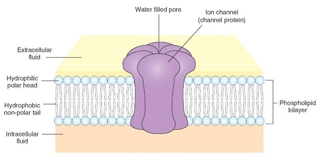
The neuronal membrane, like other cell membranes, consists of a lipid bilayer in which proteins, including ion channels, are embedded. Although three major types of lipids (phospholipids, cholesterol, and glycolipids) are present in the neuronal membrane, phospholipids are the most abundant type. Phospholipids consist of long nonpo-lar chains of carbon atoms that are bonded to hydrogen atoms. A polar phosphate group (a phosphorus atom bonded to three oxygen atoms) is attached to one end of a phospholipid molecule. All lipids in the neuronal membrane are amphiphilic; that is, they have a hydrophilic (or "water-soluble") end, as well as a hydrophobic (or "water-insoluble") end. Phospholipids have a hydrophilic "polar" end (head) and a hydrophobic "nonpolar" end (tail). The lipids in the neuronal membrane form bilayers, with their hydrophobic tails facing each other and their hydrophilic heads oriented on opposite sides (Fig. 6-1).
The lipid bilayer isolates the cytosol (cytoplasm) of the neuron from the extracellular fluid, and the proteins embedded in it are responsible for most of the membrane functions, such as serving as specific receptors, enzymes, and transport proteins. The neuronal membrane is impermeable to (1) most polar molecules (e.g., sugars and amino acids), and (2) charged molecules (even if they are very small). Cations (positively charged ions) are attracted electrostatically to the oxygen atom of water (which bears a net negative charge). Anions (negatively charged ions) are attracted to the hydrogen atom of water (which bears a net positive charge). Therefore, cations and anions contain electrostatically bound water (water of hydration). The attractive forces between the ions and the water molecules make it difficult for the ions to move from a watery environment into the hydrophobic lipid bilayer of the neuronal membrane. Examples in this category include sodium (Na+), potassium (K+), calcium (Ca2+), magnesium (Mg2+), chloride (Cl-), and hydrogen carbonate (HCO3-).
STRUCTuRE of PrOTEINS
Protein molecules are made up of different combinations of amino acids. Each amino acid has a carbon atom (called the alpha carbon) that is covalently bonded to a hydrogen (H) atom, an amino group (NH3+), a carboxyl group (COO-), and an R group (this group varies in different amino acids). In a covalent bond, two atoms share electrons. Amino acids assemble to form proteins according to instructions provided by messenger RNA in cell organelles called ribosomes. The synthesis of proteins occurs in ribosomes in the neuronal cell body. The amino acids are connected by peptide bonds to form a chain. In a peptide bond, the amino group of one amino acid joins with the carboxyl group of another amino acid. When a protein is made of a single chain of amino acids, it is called a polypeptide. The term "primary structure" describes the linear sequence of amino acids that make up the protein. Usually proteins fold up so that the exposure of hydrophobic amino acids to the solvent is minimized while the hydrophilic amino acids are exposed to the solvent. This is accomplished by secondary and tertiary structure of proteins. The secondary structure of proteins consists of alpha- helices and/or beta-sheets. In the formation of an alpha-helix, there is H-bonding between amide nitrogens and carbonyl carbons of peptide bonds spaced four residues apart. The orientation of H-bonding results in a helical coiling of the peptide backbone so that R-groups remain on the exterior of the helix and perpendicular to its axis. Beta-sheets consist of two or more different regions of stretches of 5-10 amino acids.
FIGURE 6-1 Schematic representation of the neuronal membrane.
The stretches of the polypeptide backbone are aligned aside one another. The tertiary structure refers to the 3-dimentional structure of the polypeptide units of a protein. When different polypeptide chains assemble to form a large protein molecule, the protein is said to acquire a quarternary structure. In a quarternary polypeptide, each polypeptide is called a subunit.
Membrane Transport-Proteins
Essential nutrients (e.g., sugars, amino acids, and nucle-otides) need to enter the neuron, whereas metabolic waste products must be removed from the neuron. In addition, the concentration of various ions has to be maintained within the neuron. Therefore, this necessitates influx of some ions and efflux of others. These functions are carried out by different membrane transport-proteins. Two types of proteins are implicated in the transport of solutes across the neuronal membrane: (1) carrier proteins and (2) channel proteins.
Carrier Proteins (Carriers or Transporters)
When a specific solute binds to a carrier protein, a reversible conformational change occurs in the protein which, in turn, results in the transfer of the solute across the lipid-bilayer of the membrane. A hypothetical scheme of how solutes are transported across neuronal membranes is as follows. When the carrier protein is in one conformational state, its binding sites may be exposed to the extracellu-larly located solute molecules. The binding of the solute to the carrier protein results in a change in the conforma-tional state of the protein so that the solute is now exposed to the cytoplasmic side of the neuronal membrane. The solute then dissociates from its binding site on the carrier protein and enters the interior of the neuron.
Channel Proteins
The channel proteins span the neuronal membrane and contain water-filled pores (Fig. 6-1). The inorganic ions of suitable size and charge (e.g., Na+ and K+) can pass through the pore when it is in the open state and, thus, pass through the membrane. Ion channels are discussed in the "Electro-physiology of the Neuron" section.
Transport of Solutes Across Cell Membrane
Simple Diffusion
The categories of substances that pass through the neuro-nal membrane by simple diffusion include: (1) all lipid-soluble (hydrophobic or nonpolar) substances (e.g., oxygen molecules), and (2) some polar (lipid-insoluble or water-soluble) molecules, provided they are small and uncharged (e.g., carbon dioxide, urea, ethanol, glycerol, and water molecules). The rate of transport of solutes during simple diffusion is proportional to the solute concentration; the solutes move from regions of high concentration to regions of low concentration (i.e., "downhill"). Such a difference in concentrations of the solute is called a concentration gradient (Fig. 6-2). No metabolic energy is needed when the molecules are transported across the cell membrane by simple diffusion.
Passive Transport (Facilitated Diffusion)
All channel proteins and some carrier proteins embedded in the neuronal membrane mediate passive transport (Fig. 6-2). The solutes are transported across the neuronal membrane passively, so no metabolic energy is needed for the transport of the molecules. If the molecule to be transported is uncharged, then the concentration gradient of the solute determines the direction in which it moves; it diffuses from the side where its concentration is higher to the side where its concentration is lower.
FIGURE 6-2 Transport of solutes across the neuronal membrane. (A) Simple diffusion. (B, C) Passive transport (facilitated diffusion) occurs either by channel-mediated (B) or carrier-mediated (C) diffusion. (D) Active transport occurs by specific carrier proteins, against the (E) electrochemical gradient. Active transport requires coupling of a carrier protein to a source of metabolic energy (e.g., hydrolysis of adenosine triphosphate).
Active Transport
Active transport is always mediated by specific carrier proteins and requires coupling of the carrier protein to a source of metabolic energy (e.g., hydrolysis of adenosine triphosphate [ATP]) (Fig. 6-2). If the solute molecule has an electrical charge, the determinants of its movement are the concentration gradient and the electrical potential between the two sides. The combination of the concentration and electrical potential gradients is called the electrochemical gradient. There are many negatively charged organic molecules (e.g., proteins, nucleic acids, carboxylic groups, and metabolites carrying phosphate) within the cell that are unable to cross the neuronal membrane. These are called fixed anions, and they change the charge inside the neuronal membranes to negative, as compared to the outside of the cell. Therefore, entry of positively charged ions (cations) will be permitted, whereas negatively charged ions (anions) will not be able to enter. Under these conditions, some carrier proteins transport certain solutes by active transport (i.e., the solute is moved across the neuronal membrane against its electrochemical gradient, or "uphill"). Two examples of this type of carrier protein are the sodium-potassium (Na+-K+) and calcium pumps.
Sodium-Potassium Ion Pump
The sodium-potassium ion pump (Na+-K+ pump), also known as Na+-K+ ATPase, is located in the membranes of neurons as well as other cells. The Na+-K+ ATPase consists of a small glycoprotein (regulatory P subunit) and a large multi-pass transmembrane unit (catalytic a subunit). Three Na+ ions bind on the cytoplasmic side of the trans-membrane unit of the ATPase, a conformational change occurs, and ATP binds to the cytoplasmic side of the trans-membrane unit. In this new conformation, the protein has low affinity for Na+ ions, which dissociate and diffuse into the extracellular fluid. The new conformation has high affinity for K+, and two K+ ions bind at the extracellular site of the transmembrane unit. The bound phophate dissociates, and the ATPase reverts to its original conformation exposing K+ ions to the cytoplasmic side. In this conformation, the ATPase has low affinity for K+ ions, two of which dissociate and diffuse into the interior of the cell. The cycle is then repeated.. With each cycle, one molecule of ATP is hydrolyzed, and the energy generated is used to move Na+ and K+ ions across the neuronal membrane. There are three binding sites for Na+ and two binding sites for K+ on the Na+-K+ ATPase. Therefore, three Na+ ions are transferred out of the neuron for every two K+ ions that are taken in. A net outward ionic current is generated because of this unequal flow of Na+ and K+ ions across the neuronal membrane. Because a current is generated, the Na+-K+ pump is said to be electrogenic. Because the Na+-K+ pump drives more positive charges (Na+) out of the neuron than it brings into the neuron (K+), the inside of the neuron becomes more negative relative to the outside. It should be noted that the contribution of the Na+-K+ pump in making the inside of the neuron more negative relative to the outside is only about 10%; the major factor responsible for the negativity of the interior of the neuron is the presence of fixed anions.
Because the extracellular concentration of Na+ ions is high, these ions leak into the neuron. The Na+-K+ ATPase pumps them out, and, thus, maintains the high extracellular concentration of Na+. It is necessary to keep the extracellular concentration of Na+ ions high to prevent water from entering the neuron; a high intracellular concentration of solutes (fixed anions and accompanying cations) tends to pull water into the neuron. Thus, Na+-K+ ATPase plays an important role in maintaining (1) the ionic concentrations inside and outside the neuron and (2) the osmotic balance of the neuron.
Calcium Pump
The neuronal plasma membrane contains a calcium pump, which is an enzyme that actively transports Ca2+ out of the cell so that the concentration of this ion remains small inside the resting cell. One Ca2+ is transported for each ATP hydrolyzed.
Intracellular and Extracellular Ionic Concentrations
As mentioned earlier, the concentration of various ions needs to be maintained within the neuron, necessitating flow of some ions into and out of the cell. Table 6-1 shows the differences in the intra-cellular and extracellular concentrations of major ions. The concentration of Na+ ions is much greater (approximately 10 times) outside the neuron compared with the concentration inside the neuron. On the other hand, the concentration of K+ ions is greater (approximately 20 times) inside the cell than outside. The presence of many negatively charged fixed ions makes the interior of the cell more negative relative to outside.
TABLE 6-1 Approximate Neuronal Intracellular and Extracellular Concentrations of Some Important Ions
|
Ion |
Extracellular Concentration (mM) |
Intracellular Concentration (mM) |
|
Cations |
|
|
 |
150 |
15 |
 |
5 |
100 |
 |
2 |
0.0002 |
|
Anions |
||
|
Cl- |
150 |
13 |
|
A- (fixed anions; organic acids and proteins) |
— |
385 |
mM = millimolar concentration.
To balance these negative charges, positively charged K+ ions are retained inside the neuron, and, as such, the intra-cellular concentration of K+ ions is much greater than the extracellular concentration. The intracellular concentration of Ca2+ is very small, and most of it is bound with proteins or stored in various organelles. Like Na+ ions, the concentration of the Cl- ions is greater outside relative to inside the neuron. The differences in intracellular and extracellular concentrations of different ions are maintained by Na+-K+ and Ca2+ pumps.
Electrophysiology of the Neuron
An understanding of the electrophysiology of neurons is likely to be facilitated if the student bears in mind the following terms.
Terminology
Ions: Ions are atoms or molecules that have an electrical charge.
Anions: Anions are negatively charged ions (e.g., Cl-). Cations: Cations are positively charged ions (e.g., Na+, K+, and Ca2+).
Influx of ions: The flow of ions into the neuron is influx. Efflux of ions: The flow of ions out of the neuron is efflux.
Electrical Charge-Related Terms
Anode: An anode is the positive terminal of a battery; negatively charged ions (e.g., Cl-) move toward an anode.
Cathode: A cathode is the negative terminal of battery; positively charged ions (e.g., Na+) move toward a cathode.
Electrical current: The movement of electrical charge is electrical current. The flow of electrical current (I) depends on electrical potential and electrical conductance. It is measured in amperes (amps).
Electrical potential (voltage): The difference in charge between the anode and the cathode is its electrical potential. More current flows when this difference is increased. Electrical potential is measured in volts (V). Electrical conductance: The ability of an electrical charge to move from one point to another is determined by its electrical conductance (g). It is measured in siemans (S). Electrical resistance: The inability of an electrical charge to move from one point to another is determined by electrical resistance (R). It is measured in ohms (Q). Electrical resistance is the inverse of electrical conductance, and this relationship is expressed as R = 1/g.
Ohms law: This law describes the relationship between electrical current (I), electrical conductance (g), and electrical potential (V). According to this law, current is the product of the conductance and potential difference (I = gV).
Current Flow-Related Terms
Direction of current flow: In an ionic solution, cations as well as anions carry electrical current. Conventionally, current flow is defined as the direction of net movement of positive charge. Therefore, cations move in the same direction as the current. Anions move in the opposite direction as the current.
Inward current: Positively charged ions (e.g., Na+) flowing into the neuron is inward current. Outward current: Positively charged ions (e.g., K+) moving out of the neuron or negatively charged ions (e.g., Cl-) moving into the neuron is outward current. Leakage current: The current due to the flow of ions through the nongated ion channels (discussed later) is leakage current.
Membrane Potential-Related Terms
Excitable membrane: Cells capable of generating and conducting action potentials have excitable membranes. Membrane potential (Vm): The electrical potential difference across the neuronal membrane (i.e., between the interior and the exterior of the neuron) at any time is called the membrane potential.
Resting membrane potential: When a neuron is not generating action potentials, it is at rest. When the neuron is at rest, its cytosol along the inner surface of its membrane is negatively charged compared with the charge on the outside. Typically, the resting membrane potential (or resting potential) of a neuron is -65 millivolts (mV) (1 volt = 1,000 mV).
Threshold potential: The level of membrane potential at which a sufficient number of voltage-gated sodium channels open and relative permeability of sodium ions is greater than that of potassium ions is its threshold potential. Action potentials are generated when the membrane is depolarized beyond the threshold potential.
Depolarization: Depolarization occurs when there is a reduction in the negative charge inside the neuron (e.g., if the resting membrane potential is changed from -65 to -60 mV by flow of positively charged ions, like Na+, into the neuron).
Hyperpolarization: Hyperpolarization occurs when there is an increase in the negative charge inside the neuron (e.g., if the resting membrane potential changes from -65 to -70 mV by flow of positively charged ions, like K+, out of the neuron or flow of negatively charged ions, like Cl-, into the neuron).


