Major Cell Groups
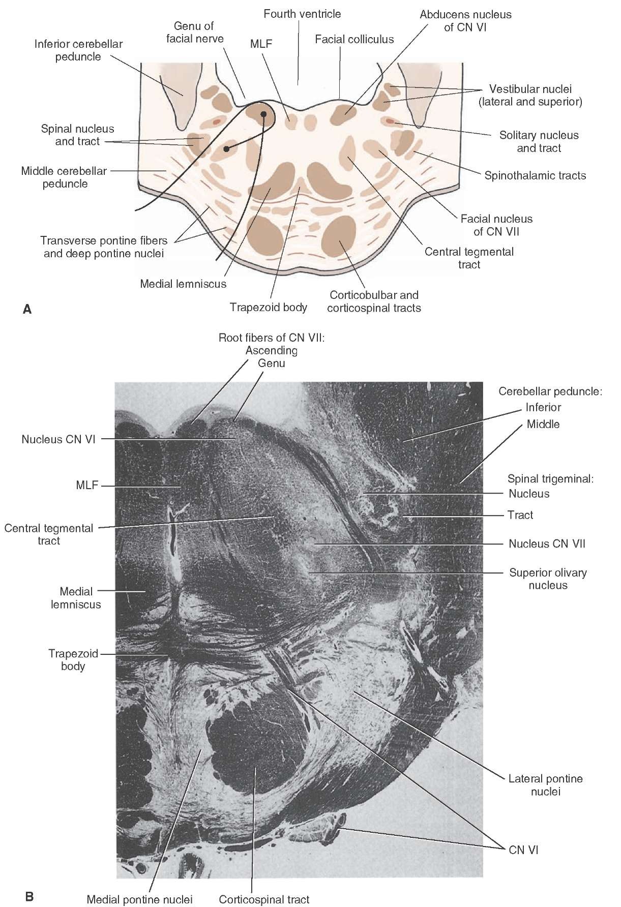
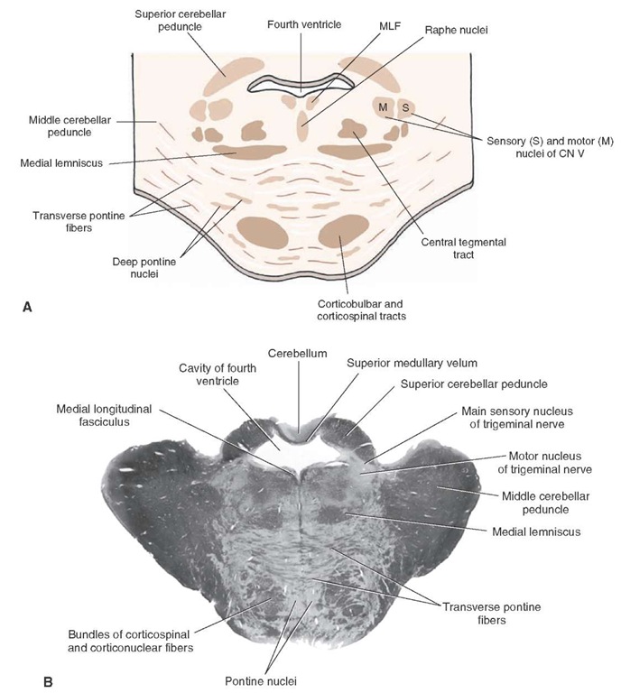
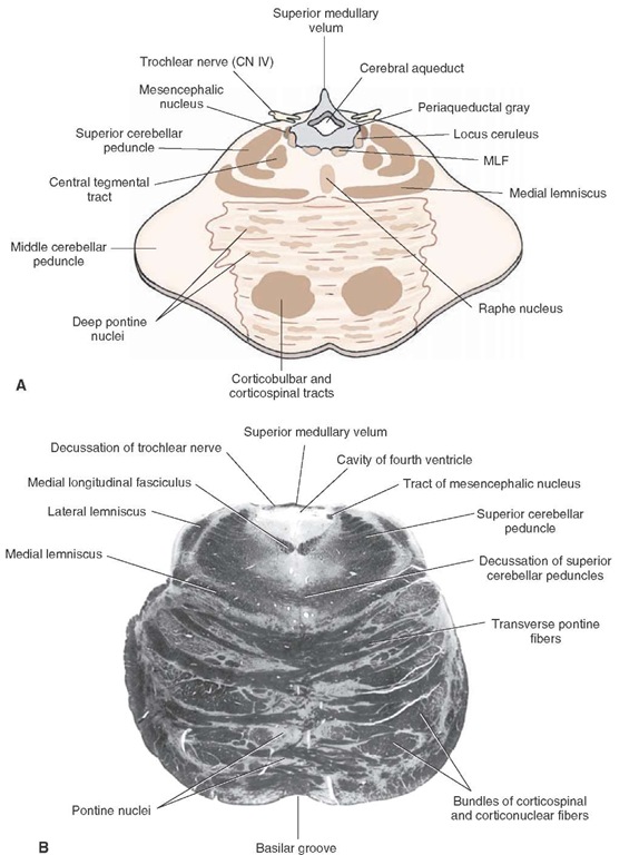
Figures 11-3 through 11-6 depict major nuclear cell groups.
Caudal Pons
A number of important nuclei, associated in part with cranial nerves, are present in the caudal pons. These include the motor nuclei of the facial (cranial nerve [CN] VII) and abducens (CN VI) nerves, the spinal nucleus of the trigeminal CN V, the superior olivary nucleus (a relay nucleus of the auditory pathway), raphe nuclei, and pontine nuclei of the basilar pons.
Rostral Pons
The principal nuclei present in the rostral half of the pons include the main sensory, mesencephalic, and motor nuclei of CN V, the superior and lateral vestibular nuclei, and the locus ceruleus. Other cell groups, such as the pontine nuclei (of the basilar aspect) and the raphe nuclei (of the tegmentum), are also present at all levels of the pons.
Basilar Aspect of the Pons
Throughout its entire length, the pons is divided into two distinct parts, a dorsal and a ventral region. The dorsal part is called the tegmentum, which is continuous with the teg-mentum of the medulla and midbrain. The ventral part is called the basilar pons. The basilar (or ventral) pons may be viewed as a rostral extension of the ventral aspect of the medulla, which contains the pyramidal tract. However, at the level of the pons, significant morphological changes can be noted. The most characteristic feature is the marked presence of massive bundles of fibers that run transversely and coalesce as the middle cerebellar peduncle, which enters the cerebellar cortex. Interspersed among the transverse fiber bundles are large numbers of cells. These cells are referred to as (deep) pontine nuclei. They are important because they give rise to the transverse pontine fibers. The pontine nuclei, which receive significant inputs from the cerebral cortex, send their axons (the transverse pontine fibers) to the cerebellar cortex. Thus, the pathway from the cerebral cortex to the cerebellar cortex constitutes a two-neuronal arc (corticopontine and pontocere-bellar fibers).
In addition to the deep pontine nuclei and transverse pontine fibers, the basilar pons contains the corticospinal and corticobulbar tracts, which follow the longitudinal axis of the brainstem and which pass at right angles to the transverse pontine fibers. At the level of the pons, numbers of corticobulbar fibers (also called corticonuclear fibers) exit and terminate mainly upon interneurons of the reticular formation situated near cranial nerve motor nuclei as well as nuclei of reticulospinal fibers.
Pontine Tegmentum
Lower (Caudal) Half of the Pons
Within the caudal aspect of the pons (Fig. 11-4), several features characteristic of the medulla are still present. The following structures are noted: the MLF and tectospinal tracts that lie in the dorsomedial aspect of the pons; the spinal nucleus and tract that lie in the lateral aspect of the pons; and the medial lemniscus, whose position at this level remains within the ventromedial aspect of the pon-tine tegmentum.
Important structures that appear at this level include:
1. Abducens nucleus (nucleus of CN VI). This nucleus lies in a dorsomedial position within the pons close to the floor of the fourth ventricle. This nucleus serves as a general somatic efferent (GSE) (lower motor neuron), which innervates the lateral rectus muscle and thus provides the anatomical basis for the lateral movement of the eye.
2. Facial nucleus (nucleus of CN VII). This nucleus lies in the ventrolateral aspect of the tegmentum. Its axons (special visceral efferent [SVE] fibers) follow an unusual course by initially passing in a dorsomedial direction. Where the axons approach the dorsal aspect of the pons, the fibers course laterally over the abducens nucleus, forming the facial colliculus. They then continue vent-rolaterally to where the fibers exit the brain to innervate the muscles controlling ipsilateral facial expression.
3. Superior and lateral vestibular nuclei. These nuclei are located in the dorsolateral aspect of the pontine tegmentum and, essentially, replace the medial and inferior vestibular nuclei that are situated more caudally in the medulla. The superior and lateral vestibular nuclei receive direct inputs from the vestibular apparatus and contribute fibers to the MLF. In addition, axons of the lateral vestibular nucleus form the lateral vestibu-lospinal tract, which innervates all levels of the spinal cord.
FIGURE 11-4 Caudal pons. (A) Cross section through the caudal aspect of the pons at the level of the facial colliculus of the pons. Note that the axons of cranial nerve (CN) VII pass dorsomedially around the dorsal aspect of the nucleus of CN VI, forming the facial colliculus, and then pass ventrolaterally to exit the brainstem. In contrast, the axons of CN VI pass ventrally to exit the brainstem in a relatively medial position. (B) Myelin-stained cross section through the caudal aspect of the pons.
4. Auditory relay structures—trapezoid body (and nucleus of trapezoid body), superior olivary nucleus, and lateral lemniscus. These nuclei are located in the ventral aspect of the pontine tegmentum. The nucleus of the trapezoid body and superior olivary nucleus receive auditory inputs primarily from the cochlear nuclei and transmit auditory signals to higher centers of the brain-stem. The trapezoid body is made up of commissural fibers, which originate from either the ventral cochlear nucleus or superior olivary nucleus. The lateral lemnis-cus represents fibers of the cochlear nuclei and superior olivary nucleus that transmit auditory signals to the inferior colliculus.
5. Superior salivatory nucleus. This nucleus (GVE) lies in a position just ventrolateral to the abducens nucleus. It gives rise to preganglionic parasympathetic axons of CN VII, which innervate submandibular, submaxillary, and pterygopalatine ganglia. Thus, this nucleus serves to mediate lacrimation, salivation, and vasodilation. This nucleus is quite small and cannot be seen without special physiological or histological manipulations.
6. Nuclei and fibers of the reticular formation.Three areas of the pontine reticular formation are noted: a midline region, containing raphe nuclei; a large-celled region situated within the medial two thirds of the reticular formation; and a small-celled region situated in the lateral aspect of the reticular formation. In brief, the raphe cells produce serotonin; these cells project to many areas of the central nervous system. The large-celled region gives rise to long ascending and descending fibers contained to a considerable extent in the central tegmental tract, and the small-celled region serves as a source of inputs to the reticular formation.
Upper (Rostral) Half of the Pons
Within the rostral half of the pons, a number of structures appear for the first time. These include:
1. Main sensory (trigeminal) nucleus (CN V). The main sensory nucleus of CN V (general sensory afferent [GSA]) is a large nucleus found in the dorsolateral aspect of the tegmentum. It replaces the position occupied by the spinal trigeminal nucleus at lower levels. It contains second-order neurons for the transmission of somatosensory information from the head region as its axons project to the ventral posteromedial nucleus of the thalamus (Fig. 11-5).
2. Motor nucleus (CN V). The motor nucleus of CN V (SVE) projects its axons to the muscles of mastication and, therefore, plays a vital role in closing the jaw. It lies immediately medial to the main sensory nucleus and can be distinguished from it by the presence of trigemi-nal root fibers that pass between it and the main sensory nucleus (Fig. 11-5).
3. Nucleus locus ceruleus. This nucleus can be found in the dorsolateral aspect of the tegmentum of the upper pons at the level of the motor nucleus of CN V and the region slightly rostral to it (Fig. 11-6). The locus ceruleus consists of norepinephrine-containing neurons, which project to different cell groups in the brainstem; fore-brain, including the cerebral cortex; and cerebellum.
4. Mesencephalic nucleus of CN V. The mesencephalic nucleus extends from the level of the rostral pons into the midbrain. It is located in the ventrolateral aspect of the gray matter surrounding the rostral end of the fourth ventricle and the beginning of the cerebral aqueduct (Fig. 11-6). It receives mainly muscle spindle afferents from the jaw and related areas of the face (i.e., masseter and temporalis muscles), which serve as afferents for reflex closing of the jaw.
5. Superior cerebellar peduncle. At the rostral aspect of the pons, the superior cerebellar peduncle, which contains the large majority of cerebellar efferent fibers to the midbrain and thalamus, forms the lateral wall of the fourth ventricle (Fig. 11-5). The superior cerebellar peduncle on each side is attached by the superior medullary velum, which forms the roof of the fourth ventricle. At the rostral border of the pons (Fig. 11-6), the superior cerebellar peduncle enters the pons and courses towards the midline. At the level of the caudal mid-brain, these fibers will ultimately cross over to the opposite side of the brainstem.
The Cerebellum
Damage to different parts of the cerebellum will cause such deficits as loss of balance, tremors, lack of coordination of muscles, and reduced muscle tone. The cerebellum is attached to the brainstem by three pairs of peduncles (see Fig. 1-9). The inferior cerebellar peduncle extends from the dorsolateral aspect of the upper medulla into the cerebellum. It contains fibers that arise from the spinal cord and lower brain-stem. The middle cerebellar peduncle is attached to the lateral aspect of the pons and, therefore, enters the cerebellum from a lateral position. It comprises mainly the second limb of a disynaptic pathway linking the cerebral cortex with the cerebellar cortex. The superior cerebellar peduncle passes from the rostro-medial aspect of the cerebellum in a rostral direction and enters the brainstem at the level of the upper pons, where it targets mainly the red nucleus and ventrolateral nucleus of the thalamus (which, in turn, projects to motor cortex). Thus, the inferior and middle cerebellar peduncles generally contain cerebellar afferent fibers, whereas most fibers present within the superior cer-ebellar peduncle constitute cerebellar efferent fibers.
FIGURE 11-5 Middle pons. (A) Cross section through the middle of the pons at the level of the main sensory and motor nuclei of the trigeminal nerve. CN = cranial nerve. (B) Myelin-stained section through the same level of the pons at the level of the main sensory and motor nuclei of the trigeminal nerve.
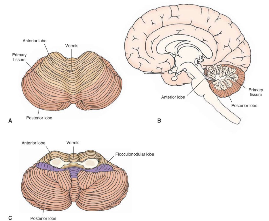
The cerebellum consists of two hemispheres continuous with a median vermal region (Fig. 11-7). The cerebellar hemispheres are divided into three lobes. The lobe closest to the midbrain is called the anterior lobe, which receives inputs mainly from the spinal cord and is separated from the posterior lobe (which is the largest lobe) by the primary fissure. The posterior lobe recieves inputs from the cerebral cortex and various regions of the brainstem, such as the reticular formation and inferior olivary nucleus. The smallest lobe is called the flocculonodular lobe and mainly receives inputs from the vestibular apparatus or vestibular neurons. It is located most caudally and is separated from the posterior lobe by the posterolateral fissure.
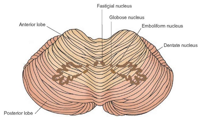
Internally, the cerebellum consists of a cortex, white matter that lies immediately deep to the gray matter, and a series of nuclei referred to as deep cerebellar nuclei. The most lateral and largest of the nuclei is called the dentate nucleus, and its neurons target the ventrolateral nucleus of the thalamus (Fig. 11-8). The most medial nucleus is called the fastigial nucleus, and its main projection targets include the reticular formation and vestibular neurons. Between the dentate and fastigial nuclei lie two smaller nuclei, the emboliform and globose nuclei. In humans, the globose nucleus lies medial to the emboliform nucleus, and the emboliform nucleus is situated close to the hilus of the dentate nucleus. However, in lower forms of animals, such as the cat, these two nuclei are fused, and the structure is referred to as the interposed nucleus. Its main efferent target is the red nucleus of the midbrain. Thus, the overall importance of these nuclei is that their axons constitute the primary means by which information exits the cerebellum.
FIGURE 11-6 Rostral pons. (A) Diagram of a cross section through the upper pons at the level of the locus ceruleus and mesencephalic nucleus of the trigeminal nerve. CN = cranial nerve; MLF = medial longitudinal fasciculus. (B) Myelin-stained section through the same level of the pons as the level of the nucleus ceruleus and mesencephalic nucleus of the trigeminal nerve.
Clinical Considerations
With respect to the pons, different syndromes associated with distinct regions of the pons can be identified. The more common ones are described in the following sections.
Caudal Tegmental Pontine Syndrome
Caudal tegmental pontine syndrome, caused by occlusion of circumferential branches of the basilar artery, impacts upon structures situated within the caudal aspect of the pontine tegmentum. The major structures thus affected include the MLF, motor nuclei of CN VI and CN VII, spinal nucleus of CN V, spinothalamic tracts, and descending pathways from the hypothalamus. Therefore, deficits seen from this vascular occlusion would include ipsilateral facial nerve palsy (CN VII), lateral gaze palsy (CN VI), and Horner’s syndrome (resulting from loss of descending sympathetic input to the lower brainstem). The exact nature of these deficits is a function of the extent of damage incurred by the vascular occlusion.
FIGURE 11-7 Cerebellum. Superior (A), midsagittal (B), and inferior (C) views of the cerebellum illustrating anterior, posterior, and flocculonodular lobes as well as hemispheres and vermal regions.
FIGURE 11-8 Cerebellum and deep cerebellar nuclei. This view of the cerebellum illustrates the positions occupied by the deep cerebellar nuclei. The largest nucleus, the dentate nucleus, is located laterally. The fastigial nucleus is situated medially, and the globose and emboliform (interposed) nuclei are located in an intermediate position.
