Internal Nuclei of the Brainstem
Reticular Formation. The internal core of the entire brain-stem contains mainly a complex set of neuronal groups (coupled with related fiber bundles) that are collectively referred to as the reticular formation.In brief, it appears to be involved in such processes as modulation of sensory transmission to the cortex, regulation of motor activity, autonomic regulation, sleep and wakefulness cycles, and modulation of emotional behavior.
Major Nuclei of the Medulla. In the lower levels of the medulla, the principal nuclei of the caudal (or lower) medulla include the nucleus gracilis and nucleus cuneatus, which receive first-order somatosensory fibers from the fasciculus gracilis and fasciculus cuneatus, respectively, and the spinal nucleus of CN V, which receives first-order pain and temperature signals from the region of the head (Fig. 10-2).
In the upper levels of the medulla, numerous important nuclei are present. The spinal nucleus is present at this level, as it is at lower levels of the medulla. Other sensory neurons include the cochlear nuclei, which receive first-order auditory fibers (CN VIII), and vestib-ular nuclei, which also receive first-order vestibular inputs (CN VIII). In addition, a number of different cranial nerve nuclei appear. These include somatomotor nuclei (hypoglossal nucleus [CN XII] and nucleus ambig-uus of CN X and CN IX) and sensory and motor auto-nomic nuclei (the solitary nucleus [CN IX and X] and dorsal motor nucleus of CN X)
Levels of the Medulla
In describing the anatomy of the medulla, we can distinguish at least two levels. The caudal half, which includes the "closed" medulla, contains two decussations: (1) the pyramidal decussation and (2) a second level that contains the decussation of the medial lemniscus. The upper half of the medulla, which includes the "open" medulla, contains the inferior olivary nucleus. The following sections consider the neuroanatomical organization of the medulla on the basis of the structures contained at each of these levels.
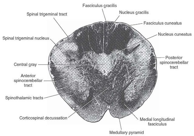
Level of the Pyramidal Decussation. At the level of the pyramidal decussation, the general organization of the medulla is very similar to that of the cervical spinal cord. For example, the dorsal aspect of this level of the medulla contains the fasciculus gracilis and fasciculus cuneatus (Figs. 10-3 and 10-4). The spinocerebellar and spinothalamic tracts also lie in the same general lateral position they occupy within the spinal cord. Some anterior horn cells can generally be seen at this level as well.
There are major new features found at this level of medulla that are not present in the spinal cord. The most obvious change is the presence of the pyramidal decussation. As indicated earlier, about 90% of the fibers of each pyramid cross over to the contralateral side of the brain in their descent into the spinal cord as the lateral corticospinal tract. Approximately 8% of the fibers continue to descend uncrossed into the spinal cord as the anterior corticospinal tract and are situated in a ven-tromedial position; ultimately, these fibers cross over to the opposite side of the spinal cord. The remaining 2% of the corticospinal tract supplies the ipsilateral spinal cord grey matter. A second change is the presence of the spinal nucleus of the trigeminal nerve. It replaces the substantia gelatinosa, in that it is situated in the dorso-lateral aspect of the medulla, which is the approximate position occupied by the substantia gelatinosa in the dorsal horn of the spinal cord. The anatomical similarities between the substantia gelatinosa and the spinal nucleus of CN V are matched by their functional similarities. Both structures mediate pain and temperature modalities of sensation. The difference between the two relates to the fact that the substantia gelatinosa mediates these sensations from the body, whereas the spinal nucleus mediates sensations associated with the region of the anterior two thirds of the head, oral and nasal cavities, and the cutaneous surfaces of the ear and external auditory meatus.
FIGURE 10-4 Photograph of a cross section of the human medulla at the level of the decussation of the pyramids (Weigert myelin stain). Note that the fasciculus gracilis and fasciculus cuneatus are still clearly present at this level, and note the positions occupied by the ascending sensory axons.
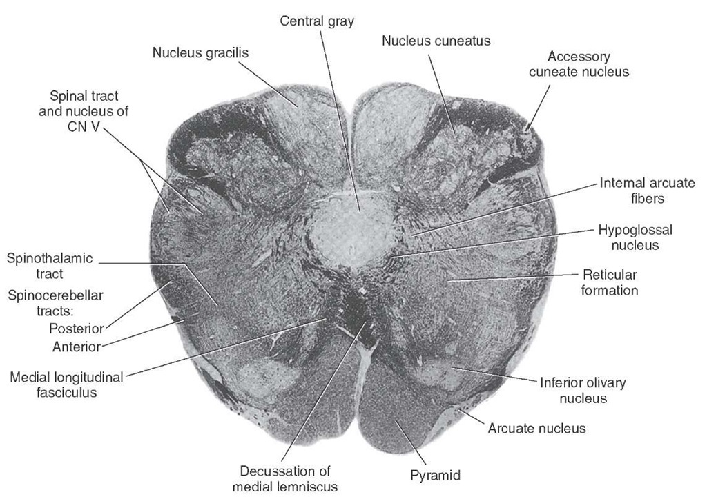
Level of the Decussation of the Medial Lemniscus. At a level slightly rostral to that of the pyramidal decussation, a second major decussation called the decussation of the medial lemniscus is present (Figs. 10-3 and 10-5). Whereas the pyramidal decussation involves fibers from sensorimotor cortex that are specifically involved in motor functions, the dec-ussation of the medial lemniscus is part of an overall sensory pathway that contains fibers that arise from the nucleus gracilis and nucleus cuneatus (located immediately lateral to the nucleus gracilis) and mediates somesthetic impulses from the body to the thalamus and, ultimately, to the postcentral gyrus. Axons of the medial lemniscus initially pass ventrally in an arc (where fibers arising from the nucleus gracilis emerge prior to those of the nucleus cunea-tus and collectively are called internal arcuate fibers) and then cross over to the contralateral side via the decussa-tion of the medial lemniscus. These fibers then turn rostrally as they ascend toward the forebrain where they terminate in the ventral posterolateral (VPL) nucleus of the thalamus.
At this level of the medulla, another nucleus, called the accessory cuneate nucleus, is attached to the lateral edge of the nucleus cuneatus (Fig. 10-5). This structure receives inputs from muscle spindles and Golgi tendon organs associated with the skeletal muscles of the upper limbs that are conveyed by first-order fibers passing in the fasciculus cuneatus. Axons of the accessory cuneate nucleus, called external arcuate fibers, form the cune-ocerebellar tract as they pass ipsilaterally into the inferior cerebellar peduncle. Thus, they mediate unconscious proprioception from the upper limbs to the cerebellum and represent the upper limb equivalent of the dorsal spinocerebellar tract.
Other structures that are prominent at the level of the decussation of the medial lemniscus are also present more caudally within the medulla. These include the pyramidal, spinocerebellar, and spinothalamic tracts as well as the spinal nucleus and tract of the trigeminal nerve. Rostral Half of the Medulla. At this level of the medulla, a number of new structures make their appearance, including important cranial nerve nuclei.On developmental principles, the arrangement of sensory and motor nuclei, especially those of cranial nerves, is reviewed in the following paragraphs and is depicted in Figure 10-6.
FIGURE 10-5 Photograph of a cross section of the human medulla at the level of the decussation of the medial lemniscus (Weigert stain). Note that the nucleus gracilis and nucleus cuneatus are quite prominent at this level and that the accessory cuneate nucleus can be seen situated just lateral to the cuneate nucleus. In this section, one can see that internal arcuate fibers, which arise from the dorsal column nuclei, pass in a ventromedial direction to the opposite side to form the medial lemniscus.
FIGURE 10-6 The arrangement of sensory and motor nuclei of cranial nerves. The key point is that the region derived from the basal plate is located medially and contains motor nuclei, while the region derived from the alar plate is located more laterally and contains sensory nuclei. The region in between these two regions, which is formed from the sulcus limitans, contains neurons that relate to autonomic functions. SSA = special sensory afferent neurons; GSA = general somatic afferent neurons; SVA =, special visceral afferent neurons; GVA = general visceral afferent neurons; GSE = general somatic efferent fibers.
In brief, the locations of neuronal cell groups associated with cranial nerve function can be determined by knowing the following medial-to-lateral arrangement. Cell groups located in the most medial position, derived from the basal plate, are classified as general somatic efferent (GSE) neurons; they innervate skeletal muscle of somite origin. The clearest example of such a cell group that is situated at this level of the brainstem is the hypoglossal nucleus, which can be easily seen in Figures 10-7 and 10-8 and lies just above the MLF in a dorsomedial position within the medulla. Axons of the hypoglossal nucleus innervate the intrinsic and extrinsic muscles of the tongue and thus control its movements.
The next column includes those cell groups that are derived from mesenchyme of the branchial arches and are classified as special visceral efferent (SVE) neurons, which also innervate skeletal muscle (Fig. 10-6). An example of this type of cell group is the nucleus ambiguus, which gives rise to axons of CN IX and X. Axons of the nucleus ambig-uus travel, in part, in the vagus nerve and innervate the muscles of the larynx and pharynx, thus controlling phona-tion, gagging, and swallowing. Drawing a line from the dorsolateral tip of the inferior olivary nucleus to the hypoglossal nucleus can identify the position of the nucleus ambiguus. The nucleus ambiguus is located at the mid-point of this line.
Continuing in a lateral direction, the third column includes those cell groups that comprise the general visceral efferent (GVE) neuron groups (Fig 10-6). These represent preganglionic parasympathetic fibers whose axons innervate autonomic ganglia. Postganglionic para-sympathetic neurons are present either in these ganglia or within the wall of the target organs, and their axons innervate smooth muscles and glands.
Two groups of such neurons can be detected within the medulla. The first group of neurons includes the dorsal motor nucleus of the vagus nerve (CN X), whose axons contribute to the vagus nerve. The dorsal motor nucleus lies dorsolateral to the hypoglossal nucleus just medial to the sulcus limitans at the rostral end of the closed medulla.The second group of neurons constitutes the inferior salivatory nucleus, whose axons form part of CN IX and which contribute to the process of salivation. Axons of these neurons form preganglionic parasympa-thetic fibers that innervate the otic ganglion, from which postganglionic parasympathetic fibers innervating the parotid gland arise.
Immediately lateral to the sulcus limitans lies the general visceral afferent (GVA) column (Fig. 10-6). Cells in this region, which are derived from the alar plate, receive afferent signals from visceral organs, such as blood vessels, that are mediated via branches of the glossopharyngeal (CN IX) and vagus (CN X) nerves. The principal nucleus that receives and processes such information is the caudal portion of the solitary nucleus, which lies in a position slightly ventrolateral to the dorsal motor nucleus of the vagus nerve. Superimposed on this column are the special visceral afferent (SVA) neurons (Fig. 10-6), which includes the rostral half of the solitary nucleus (gustatory portion). The reason for the proximity of these two groups of separately classified cell types of neurons is that the primary neuron for both functions at this level of the brain is the solitary nucleus. Thus, different groups of cells within the solitary nucleus mediate two distinctly separate processes, one associated with GVA functions (i.e., cardiovascular and respiratory reflexes) and the other associated with SVA functions (i.e., taste).
The final categories of cell groups include general sensory afferent (GSA) neurons and special sensory afferent (SSA) neurons. They both lie in a most lateral position within the medulla. The primary GSA neuronal cell group found at this level of the brainstem is the spinal nucleus of the trigeminal nerve. Within the medulla, separate groups of SSA neurons can be detected. One such group is the vestibular nuclei. There are four vestibular nuclei that receive direct inputs from the vestibular apparatus: the inferior, medial, lateral, and superior nuclei. The inferior and medial vestibular nuclei are located in the rostral medulla at the approximate level of the rostral portions of the inferior olivary nucleus. The superior and lateral vestibular nuclei lie in a more rostral position at the transition region between the medulla and pons. The medial and inferior vestibular nuclei are best seen in the rostral open medulla, whereas the superior and lateral vestibular nuclei are best seen in the junction between the pons and medulla just lateral to the fourth ventricle and medial to the inferior cerebellar peduncle.
The other group of SSA neurons is the cochlear nuclei, which lie on the dorsolateral shoulder of the inferior cer-ebellar peduncle at the rostral aspect of the medulla. These neurons receive inputs from primary auditory fibers and, thus, constitute part of a complex auditory pathway that eventually transmits auditory signals to the cerebral cortex.
This discussion has indicated how nuclei related to cranial nerve function are organized within the lower brain-stem. Thus, when taken together with the analysis of the structures of the lower medulla considered earlier in this topic, it is now possible to reconstruct the organization of the rostral half of the medulla. As is shown in Figure 10-6, the structures seen in more caudal levels of the medulla that are present at more rostral levels are again represented in the same general locations as noted in sections of the lower medulla.
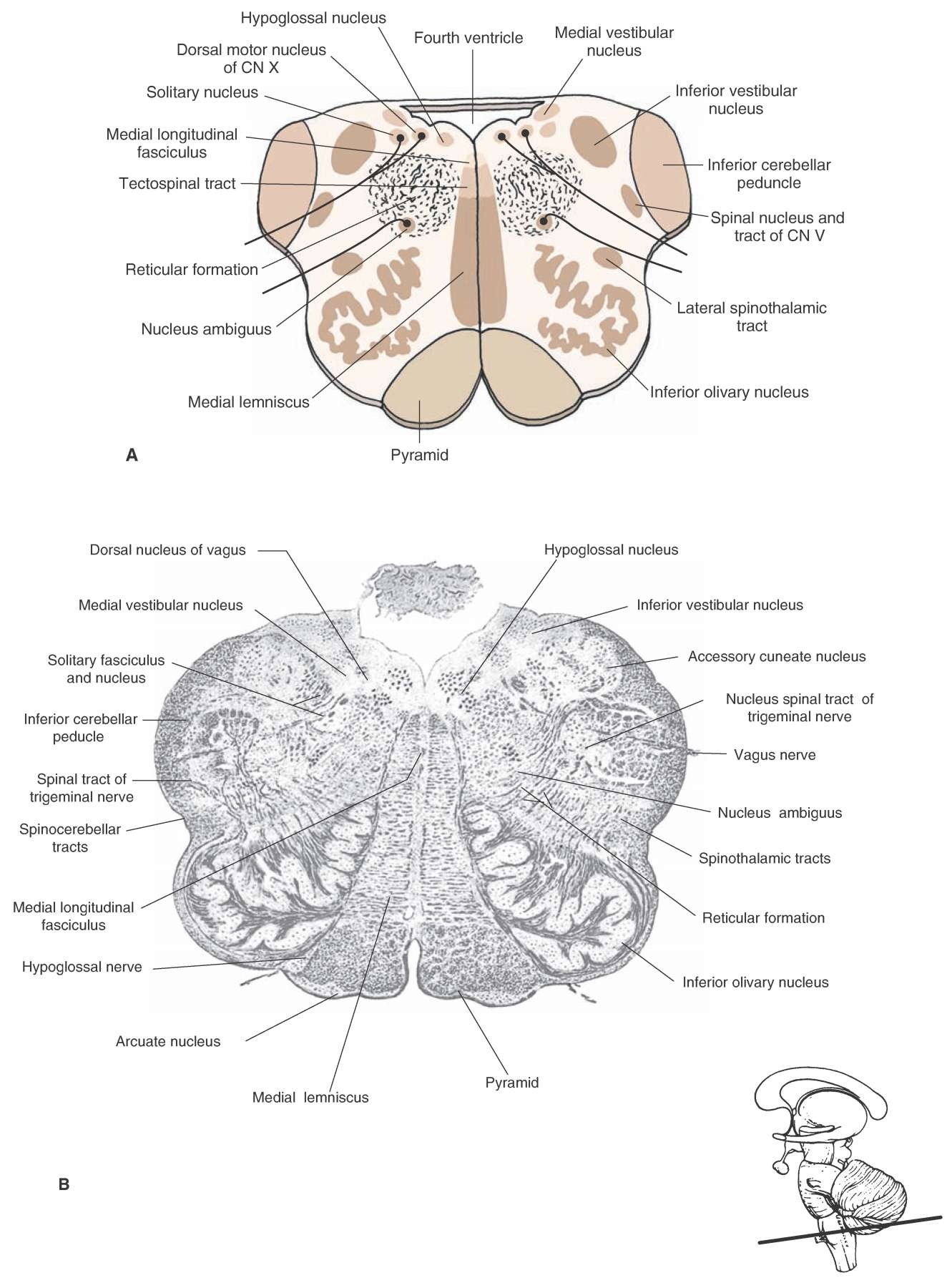
Structures Situated in the Medial Medulla. The arrangement of structures when going from a ventral to a dorsal level is as follows. On the ventromedial aspect of the rostral half of the medulla are the pyramids (Figs. 10-7 and 10-8). Dorsal to them, lies the medial lemniscus, and dorsal to the medial leminiscus lies two descending pathways: (1) the tectospi-nal tract, which contains descending fibers from the superior colliculus to the cervical cord that are presumed to mediate postural adjustments to visual stimuli; and (2) the MLF, whose descending component mediates head movements in response to postural changes in concert with the position of the eyes. Just dorsal to the position of these two pathways is the hypoglossal nucleus whose action is to protrude the tongue.
FIGURE 10-7 Central medulla. (A) Cross-sectional diagram of the medulla through the level of the inferior olivary nucleus. CN = cranial nerve. (B) Photograph of a cross section through the same level of medulla (Weigert myelin stain). Insert at bottom right indicates the level at which the section was taken.
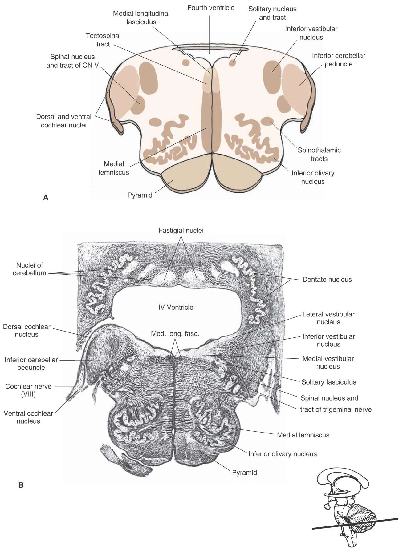
FIGURE 10-8 Upper medulla. (A) Cross-sectional diagram of the upper medulla just caudal to the pons. CN = cranial nerve. (B) Photograph of a cross section through the same level of medulla (Weigert myelin stain). Insert at bottom right indicates the level at which the section was taken.
Structures Situated in the Lateral Medulla. On the lateral side, the inferior olivary nucleus is most conspicuous because it is situated in a ventrolateral position just lateral to the pyramids (Figs. 10-7 and 10-8). The inferior olivary nucleus consists of many groups of cells, which project to the con-tralateral cerebellar cortex. The inferior olivary nucleus receives inputs from the spinal cord and red nucleus. The fibers from the spinal cord reach the inferior olivary nucleus via spino-olivary fibers,whereas fibers from the red nucleus reach the inferior olivary nucleus via rubro-olivary fibers contained in a larger bundle, the central tegmental tract.The primary function of the inferior olivary nucleus appears to be that of an important relay for the transmission of afferent signals to the cerebellum that are required for smooth, coordinated movements to be made.
Situated dorsally to the inferior olivary nucleus is the reticular formation, which contains many different groups of nerve cells as well as ascending and descending fibers. Within the core of the brainstem that forms the reticular formation lie several of the structures previously mentioned that are associated with cranial nerves. Two of these structures include the nucleus ambiguus and the solitary nucleus. Located just under the dorsolateral surface of the brainstem are two vestibular nuclei, the medial and inferior vestibular nuclei. The inferior vestibular nucleus can be easily recognized by its stippled appearance, which is due to the presence of descending fibers from the lateral vestibular nucleus that form the lateral vestibulospinal tract (Fig. 10-7B). The vestibular nuclei receive sensory information from the vestibular apparatus and transmit this information to the cerebellum; CN III, IV, and VI; and the spinal cord.
In its ventral half, the far lateral aspect of the medulla contains the ascending dorsal and ventral spinocerebellar and spinothalamic pathways as well as the descending rubrospinal tract. In a more dorsal position, the spinal nucleus and tract of CN V can be easily seen, and dorsola-teral to these structures is the inferior cerebellar peduncle, which is quite prominent at this level. At the most rostral aspect of the medulla, the primary afferent fibers of CN VIII terminate upon two additional nuclear groups, the ventral and dorsal cochlear nuclei, which lie off the dorsolateral shoulder of the inferior cerebellar peduncle and constitute part of the relay system for the transmission of auditory impulses from the periphery to the cerebral cortex (Fig. 10-8). In the human brain, the dorsal cochlear nucleus is small, whereas the ventral cochlear nucleus is large, extending into the junction between the medulla and pons.


