Gross Anatomical View and Internal Organization
Gross Anatomical View
The purpose of this topic is to begin to develop an understanding of the organization of the brainstem by considering the neuroanatomy of the medulla. Knowledge of the anatomy of the principal neural cell groups and pathways of the medulla is essential in acquiring an appreciation of their functions and insight into the clinical disorders resulting from damage to particular areas of this region of the brain-stem. One may view the medulla as an extension of the spinal cord, in that it contains a number of the fiber tracts present in the spinal cord and, like the spinal cord, also includes sensory, motor, and autonomic neurons. Thus, the principal pathways include long ascending and descending fiber systems that begin or terminate in the spinal cord and that pass through the medulla. Several other pathways mediating sensory, motor, and autonomic functions arise from the medulla and, to a considerable extent, are linked to cranial nerves. In addition, one of the three cerebellar peduncles, which connect the cerebellum to the brainstem, includes the inferior cerebellar peduncle. This massive fiber bundle represents an important conduit for transmission of information from the spinal cord and medulla to the cerebellum and is attached to the cerebellum from the dorsolateral aspect of the rostral medulla.
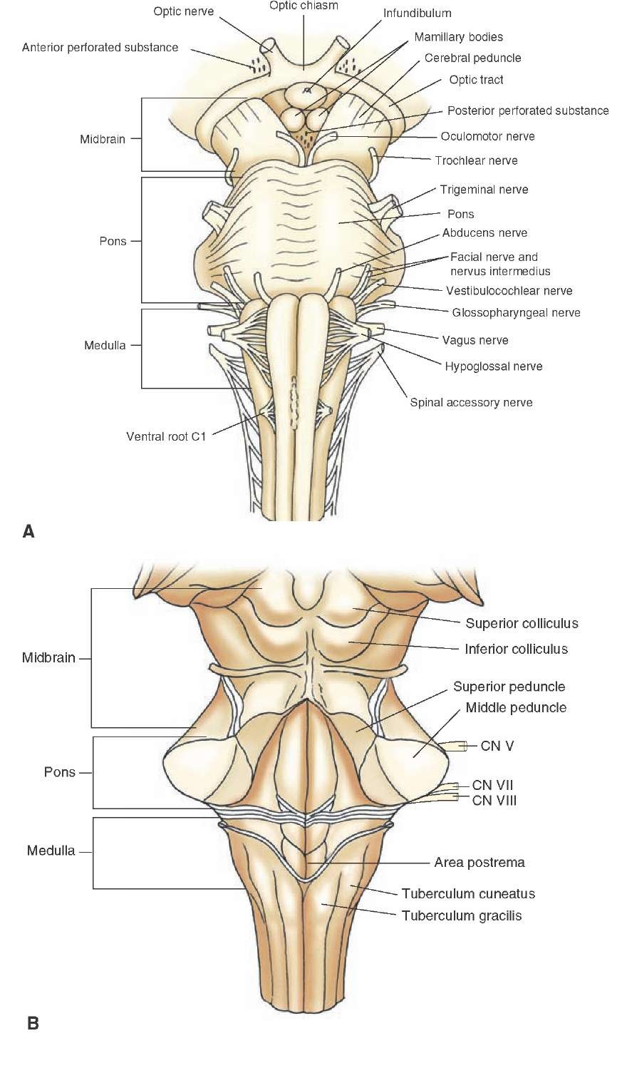
FIGURE 10-1 Different views of brainstem. (A) Anterior view of the medulla. (B) Posterior view of the medulla. Note the position at which the fourth ventricle begins (the "open" part of the medulla) at the level of the area postrema. In this illustration, the cerebellum has been removed in order to see the structures situated on the dorsal surface of the medulla. The midbrain and pons are included for purposes of orientation. Cranial nerves (CN) VII-X and XII are presented to illustrate their medial or lateral positions along the neuraxis of the medulla.
The medulla, or myelencephalon, is located between the pons and spinal cord. At its caudal end, it is continuous with the spinal cord. At its rostral end, the medulla is continuous with the pons (Fig. 10-1). Within the caudal half of the medulla, one can observe the positions of the fasciculus gracilis and fasciculus cuneatus, which ascend from the spinal cord (Fig. 10-1B). These tracts mediate conscious proprioception, vibratory sensation, and some tactile sensation from the body to the brain. As these structures pass in a rostral direction, they form two swellings (seen on the dorsal surface) referred to as the tubercu-lum gracilis and tuberculum cuneatus (or gracile and cuneate tubercles [Figs. 10-1 to 10-3]). They reflect the underlying nucleus gracilis and nucleus cuneatus (the latter lies immediately lateral to the nucleus gracilis); these nuclei receive inputs from the fasciculus gracilis and cuneatus, respectively, and thus constitute a relay system for the transmission of conscious proprioception, vibratory sensation, and tactile impulses to higher regions of the brain. The gracile and cuneate tubercles extend rostrally to a level where the fourth ventricle begins. The rostral third of the medulla, which contains a portion of the fourth ventricle, is known as the "open" part of the medulla (described later in the "Levels of the Medulla" section). The part of the medulla where the central canal is present is referred to as the "closed" part of the medulla (described later). At the rostral end of the medulla, the dorsolateral surface is expanded to form the inferior cerebellar peduncle, which contains proprioceptive and vestibular fibers which project to the cerebellum.
Along the medial aspect of the ventral surface of the medulla, there is a swelling referred to as the pyramid, which is also present throughout the rostro-caudal extent of the medulla. The pyramid is composed of numerous nerve fibers that arise from the precentral, postcentral, and premotor regions of the cerebral cortex. These nerve fibers pass to the spinal cord as the corticospinal tracts or terminate within the medulla as corticobulbar fibers.Similarly, the corticobulbar tract mediates voluntary control over movements of the head region.
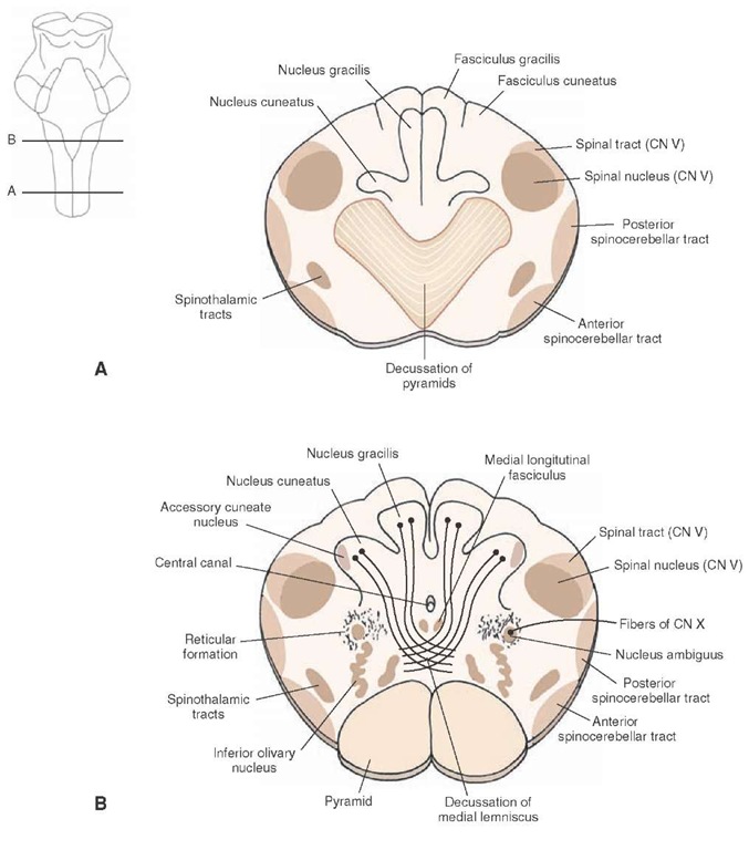
At the caudal end of the medulla immediately above the rostral end of the spinal cord, the pyramid is considerably smaller because, at this level, 90% of the fibers are crossed in a dorsolateral direction to the opposite side in a series of bundles called the decussation of the pyramids. The decussated axons then descend into the spinal cord as the lateral corticospinal tract (Fig. 10-3). The remaining (uncrossed) component of the corticospinal tract passes ipsilaterally into the ventral white matter of the spinal cord as the ventral corticospinal tract.
FIGURE 10-2 Ascending and descending pathways of fiber tracts and associated nuclei. Note the most prominent ascending pathways (the dorsal column-medial lemniscus) and descending pathways (the medial longitudinal fasciculus, corticospinal and corticobulbar tracts, and descending tract of cranial nerve [CN] V) that traverse the medulla. In the lower half of the medulla, the nucleus gracilis and nucleus cuneatus are shown. At more rostral levels, the relative positions of the nucleus ambiguus (CN IX and X); spinal nucleus of CN V and its rostral extension in the pons; the main sensory nucleus of CN V; hypoglossal nucleus (CN XII); inferior olivary nucleus; inferior (I), medial (M), lateral (L), and superior (S) vestibular nuclei; and cochlear nuclei (CN VIII) are depicted.
At the level of the rostral half of the medulla, another swelling located just lateral to the pyramids is referred to as the inferior olivary nucleus.
Internal Organization
Major Fiber Tracts and Associated Nuclei
Figures 10-2 and 10-3 depict major fiber tracts and their associated nuclei.
Pyramidal (Corticospinal) Tract. The pyramidal tract is situated in a ventromedial position throughout most of the medulla but changes position at its caudal end. Here, approximately 90% of the fibers, en route to the spinal cord, pass in a dorsolateral direction while crossing the midline to reach the dorsolateral aspect of the caudal medulla. The fibers, which cross to the contralateral side of the brainstem, form the decussation of the pyramid and descend into the dor-solateral aspect of the lateral funiculus of the spinal cord as the lateral corticospinal tract.
Medial Lemniscus. The medial lemniscus originates from the dorsal column nuclei.These fibers collectively pass ventrally for a short distance in an arc-like trajectory and are referred to as internal arcuate fibers. These second-order somatosensory fibers then cross to the opposite side of the medulla as the decussation of the medial lemniscus. The medial lemniscal fibers then pass rostrally in a medial position throughout the remainder of their trajectory through the medulla and continue into the pons. The function of the medial lemniscus is to transmit information associated mainly with conscious proprioception and vibratory stimuli to the thalamus (Fig. 10-2).
The Medial Longitudinal Fasciculus. The medial longitudinal fasciculus (MLF) tract is located in a dorsomedial position within the medulla and consists of both ascending and descending axons (Fig. 10-2 and Fig. 10-3B). Ascending axons arising from the lateral, medial, and superior ves-tibular nuclei project to the pons and midbrain; they provide information about the position of the head in space to cranial nerve nuclei that mediate control over the position and movements of the eyes. The descending fibers, which arise from the medial vestibular nucleus and pass caudally to cervical levels of the cord, are often called the medial vestibulospinal tract. This tract serves to adjust changes in the position of the head in response to changes in vestibu-lar inputs.
FIGURE 10-3 Cross-sectional diagrams of the lower medulla. (A) Level of the decussation of the pyramids. (B) Level of the decussation of the medial lemniscus. CN = cranial nerve.
Descending Tract of Nerve V. The descending fibers of the trigeminal nerve extend caudally as far as the second cervical segment and occupy a far lateral position within the medulla immediately lateral to the spinal nucleus of the trigeminal nerve (cranial nerve [CN] V [Figs. 10-2 and 10-3]). The spinal nucleus of CN V is present throughout the entire length of the medulla, extending from the level of the lower pons caudally to the spinal cord-medulla border, where it becomes continuous with the substantia gelatinosa. In the middle of the pons, the spinal nucleus is replaced by the main sensory nucleus of CN V.
The descending fibers of CN V constitute first-order axons that mediate somatosensory inputs from the head region to the brain; they synapse at different levels along the rostro-caudal axis of the spinal nucleus. Therefore, the spinal nucleus constitutes the second-order neuron, which transmits somatosensory information from the head region to the thalamus (for subsequent transmission to the cerebral cortex).Other Fiber Tracts. Several other tracts, shown in part in Figure 10-3, reflect pathways that mediate sensory and motor functions. Sensory tracts include the spinothalamic and spinocerebellar pathways. The spinothalamic and spinocerebellar fibers are situated laterally within the medulla and, thus, retain the same general positions that they occupied within the lateral funiculus of the spinal cord. The lateral spinothalamic tract mediates pain and temperature sensation from the contralateral side of the body to the thalamus, and the spinocerebellar tracts mediate unconscious proprioception (i.e., from muscle spindles and Golgi tendon organs) to the cerebellum. Tracts mediating motor functions in addition to the corticospinal tract include the tectospinal tract, which follows the MLF to cervical levels and mediates postural movements from the superior colliculus; the rubrospinal tract, which passes in a ventrolateral position in the medulla from the red nucleus to all levels of the spinal cord and facilitates spinal cord flexor motor neuron activity; and the lateral and medial reticulospinal tracts, which arise from the medulla and pons, respectively, pass to different levels of the spinal cord, and modulate muscle tone.

![Ascending and descending pathways of fiber tracts and associated nuclei. Note the most prominent ascending pathways (the dorsal column-medial lemniscus) and descending pathways (the medial longitudinal fasciculus, corticospinal and corticobulbar tracts, and descending tract of cranial nerve [CN] V) that traverse the medulla. In the lower half of the medulla, the nucleus gracilis and nucleus cuneatus are shown. At more rostral levels, the relative positions of the nucleus ambiguus (CN IX and X); spinal nucleus of CN V and its rostral extension in the pons; the main sensory nucleus of CN V; hypoglossal nucleus (CN XII); inferior olivary nucleus; inferior (I), medial (M), lateral (L), and superior (S) vestibular nuclei; and cochlear nuclei (CN VIII) are depicted. Ascending and descending pathways of fiber tracts and associated nuclei. Note the most prominent ascending pathways (the dorsal column-medial lemniscus) and descending pathways (the medial longitudinal fasciculus, corticospinal and corticobulbar tracts, and descending tract of cranial nerve [CN] V) that traverse the medulla. In the lower half of the medulla, the nucleus gracilis and nucleus cuneatus are shown. At more rostral levels, the relative positions of the nucleus ambiguus (CN IX and X); spinal nucleus of CN V and its rostral extension in the pons; the main sensory nucleus of CN V; hypoglossal nucleus (CN XII); inferior olivary nucleus; inferior (I), medial (M), lateral (L), and superior (S) vestibular nuclei; and cochlear nuclei (CN VIII) are depicted.](http://what-when-how.com/wp-content/uploads/2012/04/tmp14113_thumb.jpg)

