The Basilar Artery
The two vertebral arteries join at the caudal border of the pons to form the single basilar artery (Fig. 4-1). The major branches of the basilar artery are described in the following sections.
The Anterior Inferior Cerebellar Artery
The anterior inferior cerebellar artery (AICA) is the most caudal branch arising from the basilar artery (Fig. 4-1). The AICA supplies the ventral and inferior surface of the cerebellum and lateral parts of the pons.
The Labyrinthine (Internal Auditory) Artery
The labyrinthine (internal auditory) artery (Fig. 4-1) is usually a branch of the AICA and supplies the cochlea and labyrinth.
The Pontine Arteries
Several pairs of pontine arteries (Fig. 4-1) arise from the basilar artery. Some pontine arteries (the paramedian arteries) enter the pons immediately and supply the medial portion of the lower and upper pons. The following structures are located in these regions: the pontine nuclei, cor-ticopontine fibers, the corticospinal and corticobulbar tracts, and portions of the ventral pontine tegmentum and medial lemniscus. Some pontine arteries (the short circumferential arteries) travel a short distance around the pons and supply a wedge-shaped area in the ventrolateral pons. The long circumferential arteries supply most of the tegmentum of the rostral and caudal pons and lateral portions of the midbrain tegmentum.
The Superior Cerebellar Artery
The superior cerebellar artery (Fig. 4-1) arises just caudal to the bifurcation of the basilar artery and supplies the rostral level of the pons, caudal part of the midbrain, and superior surface of the cerebellum. This artery supplies the following structures: portions of the superior and middle cerebellar peduncles, the medial and lateral lemniscus, part of the spinal trigeminal nucleus and tract, the spi-nothalamic tract, and superior cerebellar peduncle.
The Posterior Cerebral Arteries
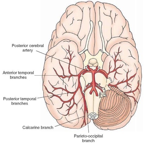
The posterior cerebral arteries arise at the terminal bifurcation of the basilar artery (Fig. 4-1). Branches of the posterior cerebral arteries (Fig. 4-4) supply most of the midbrain, thalamus, and subthalamic nucleus. The major branches arising from this artery after it passes around the midbrain (anterior and posterior temporal and parieto-occipital branches) supply the temporal lobes and medial and inferior occipital lobes of the cerebral cortex. The branch called the calcarine artery supplies the primary visual cortex.
FIGURE 4-4 Major branches of the posterior cerebral artery. These include the anterior and posterior temporal branches, the parieto-oc-cipital branch, and the calcarine branch.
Cerebral Arterial Circle (Circle Of Willis)
The cerebral arterial circle (Fig. 4-1) surrounds the optic chiasm and the infundibulum of the pituitary. It is formed by the anastomosis of the branches of the internal carotid artery and the terminal branches of the basilar artery. The anterior communicating artery connects the two anterior cerebral arteries, thus forming a semicircle. The circle is completed as the posterior communicating arteries arising from the internal carotid arteries at the level of the optic chiasm travel posteriorly to join the posterior cerebral arteries that are formed by the bifurcation of the basilar artery. The circle of Willis is patent in only 20% of individuals. When it is patent, this arterial system supplies the hypothalamus, hypophysis, infundibulum, thalamus, caudate nucleus, putamen, internal capsule, globus pal-lidus, choroid plexus (lateral ventricles), and temporal lobe. When the blood flow in either the internal carotid arteries or vertebro-basilar system is reduced, collateral circulation in the circle of Willis provides blood to the deprived brain regions.
Arteries of the Dura
Meningeal arteries arise from the internal carotid artery as it passes through the cavernous sinus. The primary arterial supply to the dura is provided by the middle meningeal artery. The anterior meningeal arteries supply the dura located in the anterior fossa, and the posterior meningeal arteries supply the dura located in the posterior fossa.
Venous Drainage of the Brain
The brain is drained by a system of veins that empty into the dural sinuses. The sinuses empty into the right and left internal jugular veins.
The Sinuses
The Superior Sagittal Sinus
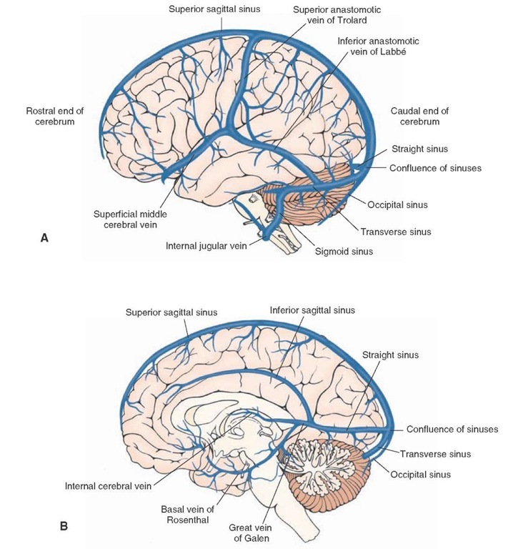
The superior sagittal sinus (Fig. 4-5, A and B) lies along the superior border of the falx cerebri and empties into the confluence of sinuses.
The Inferior Sagittal Sinus
The inferior sagittal sinus (Fig. 4-5B) lies in the inferior border of the falx cerebri. The great cerebral vein of Galen joins the inferior sagittal sinus to form the straight sinus (Fig. 4-5B) that runs caudally and empties into the caudal end of the superior sagittal sinus at the level of the confluence of sinuses.
The Transverse Sinuses
The transverse sinuses (Fig. 4-5, A and B) originate on each side of the confluence of sinuses. Each transverse sinus travels laterally and rostrally and curves downward to form the sigmoid sinus that empties into the internal jugular vein on the same side (Fig. 4-5A).
The Confluence of Sinuses
At the confluence of sinuses, the superior sagittal, straight, transverse, and occipital sinuses join (Fig. 4-5, A and B). The occipital sinus ascends from the foramen magnum.
FIGURE 4-5 Major dural sinuses and veins. (A) Major dural sinuses shown here include the superior sagittal, straight, occipital, transverse, and sigmoid sinuses. The superior sagittal, straight, transverse, and occipital sinuses join at the confluence of sinuses. Major veins include the superficial middle cerebral vein, the superior anastomotic vein of Trolard, and the inferior anastomotic vein of Labbe. (B) Major dural sinuses shown here include the superior sagittal, inferior sagittal, straight, occipital, and transverse sinuses, and their confluence. Major veins shown here include the great vein of Galen, the basal vein of Rosenthal, and the internal cerebral vein.
The Cavernous Sinuses
The cavernous sinuses are located on each side of the sphenoid bone. Ophthalmic and superficial middle cerebral veins drain into these sinuses.
The Sphenoparietal Sinuses
The sphenoparietal sinuses are located below the sphenoid bone and drain into the cavernous sinus.
The Cerebral Veins
The cerebral veins are usually divided into the superficial cerebral veins and the deep cerebral veins.
The Superficial Cerebral Veins
There are three major veins in the group of the superficial cerebral veins (Fig. 4-5 A). (1) The superficial middle cerebral vein, which runs along the lateral sulcus, drains the temporal lobe and empties into the cavernous sinus.
(2) The superior anastomotic vein of Trolard is the largest superficial vein; it travels across the parietal lobe, drains into the superior sagittal sinus, and connects the superficial middle cerebral vein with the superior sagittal sinus.
(3) The inferior anastomotic vein of Labbe connects the superficial middle cerebral vein with the transverse sinus. It is the largest vein draining into the transverse sinus. It travels across the temporal lobe.
The Deep Cerebral Veins
Three major veins are included in the group of the deep cerebral veins (Fig. 4-5B): (1) the great cerebral vein of Galen, (2) the basal vein of Rosenthal, and (3) the internal cerebral vein. The great cerebral vein of Galen is a short vein (about 2 cm long) and is formed by the union of two internal cerebral veins at the level of the splenium of corpus callosum. It courses caudally and joins the inferior sagittal sinus to form the straight sinus, which then empties into the confluence of sinuses. The basal vein of Rosenthal receives blood from the orbital surface of the frontal lobe, anterior part of corpus callosum, rostral parts of the cingulate gyrus, the insula, the opercular cortex (a portion of the motor area for speech), and the ventral parts of the corpus striatum. The basal vein empties into the great cerebral vein of Galen. The internal cerebral veins receive venous blood from the thalamus, striatum, caudate nucleus, internal capsule, choroid plexus, and hippocampus. The cerebellum and medulla are drained by a network of veins that empty into the great cerebral vein of Galen as well as the straight, transverse, and superior and inferior petrosal sinuses.
The Spinal Cord Arteries
Posterior Spinal Arteries
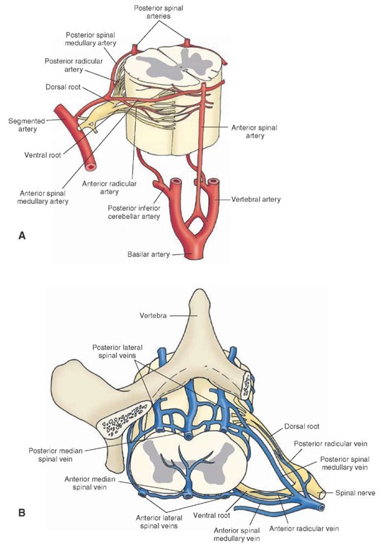
As stated earlier, in a majority of cases (75%), the PSAs arise from the PICA (Fig. 4-6A). They descend on the dorsolateral surface of the spinal cord slightly medial to the dorsal roots.
FIGURE 4-6 Vascular supply of the spinal cord. (A) Major arteries supplying the spinal cord. Note especially the two posterior and one anterior spinal arteries. The vertebral, posterior inferior cerebellar, and basilar arteries and the dorsal and ventral roots of the spinal nerve are shown for orientation purposes. (B) Major veins draining the spinal cord. The vertebra and spinal nerve are shown for orientation purposes.
Anterior Spinal Artery
Two small branches arise from the vertebral arteries as they ascend on the anterolateral surface of the medulla. These branches unite to form one single anterior spinal artery (ASA [Fig. 4-6A]) that courses along the midline of the ventral surface of the spinal cord.
The Spinal Medullary and Radicular Arteries
The posterior and anterior spinal medullary and radicular arteries arise from the segmental arteries and communicate with the PSAs and ASA (Fig. 4’6A). These arteries provide blood supply to the thoracic, lumbar, and sacral regions of the spinal cord.
Veins
In the spinal cord, on the ventral side, the anteromedian spinal vein is located in the midline and two anterolateral spinal veins are located along the line of attachment of the ventral roots (Fig. 4-6B). On the dorsal side, the posteromedian spinal vein is located in the midline and two posterolateral spinal veins are located along the line of attachment of the dorsal roots. The posteromedian and posterolateral veins are drained by posterior spinal medullary and radicular veins. The anterior median and anterolateral spinal veins are drained by anterior spinal medullary and radicular veins.
Clinical Case
History
Stan is a 78-year-old man who was brought to the local emergency room (ER) because his family noted that he was suddenly not using his left arm and leg. In addition, he began to show some behavioral changes. When asked about his inability to move his right side, he said/’l am fine. What is wrong with you?" Although normally very cooperative, he appeared to be more withdrawn and irritable. The morning that he was brought to the ER, he only shaved the right half of his face, combed the right side of his hair, and wore a sock and shoe on the right foot.
Examination
A neurologist examined Stan and found a loud bruit (pronounced as"bru-ee"; a rumbling sound) over the right carotid artery in his neck. When asked to show his left hand, Stan ignored the question. When his hand was lifted and he was confronted with the question of whether, indeed, the hand was his; he denied it and insisted that the hand belonged to the doctor. He did not blink when a hand was waved over the left lateral aspect of his visual fields, and when asked to draw a clock, he put all of the numbers on the dock on the right side. He denied any problem with the clock that he drew.The left side of his face drooped, excluding hisforehead, which was symmetrical,and although there was minimal movement on the left side of his body, he moved it very infrequently. When the lateral aspect of the plantar surface of his left foot was scratched, the great toe dorsiflexed and the other toes fanned upwards. When the same maneuver was performed on the other side, the toes deviated downward.
Explanation
Stan has a classic example of a right parietal stroke, which most often involves the superior parietal lobule. The artery infarcted is the right middle cerebral artery. Because the middle cerebral artery continues in nearly a straight line from the internal carotid artery, it is a common route for small emboli originating from the internal carotid artery.The bruit noted is most likely the result of a thrombus (clot) occluding the lumen of the carotid artery. When blood flows across the thrombus, a bruit is heard.
The parietal lobe is responsible for primary and secondary sensory information.One of the types of sensory information provided by this region is the ability to localize objects in space. People with lesions in the right parietal lobe live in "right-sided worlds"and ignore the left side of their bodies and all objects in space. Behavioral changes, including dulling of affect, accompany these deficits. These patients may become less cooperative, and it is not unknown for them to have several automobile accidents, colliding with objects on their left sides, before their deficits are recognized. Because the fibers from the lateral fields of the optic tract run through the parietal lobe of the contralateral side,a visual defect often accompanies strokes in this region,and patients don’t blink in response to hand-waving in this field. The motor strip may also be affected by an infarct of this artery, so genuine motor weakness with a Babinski sign (the up-going toe) would be present, signifying upper motor neuron weakness.
SUMMARY TABLE
Major Arteries of the Brain
|
Major Branches |
Major Sub-branches |
Major Brain Structures Supplied |
|
|
Internal Carotid Artery |
|
|
Ophthalmic |
Retina and cranial dura |
|
|
Posterior communicating |
Hypophysis, infundibulum, parts of hypothalamus, thalamus, and hippocampus |
|
|
Anterior choroidal |
Choroid plexus, optic tract, parts of internal capsule, hippocampal formation, globus pallidus.and lateral portions of thalamus |
|
|
Anterior cerebral |
Medial aspect of cerebral hemisphere, including frontal and parietal lobes, postcentral gyrus, and precentral gyrus |
|
|
Anterior communicating |
Connects the anterior cerebral arteries on the both sides |
|
|
Medial striate artery (recurrent artery of Heub-ner) |
Anteromedial part of head of caudate nucleus and parts of the internal capsule, putamen, and septal nuclei |
|
|
Orbital branches |
Orbital and medial surfaces of frontal lobe |
|
|
Frontopolar branches |
Medial portions of frontal lobe and lateral parts of the convexity of the hemisphere |
|
|
Callosomarginal |
Paracentral lobule and portions of cingulate gyrus |
|
|
Pericallosal artery |
Precuneus (portion of parietal lobe caudal to paracentral lobule and proximal to occipital cortex) |
|
|
Middle cerebral |
Lateral convexity of cerebral hemisphere, including parts of temporal, frontal, parietal, and occipital lobes; important neural structures supplied by middle cerebral artery include: Broca’s area of speech, prefrontal cortex, and primary and association auditory cortices including Wernicke’s area and supramarginal and angular gyri (association cortex) |
|
|
Lenticulostriate branches |
Putamen, caudate nucleus, and anterior limb of internal capsule |
|
|
Orbitofrontal |
Parts of frontal lobe |
|
|
Precentral (pre-Rolandic) and central (Rolandic) branches |
Primary sensory and motor cortex |
|
|
Anterior and posterior parietal branches |
Parts of parietal lobe |
|
|
Angular branch |
Angular gyrus |
|
|
Anterior, middle,and posterior temporal branches |
Parts of temporal lobe and lateral portions of occipital lobe |
|
|
Major Brain Structures Supplied |
||
|
Vertebral Artery |
||
|
Anterior spinal |
Medial structures of medulla including pyramids, medial lemniscus, medial longitudinal fasciculus, hypoglossal nucleus,and inferior olivary nucleus |
|
|
Posterior inferior cerebellar (PICA) |
Lateral medulla: spinothalamic tract, dorsal and ventral spinocerebellar tracts, descending sympathetic fibers, descending tract and spinal nucleus (CN V),dorsal motor nucleus (CN X),and nucleus ambiguus (CN IX and X) |
|
|
Posterior spinal (PSA) |
Caudal medulla:fasciculus gracilis and cuneatus,gracileand cuneate nuclei,spinal trigeminal nucleus,dorsal and caudal portions of inferior cerebellar peduncle, and portions of the solitary tract and dorsal motor nucleus of the vagus (CN X) |
|
|
Basilar Artery |
||
|
Anterior inferior cerebellar (AICA) |
— |
Ventral and inferior surface of cerebellum and lateral parts of the pons |
|
Labyrinthine (internal auditory) |
— |
Cochlea and labyrinth |
|
Pontine arteries |
Paramedian branches |
Medial portion of lower and upper pons: pontine nuclei, corticopontine fibers, corticospinal and corticobulbar tracts,and portions of ventral pontine tegmentum and medial lemniscus |
|
Short circumferential branches |
A wedge-shaped area in the ventrolateral pons |
|
|
Long circumferential branches |
Most of tegmentum of the rostral and caudal pons and lateral portions of midbrain tegmentum |
|
|
Superior cerebellar |
Rostral level of pons, caudal part of midbrain, and superior surface of cerebellum including portions of superior and middle cerebellar peduncles, medial and lateral lemniscus, part of spinal trigeminal nucleus and tract, and spinothalamic tract |
|
|
Posterior cerebral |
— |
Most of midbrain, thalamus, and subthalamic nucleus |
|
Anterior and posterior temporal; parietooccipital |
Temporal lobes and medial and inferior occipital lobe |
|
|
Calcarine |
Primary visual cortex |
CN = cranial nerve.


