Audition, or hearing, like vision, is one of the most important sensations. Much of human communication depends on a properly functional auditory system. For example, loss of hearing at birth can have the devastating effect of the child’s inability to learn to speak. In adults, loss of hearing can also be socially debilitating.
Sound is produced by vibrations that result in the alternate compression and rarefaction of air. The pressure waves generated in the air in this manner reach the ear and are transduced into neural signals that are processed in the brain. In the first part of this topic, the different properties of the auditory system that regulate the sensation of hearing are described. The second part of the topic covers the vestibular system, which regulates the movement and position of the head in space in response to signals associated with angular and linear acceleration of the head as well as the gravitational pull exerted on it.
Auditory System
Physics Of Sound
Auditory receptors respond to vibrations of the air that radiate from the sound source as pressure waves. The pitch (highness or lowness) of the sound is dependent on the frequency of the pressure wave generated by the sound. The frequency of the pressure wave is measured in cycles per second or Hertz (Hz). Young humans can detect a frequency range of 20 Hz to 20 kHz. In older individuals, the range of detectable frequencies decreases to 50 Hz to 17 kHz.
The magnitude of sound is expressed as intensity. The range of sound intensities is large. For instance, a sound that is barely audible is about 20 jiPa (micro-Pascals, which is the unit for measuring pressure), while a painful sound level is about 2 x 10⁸ Pa. Because of this wide range of intensities, it would be tedious to express sound intensity in absolute units. The sound intensity is, therefore, expressed in terms of the logarithm of the actual sound intensity. The convention is to describe sound intensity as the logarithm of the ratio of a measured pressure to a reference pressure and is expressed in decibels. The decibel (dB) is a logarithmic ratio expressed by the following equation:
Sound pressure (dB) = 20 log10 Pt / Pr; where Pt = pressure of the test sound, and Pr = reference pressure 20 mPa (or 0.0002 dynes/cm2).
Components of the Ear
External Ear
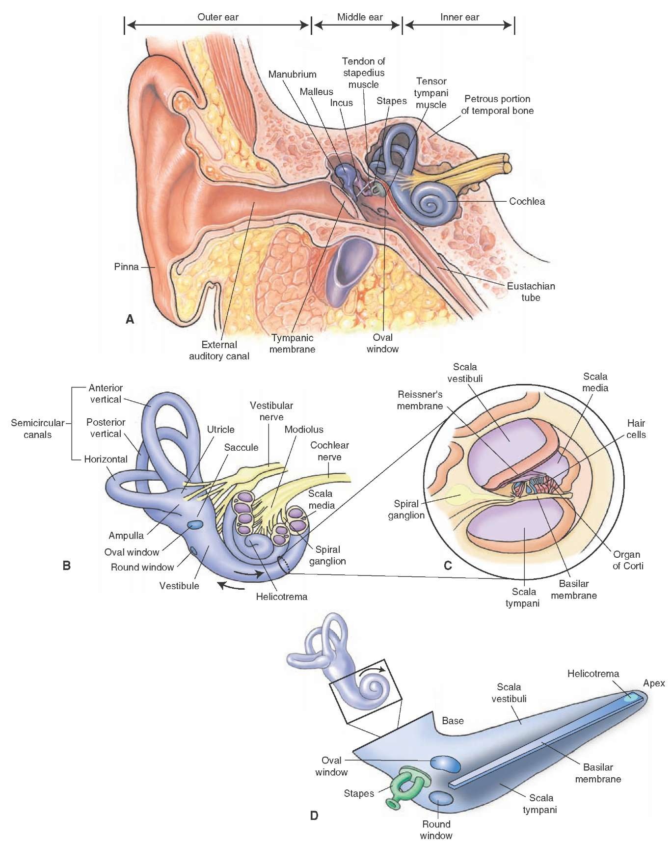
The external ear, called the pinna or auricle, directs the sound vibrations in the air to the external auditory canal (Fig. 17-1A). The sound waves travel through this auditory canal and vibrate the tympanic membrane located at the end of the canal.
Middle Ear
Three small bones (ossicles), which articulate with each other, are suspended in the cavity of the middle ear. These ossicles are the malleus (the cartilaginous process called manubrium of this bone is attached to the tympanic membrane), the incus, and the stapes. The stapes resembles a stirrup, and its footplate is bound to the oval window by an annular ligament. The middle ear is connected to the nasopharynx through the eustachian tube, which helps to equalize air pressure on the inner and outer surfaces of the tympanic membrane and to drain any fluid in the middle ear into the nasopharynx (Fig. 17-1A). A small muscle, the tensor tympani, inserts on the manubrium of the malleus; it is innervated by a branch of the trigeminal nerve (cranial nerve [CN] V). Another small muscle, the stape-dius, inserts on the stapes ossicle and is innervated by a branch of the facial nerve (CN VII). Contraction of these muscles restricts the movement of the tympanic membrane and the footplate of the stapes against the oval window, respectively, and, thus, reduces the deleterious effects of loud noises on the delicate middle and inner ear structures. Therefore, the function of the middle ear and its components is to convert the sound waves in the air to waves in the fluid located in the inner ear. If the airwaves bypass the middle ear and reach the oval window directly, only about 3% of the sound would enter the inner ear. The pressure transmitted to the oval window is amplified because (1) the area of the tympanic membrane is much greater than that of the oval window, and (2) greater mechanical efficiency is provided by the ossicles (malleus and incus) because they act as levers.
Inner Ear
The inner ear consists of the bony labyrinth, the membranous labyrinth, and the cochlea containing the organ of Corti.
The bony labyrinth consists of a complex series of cavities in the petrous portion of temporal bone. The bony labyrinth surrounds the inner membranous labyrinth and has three regions: a central region called the vestibule, three semicircular canals, and the cochlea (Fig. 17-1, A and B). The vestibule (including the saccule and utricle within it) and semicircular canals represent peripheral components of the vestibular system, which is specialized to respond to movements. The cochlea is involved in hearing and is specialized to respond to airborne sounds. All of the cavities of the bony labyrinth are filled with a fluid called the perilymph (a clear extracellular-like fluid containing high Na+ [sodium] and low K+ [potassium] concentrations). The three semicircular canals include anterior (superior), posterior, and lateral (horizontal) canals. The cochlea, shaped like a conical snail shell, has a central tubular conical axis, called the modiolus, which is surrounded by a spiral cochlear canal of two-quarter and three-quarter turns. The cochlear canal is divided into an upper scala vestibuli and a lower scala tympani (Fig. 17-1C). The scalae tympani and ves-tibuli communicate with each other at the modiolar apex via a narrow slit called the helicotrema (Fig. 17-1D). Within the bony spiral canal of the cochlea lies the coch-lear duct, which is a component of the membranous labyrinth. The cochlear duct is a spiral tube that runs along the outer wall of the bony cochlea and ends at its apex (cupula) (Fig. 17-2). The cochlear duct (also known as the scala media) is flanked by two channels within the cochlea: the scala tympani (it follows the outer contour of the cochlea) and the scala vestibuli (it follows the inner contour of the cochlea). The scala tympani ends at the round window (the secondary tympanic membrane), which separates this space (known as the tympanic cavity) from the middle ear. As mentioned earlier, the stapes (a stirrup-shaped small bone in the middle ear) is bound to the oval window by an elastic ligament. Vibrations of the stapes in response to sound waves are transmitted to the scala vestibuli via the oval window.
FIGURE 17-1 Components of the ear. (A) Note the location of the external, middle, and inner ear. The middle ear contains three small bones (the malleus, the incus, and the stapes). (B) The inner ear consists of a bony labyrinth that contains the vestibule, three semicircular (anterior, posterior, and lateral) canals, and the cochlea. (C) A cross section of cochlea reveals that the scala media is bounded by the vestibular (Reissner’s) and basilar membranes, and the organ of Corti lies within the scala media (cochlear duct). (D) A diagram of an uncoiled cochlea.
FIGURE 17-2 The membranous labyrinth is located within the cavities of the bony labyrinth (shown in beige). The cochlear duct is shown in blue, and the remainder of the membranous labyrinth is shown in green.
The membranous labyrinth is located within the cavities of the bony labyrinth and is filled with endolymph (an intracellular-like fluid containing low Na+ and high K+). The membranous labyrinth includes the utricle and saccule (two small sacs occupying the bony vestibule); three semicircular canal ducts, which occupy the bony semicircular canals; and the cochlear duct (already described) (Fig. 17-2). The scala media contains endolymph; and the scalae tympani and vestibuli contain perilymph. The scala media is bound by two membranes: the vestibular membrane (also known as Reissner’s membrane) and the basilar membrane (Fig. 17-1C). These two membranes separate the endolymph contained in the scala media from the perilymph contained in the scalae tympani and vesti-buli. In the lateral wall of the scala media lies the stria vascularis, which is highly vascular.
Na+-K+ pumps in the stria vascularis pump sodium out of and potassium into the endolymph. As noted earlier, the endolymph contains a high concentration of K+ ions and a low concentration of Na+ ions.
The organ of Corti is the sensory area of the cochlear duct and is located on the basilar membrane. It contains hair cells, which are the sensory receptors for sound stimuli. It lies within the cochlear duct (the scala media) and runs along the entire length of the basilar membrane (Fig. 17-1C). The human cochlea contains two types of hair cells: inner hair cells and outer hair cells (Fig. 17-3). About 100 stereocilia and a single kinocilium (larger hair cell) are attached to the apical border of the hair cells. The stereocilia are shorter than the kinocilium, and their length increases as they approach the kinocilium. The tips of stereocilia of outer hair cells are embedded in the overlying tectorial membrane, which is composed of muco-polysaccharides embedded in a collagenous matrix (Fig. 17-3). Movement of the basilar membrane relative to the tectorial membrane results in displacement of the cilia that is necessary for generating afferent signals (see "Mechanism of Sound Conduction"). Inner hair cells are not attached to the tectorial membrane, and movement of their stereocilia is induced by movement of the endol-ymph. There are two types of supporting epithelial cells that keep the hair cells in position: the phalangeal cells and pillar cells. The outer phalangeal cells (Deiter’s cells) surround the base of the outer hair cells and the nerve terminals associated with these cells (Fig. 17-3). These cells give out a phalangeal process. This process flattens into a plate near the apical surface of the hair cell and forms tight junctions with the apical edges of adjacent hair cells and adjacent phalangeal plates. The inner phalangeal cells surround the inner hair cell completely and do not have a phalangeal process. Similarly, there are outer and inner pillar cells whose apical processes form tight junctions with each other and with neighboring hair cells. This network of tight junctions isolates the body of the hair cells from the endolymph contained in the scala media. The spiral (cochlear) ganglion, located within the spiral canal of the bony modiolus, contains bipolar neurons. The peripheral processes of these bipolar neurons in the spiral ganglion innervate the hair cells; they form the postsynaptic afferent terminals at the base of the hair cell (Fig. 17-3). The central processes of the bipolar cells in the spiral ganglion form the cochlear division of CN VIII. The outer hair cells receive efferent fibers that arise from the superior olivary nucleus (the olivocochlear bundle). This bundle provides a basis by which the central nervous system can modulate auditory impulses directly at the level of the receptor.
FIGURE 17-3 The hair cells. Note the two types of hair cells: inner and outer hair cells with stereo-cilia and one kinocilium present on the apical border. The tips of the cilia on only the outer hair cells are embedded in the tectorial membrane. The peripheral processes of the bipolar neurons in the spiral ganglion form the postsynaptic afferent terminals at the base of the hair cell.
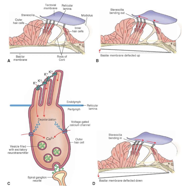
FIGURE 17-4 Mechanism of sound conduction. (A) The tips of the stereocilia on the hair cells are embedded in the tectorial membrane, and the bodies of hair cells rest on the basilar membrane. (B) An upward displacement of the basilar membrane creates a shearing force that results in lateral displacement of the stereocilia. (C) Mechanical displacement of the stereocilia and kinocilium in a lateral direction causes depolarization of the hair cell. (D) A downward displacement of the basilar membrane creates a shearing force that results in hyperpolarization of the hair cell.
Mechanism of Sound Conduction
Air pressure waves cause the tympanic membrane to vibrate, resulting in oscillatory movements of the footplate of the stapes against the oval window. Because perilymph is a noncompressible fluid and the scalae tympani and ves-tibuli form a closed system, oscillatory movements of the stapes against the oval window result in pressure waves in the perilymph present in the scalae tympani and vestibuli. The oscillatory movement of perilymph results in vibration of the basilar membrane.
As mentioned earlier, the tips of the stereocilia (of the outer hair cells) are embedded in the tectorial membrane, and the bodies of hair cells rest on the basilar membrane (Fig. 17-4A). An upward displacement of the basilar membrane creates a shearing force that results in lateral displacement of the stereocilia (Fig. 17-4B). Mechanical displacement of the stereocilia and kinocilium in a lateral direction causes an influx of K+ through their membranes, the hair cell is depolarized, and there is an influx of Ca2+ (calcium) through the voltage-sensitive Ca2+ channels in their membranes. The influx of Ca2+ triggers the release of the transmitter (probably glutamate) that, in turn, elicits an action potential in the afferent nerve terminal at the base of the hair cell (Fig. 17-4C). A downward displacement of the basilar membrane creates a shearing force that results in medial displacement of the stereocilia and kino-cilium (Fig. 17-4D). Mechanical displacement of the ster-eocilia and kinocilium in a medial direction results in hyperpolarization of the hair cell that may involve opening of voltage-sensitive K+ channels and efflux (outward flow) of K+. The sensory receptors (hair cells) located in the basal portion of the basilar membrane respond to high frequencies of sound, whereas the sensory receptors located in the apical aspect of the membrane respond to low frequencies (Fig. 17-5). This is called tonotopic distribution of responding receptors.


