Plate Section (Neuroanaesthesia and Neurointensive Care) Part 5


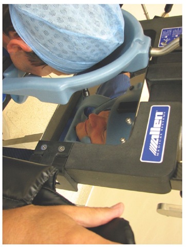
This photograph shows a patient in the park-bench position. Once the patient is draped, the airway is not easily accessible. Therefore, securing the endotracheal tube and ensuring that the neck vessels are not obstructed is of paramount importance.

Fig. 11.2. This photograph shows a patient in the park-bench position. Once the patient is draped, the airway is not easily accessible. Therefore, securing the endotracheal tube and ensuring that the neck vessels are not obstructed is of paramount importance.

Operating theatre arrangements for awake craniotomy. Note that the surgical drapes are placed to allow the anaesthetist access to the patient's airway and face for intraoperative testing and airway management.


Fig. 12.2. Operating theatre arrangements for awake craniotomy. Note that the surgical drapes are placed to allow the anaesthetist access to the patient’s airway and face for intraoperative testing and airway management.

An example of a mirror system that allows inspection of the eyes and nose during surgery in the prone position.

Fig. 15.9. An example of a mirror system that allows inspection of the eyes and nose during surgery in the prone position

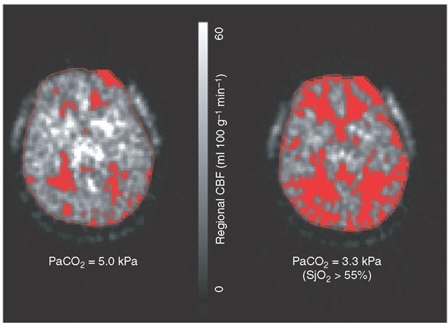
Positron emission tomography imaging showing the effect of hyperventilation in the first 24 h following TBI on regional CBF. Even with an acceptable SjO2, hyperventilation results in a marked increase in the volume of brain tissue (highlighted areas) below an ischaemic threshold (20 ml (100 g)-1 min-1) due to vasoconstriction from hypocapnia.

Fig. 21.6. Positron emission tomography imaging showing the effect of hyperventilation in the first 24 h following TBI on regional CBF. Even with an acceptable SjO2, hyperventilation results in a marked increase in the volume of brain tissue (highlighted areas) below an ischaemic threshold (20 ml (100 g)-1 min-1) due to vasoconstriction from hypocapnia.

 (b) CTangiogram demonstrating a lobular aneurysm arising from the anterior communicating artery (arrow).

Fig. 22.1. (b) CTangiogram demonstrating a lobular aneurysm arising from the anterior communicating artery (arrow).

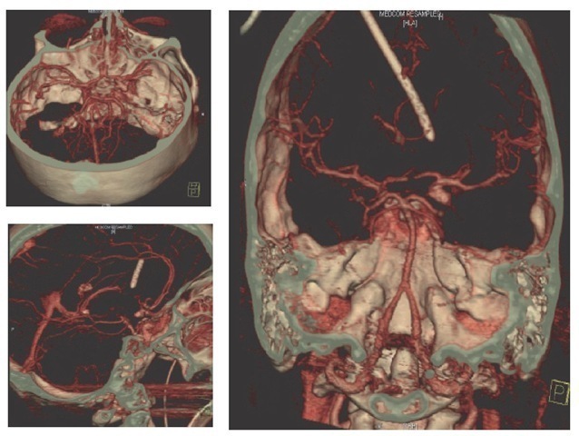
Digital subtraction angiography showing an irregular and elongated (9 mm) aneurysm at the origin of the right posterior communicating artery. The aneurysm is directed posterolaterally, with a small lobule directed medially.

Fig. 22.2. Digital subtraction angiography showing an irregular and elongated (9 mm) aneurysm at the origin of the right posterior communicating artery. The aneurysm is directed posterolaterally, with a small lobule directed medially.

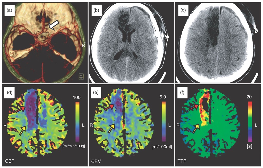
Subarachnoid haemorrhage and ischaemic changes with time. (a) Day of bleed. CT angiogram demonstrating a lobular aneurysm arising from the anterior communicating artery (arrow). Both anterior cerebral arteries originate fromthe left side with hypoplasia of the right A1. (b, c) Day 10 post-bleed. Plain CT head showing extensive infarction of the right anterior cerebral artery territory involving the genu of the corpus callosum. (d-f) Day 11 post-bleed. CT perfusion showing an established infarct in the right anterior cerebral artery territory (reduced cerebral blood flow (CBF) and cerebral blood volume (CBV), grossly delayed time to peak; red arrows). There is a large, hypoperfused salvageable area extending into the left anterior cerebral artery territory (reduced CBF with maintained CBV and moderately delayed time to peak;yellowarrows).
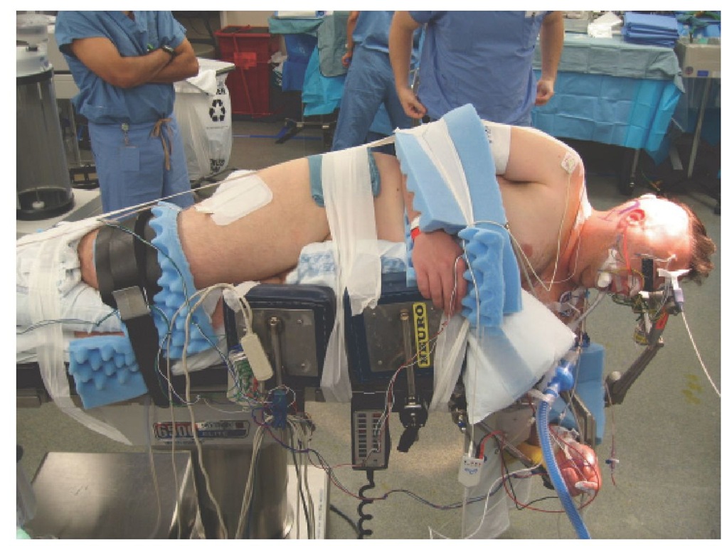
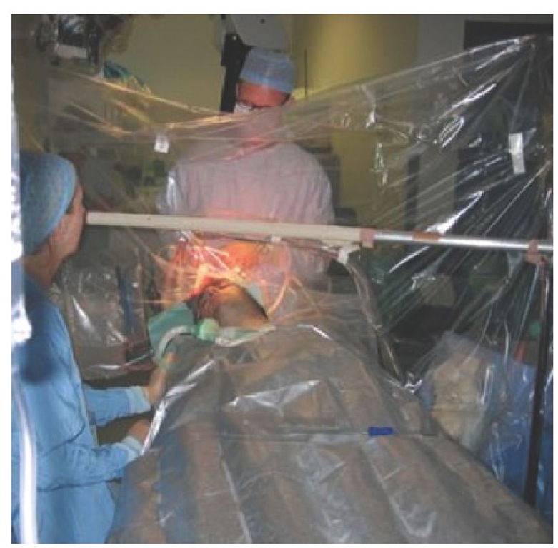
Fig. 22.3. Subarachnoid haemorrhage and ischaemic changes with time. (a) Day of bleed. CT angiogram demonstrating a lobular aneurysm arising from the anterior communicating artery (arrow). Both anterior cerebral arteries originate fromthe left side with hypoplasia of the right A1. (b, c) Day 10 post-bleed. Plain CT head showing extensive infarction of the right anterior cerebral artery territory involving the genu of the corpus callosum. (d-f) Day 11 post-bleed. CT perfusion showing an established infarct in the right anterior cerebral artery territory (reduced cerebral blood flow (CBF) and cerebral blood volume (CBV), grossly delayed time to peak; red arrows). There is a large, hypoperfused salvageable area extending into the left anterior cerebral artery territory (reduced CBF with maintained CBV and moderately delayed time to peak;yellowarrows).
Next post:

Previous post: