Till the present, an enormous number of facts concerning brain have been collected, while advancement of our understanding of the mechanisms of its activity remains insignificant. The set of known experimental data and the quantity of the scientific reports reaches a disturbing threshold, with old facts being "rediscovered" using new methods. For example, during conditional reflex elaboration, the reaction to a signal acquiring important significance usually increases, while response to the insignificant signals decreases. This change in the responses is highly specific. During learning, only those responses, whose significance was adjusted at the time of learning, are modified, whereas responses to foreign signals do not change. This axiom is true both for animal behavior [947] and for neuronal reactions [530, 19, 503], but it continues to surprise modern scientists [371, 1282, 1087].
Motivation is a leading factor in goal-directed behavior; it alters readiness to action and is the most accessible phenomenon for study of a subjective feeling. Brain evaluates the magnitudes of inner and external variables and compares present conditions with past experience and also evaluates the possible consequences of its own reactions to the alteration of these variables. Put simply, sensation affects inner environment and generates movements, which alter outer environment and thus change sensations. We usually examine brain behavior like that of any other object, but the brain itself also studies the environment and uses memory in order to control behavior.
Neuronal structure of the brain is equally complex and well-ordered. Human brain contains a number of neural centers. Certain centers are stratified, like the neocortex, hippocampus and cerebellum, while others are dispersed nuclei without strict structure. Large centers are divided into subdivisions; for example, the neocortex is separated to more than one hundred fields.
The cortex has extraordinarily specific connections between individual cells and cell classes. Brain pathways and cortical regions that are established during early development are partially genetically determined, but depend also on their synaptic inputs and behavioral instruction [1196]. In several categories of animals, such as some insects, mollusks and fishes, there are genetically specific neurons that are distinct from any other neurons, but they are identical in different creatures of the same species. Environment also plays a role in brain morphology and intelligence, particularly in humans, but the predominant influence appears to genetic (70%-80%). Monozygotic twins reared apart are more alike with respect to cognitive and morphologic properties than heterozygotic twins. Heritability does not imply inevitability because the environment can determine the relative impact of genetic variation. Between 20%-35% of the observed population differences in intellectual capability are due to differences in family environments. Also, heritability of intelligence increases with age: as twins grow older, phenotype reflects genotype more closely. A strictly environmental theory would predict the opposite [1233]. This means that morphological structure of the neural system may be predetermined. Nevertheless, this predetermined structure is not the only possible and necessary one for correct performance. If, during early individual development, brain structure is to a marked degree mechanically injured, behavior is usually recovered so completely that it is difficult to distinguish from the behavior of a normal uninjured brain. This is not the case for destructive factors affecting the whole brain, such as stress and energy deficiency. Extirpations of neural tissue are well compensated, while stress in early life leaves a negative trace and an enriched environment leaves a positive trace [1040].
Brain structure has changed dramatically during evolution, but properties of a neuron have remained conservative in the animal world. Some properties of behavior have also been conserved through evolution and these properties are, possibly, determined by the peculiarity of neurons and not by brain structure: the capability to accept signals; excitability; transportation of electrical message into chemical output and plasticity in accordance with a change of circumstances. A complication of brain structure in evolution is correlated with the property of behavior, which has been perfected during evolution. This perfection is, before all, the capability of simultaneous maintenance, in an active state, a large quantity of information ordered in time. This capability is essential for development of consciousness.
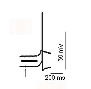
Although neurons are not complicated by evolution, they are complex themselves.Electrical signals, it was thought, are the only means of brain operation. The intracellular environment of a neuron (K+ rich and Na+ poor) has a negative membrane potential around 70 mV – and is separated from the extracellular environment by means of a microscopic neuronal lipid membrane [984]. An action potential (AP, spike, neural impulse) is a short electrical wave travelling along the cellular membrane of a neuron. Neuronal membranes contain pores, special proteins in synaptic areas (chemoreceptors) and ion channels in non-synaptic membrane. Potential-dependent (K+, Na+, Ca2+ and some others) and ligand-dependent channels control membrane permeability for corresponding ions. For example, Na+-channels contain a selectivity filter, which is made of negatively charged amino acid residues. They attract the positive Na+ ions, while the larger K+ ions cannot fit through this area. Potential-dependence means that the probability of the channel opening for the corresponding ion increases, when, as the rule, the membrane potential decreases (during depolarization). Ligand-dependent channels change the ion permeability as a result of chemical influence. In the standard scenario, depolarization at the beginning opens some sodium channels. Na+ ions penetrate the neuron and augment depolarization. As a result, the probability of the next sodium channels opening increases. There is a critical level of depolarization, the threshold, when membrane permeability for Na+ grows to maximum and the membrane potential reaches an equilibrium potential for sodium (around +40 mV): thus an AP is generated (Fig. 1.1).
Fig. 1.1. Simultaneous recording of intracellular activity of two mollusk neurons. At the top, a neuron generates excitatory postsynaptic potential (EPSP), but fails to generate an action potential (AP). Vertical arrow presentation of tactile stimulus. At the bottom, a neuron generates the AP. Bold arrow, level of AP generation. Calibrations are pointed out at the figure.
Membrane potential returns to normal level, -70 mV, when depolarization initiates opening of the potassium channels. This requires higher depolarization than the opening of sodium channels. Ions of K + leave the neuron, the membrane acquires more negative charge and membrane potential recovers. During generation of each AP, the neuron loses some K + ions and loads Na+ ions. The proportion between inner K + and outer Na+ ions is sufficient for generation of hundreds or thousands of spikes, but each neuron has a special tool for ion equilibrium recovery: protein Na+, K+-ATPase, which transmits Na+ out and K + in, spending energy as a molecule of ATP. Na+, K+-ATPase activity takes approximately half of the energy consumed by the brain [802].
Neural cells are a relatively new achievement of evolution. Basic metabolic pathways and mechanisms of any cell are preserved in a neural cell, but excitability and potential-dependent Na+ channels are qualitatively new attributes of a neuron. Quantitative distinctions of a neuron are high levels of energetic and protein metabolism and low possibilities for displacement, although motility of neuronal processes is observed. Once arisen, neural cells have conservative properties in primitive multicellular animals and human.
Although properties of neural cells feebly change during evolution, they do change during individual development. Biophysical characteristics of cell membranes are, evidently, essential for brain function, since a decrease in the rate of learning from infancy to old age is accompanied by modifications in characteristic neuronal membranes. Input resistance, the duration of action potentials, the membrane time constant of many brain neurons, the amplitude of the peak of whole cell K+ current, the excitability of identified neurons, the frequency of impulse generation, the rate of repolarization of the action potential, the synaptic efficacy and the electrical coupling between neurons all diminish with age, while the peak of whole cell Ca2+ current, the intracellular Ca2+ concentrations and the threshold for action potential generation all increase [212, 1379]. At the same time, parallels between neuronal function and development of neural tissue in the philo- and ontogenesis may be superficial.
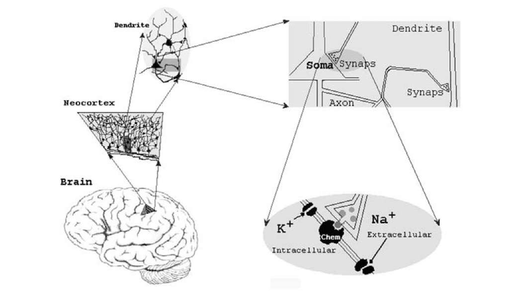
Brain contains a lot of neural centers with incompletely known functions. In their turn, neural centers consist of neurons and glial cells, connected in sometimes strictly predetermined, but sometimes unpredetermined networks. Neurons are a much more favorite object of investigation than glial cells, since neural cells produce activity similar to a digital device and this has allowed the hope to understand brain. Neurons affect each others using specific substances, called neurotransmitters. Chemical signals are impossible to transmit at large distances by means of diffusion; this is a too slow and undirected process. Signals between cells for large distances are transmitted by, action potentials. A presynaptic neuron sends an AP, as its message into its axons to all neurons to which it is connected and triggers a chemical message, a neurotransmitter, at the synaptic cleft in the next neuron (see Fig. 1.2). Postsynaptic neurons usually receive input signals through synapses to their dendrites or soma. A transmitter interacts with a chemoreceptor that is specific for this transmitter, and transitory changes in membrane conductance and excitatory or inhibitory postsynaptic potential arises (dependently on the type of the chemoreceptor). The postsynaptic potential changes the membrane potential of a neuron by the flow of positive or negative ions through the membrane. A postsynaptic potential is excitatory if it promotes generation of an action potential, while it is inhibitory, if it prevents spike generation. When many excitatory synapses unite their action on a cellular membrane, the impact to a neuron may exceed threshold, the neuron generates spikes spreading into its axon and the next postsynaptic neuron will increase its firing rate. An axon is not electrical wire. Action potential spreads along an axon in a regenerative way, as does the Bickford fuse, while local potential-dependent channels are activated.
Neurotransmitters, which are large proteins, counteract with chemorecep-tors in the cellular membrane and this counteraction leads to change in permeability of membrane to some ions (ionotropic receptors), or affect intra- cellular biochemical reactions (metabotropic receptors, through one of the G proteins). Usually, one neurotransmitter can interact with several chemore-ceptors, as, for example, GABA interacts either with GABA^ or GABA_g receptors and glutamate counteracts with a-amino-3-hydroxy-5-methyl-4- isox-azolepropionic acid (AMPA), N-methyl-D-aspartic acid (NMDA), kainite or metabotropic receptors, dopaminei and dopamine2 receptors accept the effects of dopamine (there are some more kind of these receptors) and there are S-, and k- opioid receptors. In experimental conditions, one may affect selectively to specific kind of chemoreceptors by means of selective agonists.
Fig. 1.2. Schematic representation of brain structure in different scales: whole brain; part of neocortex; morphology of cortex neurons; structure of soma, dendrite, axon and synaptic connections; potential-dependent channels (K + ,Na+) and chemosen-sitive channel (Chem).
Some neurons are connected with their neighbors interneurons while others are connected with foreign neural centers principal neurons. At any one moment, a lot of neurons in our brain are active, although not each one. A single interneuron influences thousands of postsynaptic principal cells. Interneurons having short axons usually inhibit postsynaptic cells, while principal cells with long axons excite them, though this is not a strict rule. Such a broad nonspecific dispersion of interneuronal influences indicates, rather, their impact on neuronal conditions of existence than a transmission of actual information.
Such an uncomplicated representation of neuronal activity could be explained by means of the existence of two neurotransmitters (for excitation and inhibition) and two channels in cellular membrane (Na+ and K + channels) for spike generation. However, in reality the neuron is much more complex. At present, more than one hundred neurotransmitters and several hundreds of chemoreceptors have been described. The same neurotransmitter may exert excitation or inhibition through different receptors. The receptor dictates the neurotransmitter’s effect. Facilitation and inhibition by the same neurotransmitter is probably exerted via different metabolic pathways. Sometimes, two different substances release at the same synapse. If the only function of synaptic transmission is a change in membrane potential, such vast diversity of chemical signals appears to be redundant.
Meanwhile, chemical interaction with a chemoreceptor may be continued within a neuron by means of second messengers. Second messengers are small diffusible molecules that relay a signal within a cell. The signaling molecule that activates a second messenger system does not enter the cell, but utilizes a cascade of events that transduce the signal into a cellular volume. Second messengers are synthesized as a result of external signals that are received by a transmembrane receptor protein and other membrane-associated proteins, such as G protein (GTP-binding proteins, that sense substances outside the cell and activate metabolic pathways inside the cell), etc. Their production and degradation can be localized, allowing the cell to shorten the time of signal transmission. The second messengers, cyclic adenosine monophosphate (cyclic AMP) and another cyclic nucleotide, cyclic GMP, Ca2+ and inositol trisphosphate that mobilizes Ca2+ from intracellular storages and regulates cellular reactions) unite metabolic pathways of a cell into an entire system. Second messenger the inositol trisphosphate regulates Ca2+ influx from in-tracellular stores and controls cell damage. Intracellular signaling is a very complex and diverse process, which enables cells to respond to a variety of stimuli in their interior milieu and environment.
The G protein coupled receptors are an important means for these reactions, which are the largest and most versatile protein family in the mammalian genome. G proteins activate many intracellular signaling pathways and modulate ion channel activity. There are about 1000 G protein-coupled receptors in neurons alone [1333]. Besides the G proteins, neurons may exert their influences apart from synapses through retrograde messengers, which are gases dissolved in the intra- or extracellular liquid. Little molecules of retrograde messengers penetrate in and out of neurons through lipid membrane and do not need chemoreceptors for transmission of influences between neurons. These reactions are served by energy-rich molecules, adenosine triphosphate (ATP), by means ATPases, ATP-dependent converters of energy, K+ and Ca2+ channels, and by G proteins that change production of the second and retrograde messengers and modulate cellular metabolism and gap junctions, connecting the cytoplasm of adjacent cells. Gap junctions allow small ions, molecules and electrical currents to pass freely between cells. Therefore, coupling cells via gap junctions or via synaptic connections surely has different meaning. The retrograde messengers (the cannabinoids, arachidonic acid, nitric oxide (NO) and the carbon monoxide) united cells into a temporary organ. The complexity of neuronal behavior is growing as new functions are frequently being discovered [480].
Properties of ion channels are also much more complicated than we have described above. This is partially related to the excitability of neurons. Ion channels comprise a large family of transmembrane proteins. Most ion channels are multi-subunit proteins that undergo post-translation modification. They regulate the movement of ions across the cellular membrane and can be divided into groups according to their ion specificity. Only neurons and some muscle cells are excitable and produce Na+ channels in the membranes, which are responsible for the rising phase of the AP. In order to generate spikes over and over, Na+ channel gating must be fast and reliable. In fact Na+ channel are transiently inactivated within a few milliseconds of opening. Na+ channels are heteromeric protein complexes which open pores for Na+ ions when the membrane potential decreases. There are at least nine mammalian genes encoding for Na+ channel [219, 204, 32, 38]. Voltage sensitive Ca2+ channels (five types in the central nervous system) are also exclusively expressed in excitable cells such as neurons, skeletal, cardiac, smooth muscle and endocrine cells [40]. The voltage-gated K + channels account for a large group, and they are expressed in any cell in the body and stand out with 40 different genes [480]. Electrogenesis depends also upon Cl- channels, a hyperpolarization-activated inward current, the persistent Na+ current, the low threshold Ca2+ current, a transient outward current, etc. Mammalian central neurons typically express more than a dozen of potential-dependent ion channels [93].
Functional activity of neurons correlates not only with electro-chemical processes, but also with the micro displacement of filaments, dendrite’s spines and macromolecules within the cellular membrane. Molecules and organelles move also along axonal branches with axoplasma flow, and, besides, axonal transport can be a two-way street [687]. Information is processed in the brain by both neurons and glial cells: astrocytes, microglia, etc. Astrocytes have been implicated in the dynamic regulation of neuron production, synaptic network formation, neuron electrical activity and specific neurological diseases [27].
When we are comparing the complexity of brain function with the complexity of the brain structure, we may do so with absolute satisfaction and conclude that one corresponds to the other: both are out of the range of our understanding. However, why is a neuron too complex? If its basic function is to transmit a signal to the target neuron while the only sense of this signal is exceed its input excitation over threshold, what is the purpose of so many intracellular electrical, chemical and mechanical events?
We, frequently, say "neurons send messages". This is true, but not all the truth. According to this premise, M. Stemmler and C. Koch [1178] have formulated the principle of a neuron’s operation: a neuron seeks to minimize the metabolic energy expenditure due to spiking while still transmitting as much information as possible. Here immediately arises one of the plentiful questions that we meet each time we muse upon brain. A neuron does not seek to transfer information and it does not know about the existence of other neurons and knows almost nothing about the brain. Therefore, although brain can interpret action potentials, as information, a neuron somehow uses an AP for its own needs. However, even if we have rather lofty opinion concerning the neuron’s inward world, the action of any living cell may carry out a few main senses: ingestion, aggression or flight. The ingestion function is doubtful for neurons having long axon, because proposed supplies are too extended from the cell soma. Great excitation of principal cells may damage adjacent neurons, whereas the inhibitory action of interneurons protects postsynaptic cells [797, 1390, 832, 1389]. Identifying mental and information processes has had a positive influence in neuroscience: they take us far from the idea of a homunculus. However, this may bring us to another extreme position: the association of motivation with thermostats and the association of consciousness with a computer [980].
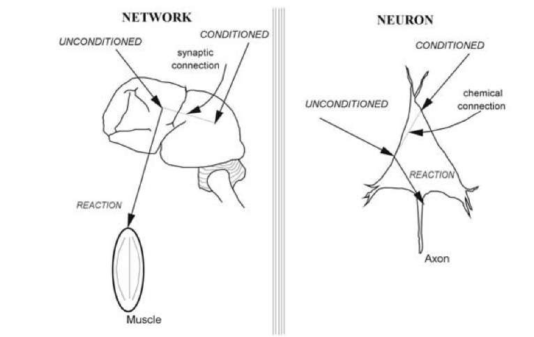
So, are neuronal signals governed solely by the structure of their anatomical connections and the distribution of their excitabilities, or a neuron itself is a self-contained system, as is represented in Fig. 1.3? Many theories consider the brain as a complex network of neurons, which are approximated by simple elements that make a summation of excitations and generate an output reaction in accordance with steady activation function. Such an idealization is far from the properties of a real neuron; however, this is a very steady superstition. 35 yeas ago, one from us discussed this problem with the great Russian mathematician Andrey Kolmogorov. He explained that when he conceives of complex networks consisting of simple neurons, he feels giddiness and inspiration. However, when he thinks that, in addition, every neuron may be complex, he feels giddy too, because he feels sick. Nevertheless, the individual neurons constituting neural networks are much more complex elements than generally accepted and are involved in cognitive functions [1126, 181, 43, 1261, 1255, 1141, 58].
Plentiful attempts have been undertaken to determine what properties are responsible for the intellectual capabilities of humans. Certainly, sometimes, the advantages of our brain over that of animals are exaggerated. In some circumstances, chimpanzee memory may be superior to human memory. Young chimpanzees have an extraordinary working memory capability for numerical recollection better even than that of human adults [578]. Surely, memorizing of instantaneous visual images is not evidence of exceptional mental power. However, taken all together, diverse lines of evidence suggest that human intellect far exceeds the animal intellect. Therefore, it has seemed that comparing different brains we can appreciate the mystery of mind. For instance, brains of some of the great scientists, writers and politicians have been thoroughly investigated in order to find out distinction what distinguishes the great minds from brains of ordinary people. Different brains are always slightly different and eminent brains had distinctive features, although these features were not the same for various eminent brains. This failure is not surprising, since we still do not know what characteristics of human brain are responsible for the superiority of our mind in the animal kingdom. Brain structure is perfected in evolution and its mass increases. Nevertheless, this is not a robust law. Large sea mammalians have larger brains than those of humans, but human brain has a larger fraction of body mass. Yet this fraction also is not an excellent indicator of brain excellence. A mouse brain is larger than human brain with respect to body mass. Humans, perhaps, take first place in the proportion of the square of brain mass to body mass, but nobody knows what this means.
Fig. 1.3. Two hypothetic mechanisms of memory: establishment of new connections in the space of brain (left) and formation of chemical specificity. Before learning, an unconditioned stimulus produced an output reaction (muscle contraction or action potential), while a conditioned stimulus does not evoke reaction. After learning, a conditioned stimulus began to produce an output reaction with the participation of a newly emerging bond (dotted lines). This bond is spatial in a network hypothesis and chemical in a neuronal hypothesis.
An absence of quantitative differences between human and animal brains is not too breathtaking. But, by the way, there are not too many qualitative features distinguishing the human organism from animals and not every distinction determines the intellectual power of humans. For instance, only do human head hairs grow endlessly. An animal could not survive in the wild, if it would need a periodical hair-cut. Nevertheless, some important distinctions concerning the neocortex do exist. The primate cortex contains more neuronal layers, with an escalation of the form and function of cortical astrocytes and with an enlarged proportion of inhibitory GABA^ergic interneurons among all other neurons [902]. The increased complexity of cortical astrocytes contrasts with the relatively limited changes to individual cortical neurons during phylogeny [683]. Frontal brain regions have rapidly expanded in recent primate evolution, consistent with their role in reasoning and intellectual function [1233]. Various cortical areas, recently appearing in evolution, differ by their morphological organization, chemical properties, function, etc. [1196]. The monkey visual cortex contains more than 30 separate areas and the organization of the human and monkey visual cortex is remarkably similar. However, human cortex contains regions that are specialized for the processing of visual letters and words. The mystery is how cells "know" which image they hold, particularly because object representation must be constantly changing after acquiring new memories [1287].
However, only one property of the human brain is prominent. Our brain is the most complicated. Could it be that the enigma of brain is a function of complexity? We were personally acquainted with the enthusiast who, in Moscow during the period 1950-1980 tried to examine this hypothesis practically. He constructed an electronic scheme in a 20 m2 room. He did not use any system and tried to create something large and complex. His model occupied more than two-thirds of the room’s volume. One could see this giant scheme its early parts, constructed of large vacuum lamps, miniature lamps (included later), resistances, thermocouples, diodes, triodes, capacitances, pentodes, transistors, microminiature modules, and analog and digital elements that were included recently, etc. At a glance, this scheme consisted of around 106 elements, and when monster was in an operative state, light pipes blinked, inductances buzzed, resisters radiated heat and relays were switched on without any visible regularity in space and time. Of course, one could not observe any intellectual activity and although this does not prove that the initial idea was poor, we know that the neural system of a nematode worm contains only 302 neurons. For example, all the behavioral steps, neuronal events and gene products leading to copulation and sperm transfer are known [149]. The knowledge now is available, but what does it mean and what does the nematode gain by its particular sexual behavior? The nematode demonstrates associative learning and exhibits social behaviors. Does brain constitute a neuronal construction, or neuronal society? Correspondingly, what is changed after learning, the construction or the chemical reactions inside of neurons (and may be glia)? It is doubtful that even if we could create a precise electronic model of the brain it would work properly.
A vast diversity of brain properties will be logically ordered, when general principles can be highlighted. Right now, it is impossible to squeeze each particular fact into united theory. On the other hand, it is high time to understand where we are at present. We will try to generate some general principles that will have an explanatory force and will be in accord with some particular facts and will not contradict others. As an example, the calcium-binding proteins calretinin, calbindin and parvalbumin have important roles: they prevent Ca2+ enhancement within cells and protect cells from damage. However, in the visual and auditory systems of the bottlenose dolphin, calretinin and calbindin are the prevalent calcium-binding proteins, whereas parvalbumin is present in very few neurons. At the same time, in both auditory and visual systems of the macaque monkey, the parvalbumin-immunoreactive neurons [479] are present in comparable or higher densities than the calretinin and calbindin-immunoreactive neurons. It is rather possible, that different calcium-binding proteins play specific, still unknown roles in dolphins and primates. However, in our consideration one may confine ourselves to the general effects of intra-cellular Ca2+.


