Structural Color by Diffraction
Diffraction can produce structural color when white light is incident on the edge of an object or on a medium that comprises a periodicity in structure or refractive index. Such a medium may be called a diffraction grating and may function in reflection or in transmission, depending on its geometry. A high periodicity is generally required for bright structural color, the spatial dimension of which can range from a few thousand to a few hundred nanometers. ]
Consider white light incident on a medium bearing a sinusoidally periodic surface profile of approximately 1-p.m pitch. Owing to the presence of the periodicity on the surface, the incident planar wave fronts of all wavelengths comprising the white light are diffracted to form a large number of series of circular wave fronts for each wavelength. These series of reflected circular wave fronts then interfere strongly with each other, constructively in certain specific directions and destructively in others. Accordingly, diffraction, especially that associated with surface periodicities, manifests itself by the production of colored bands of light in reflection or transmission at different angles from the periodic surface onto which white light is incident.
The use of diffraction gratings is widespread. Measurements of signals are frequently based on the analysis of wavelength content in an incoming light beam.[27] Such analysis requires a diffraction grating to discriminate between constituent wavelength components. Within optical devices such as interferometers, laser systems, multiplexers, and microphotonic components (integral in most modern optical communication and instrumentation systems) such as gratings perform an essential function.
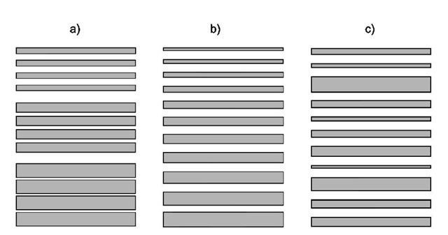
Fig. 4 Schematic diagrams to illustrate three arrangements of multilayer systems that can produce broad band reflectance: (a) a system comprising three overlapping quarter-wave filters tuned for different portions of the overall reflection band, (b) a chirped system comprising gradually changing layer thicknesses, and (c) a chaotic system in which there is little order in the arrangement of layer thicknesses.
Structural color produced from diffraction is often observed from objects such as CD or vinyl record surfaces that incorporate micron-sized periodicity. When incident white light diffracts from the periodic data tracks and grooves on their surfaces, color spectra are observed. The production of such color, however, is a side effect. One must look to natural systems for diffractive structural color that is produced for specific purposes.
Until recently, the occurrence of diffractive systems in the natural world was believed to be rather rare.[28] They are now known to be fairly common, especially in certain invertebrate phyla. In the phylum Mollusca, iridescence is observed on the outer surfaces of many mollusk shells. The surface of the shell of Pinctada margaritifera exhibits bright iridescence that results from diffraction of approximately 3.4-mm pitch from a series of shallow parallel grooves on the shell surface.[29] This iridescence is distinctly different from the ”milky” opalescence or pearlescence observed in many mother-of-pearl shells, where thick flat CaCO3 crystals cemented together with layers of protein yield interference of several colors simultaneously.[12]
Series of parallel narrow surface grooves are extensively found on the elytra of scarabaeid beetles, some of which are known to produce structural color through diffraction.[30] For instance, many of the members of the Phalacridae family of scarabs exhibit surface gratings of approximately 1 mm pitch, resulting in several bands of colored reflection. The intensity of these reflected color bands is relatively low, however, because of the heavily melanized cuticular integument that has a relatively low reflection coefficient.
The setae (hairs) of many Crustacea comprise series of juxtaposed rings having walls that are circular in cross section with a pitch of approximately 700 nm. Bright diffracted colors are visible to the eye when the specimen is tilted at an angle under a fixed light source.[28]
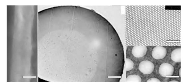
An interesting example of structural color caused by 2-D periodicity is that exhibited by certain taxa of marine polychaete worm. One of these, Aphrodita, a genus of polychaete worm commonly known as the sea mouse, has a short segmented body that is covered with hairlike structures called setae. These setae, extruded by epidermal cells, are exceptional because of their strong iridescence. They are composed of bundles of thin-walled cylinders of chitin held together by sclerotinized protein to form fine and threadlike hairs. The diameter of the cylinder centers varies between approximately 100 and 360 nm in Aphrodita;[31] in another polychaete species, Pherusa, optically similar setae exhibit a greater number of cylinder periodicities with a more uniform diameter of approximately 220-270 nm[32] (Fig. 5).
The iridescent color in these setae (shown in color in the on-line version of this article) is produced through diffraction from the periodicity of the cylinder elements. Detailed optical analysis associated with the structure of Aphrodita is presented elsewhere.[33]
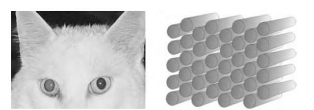
A variation of the structure found in iridescently colored polychaete setae is also found in the tapeta of the eyes of cats. However, whereas polychaete setae consist of bundles of hollow cylinders, the tapeta of cats’ eyes consist of bundles of solid cylinders or rods made of cuticular material. Within each bundle, rods of diameter 200-350 nm are spaced 300-500 nm apart and are arranged in a mutually parallel geometry that yields hexagonal symmetry in transverse cross section (Fig. 6).
Strong diffraction, specifically Bragg diffraction, arising as a result of the periodicity of these rods produces the colored eye shine normally observed with cats’ eyes.[34] This mechanism is similar to the Bragg diffraction of X-rays from a crystalline material,[35] except that the difference of scale means the effect is seen for ordinary light. Its physiological function, similar to that of tapeta in certain arthropods, is believed to be optimization of photoreception in the cats’ visual system.
Fig. 5 A single setae from the polychaete worm Pherusa, viewed by optical microscopy (left) and in cross section by electron microscopy showing increasing magnifications of transverse sections through the setae. Structural color effects are visible in the optical image (the on-line version of this figure is in color), produced from highly periodic microstructure derived through the longitudinal stacking of hollow cylinders within each seta. The images on the right show magnified regions of the central portion of the middle image. (Scale bars: left, 0.25 mm; middle, 60 mm; top right, 4 mm; bottom right, 250 nm).
Fig. 6 Curious color effects and nocturnal eye shine in cats is produced by domains of ordered 2-D lattices of rodlet bundles in the tapetum lucidum of their eyes. Each rodlet is approximately 200 nm in mean diameter.
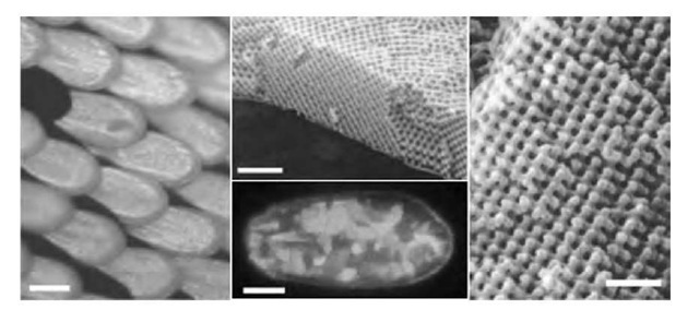
In certain Lepidoptera, rather than 2-D periodicity such as that just described for polychaete worm setae, a remarkable 3-D periodicity exists. Such 3-D structures are sometimes referred to as volume diffraction gratings or photonic solids. It is, in principle, possible for photonic solids to expel light of certain frequencies in all directions; this implies that light of these frequencies will simply not propagate in the material and that all of it will be reflected from the surface of the solid. When this occurs, the material is said to exhibit a photonic band gap, which is analogous to the familiar electronic band gap of solid-state physics, except that it applies to photons instead of electrons.[36] However, for a photonic solid of specific symmetry to exhibit a full photonic band gap, there must be a minimum refractive index contrast between the two media that make up the periodic solid. Natural structures that exhibit these photonic solids are generally composed of cuticle and air or cuticle and water, both refractive index combinations of which are insufficient to create a full photonic band gap. However, their combination and structural symmetry can be enough to produce considerable reflectivities over a significant range of angles and over a relatively small wavelength band. The selective advantage of color production using this manner of microstructure rather than a surface grating or a multilayer system, excluding physiological considerations, is that it can sometimes be incorporated into multiple neighboring grains, each oriented slightly differently, to produce a macroscopic, completely angle-independent, structural color. This effect is found in several butterflies, e.g., Parides sesostris, and is shown in color in the on-line version of this article[37] (Fig. 7).
Fig. 7 Optical micrographs showing P. sesostris scales in reflection under normal illumination (left) and a single P. sesostris scale viewed in reflection with linearly polarized white light illumination (bottom middle) with the image captured through a crossed linear analyzer. Top center and right images are electron micrographs of a fractured transverse section of a P. sesostris iridescent scale (the 3-D periodic lattice of cuticle is clearly visible). (Scale bars: left 90 mm; bottom middle 50 mm; top middle 3 mm; right 1 mm).
Structural Color by Scattering
The final category of structural color exists in addition to those that cause refraction, interference, and diffraction, producing wavelength-selective optical scattering.
The phenomenon of blue scatter is fairly widespread in nature. This color effect, associated with the selective scattering of shorter-wavelength light by small particles, can be observed in the laboratory by using white light to illuminate droplets suspended in liquids (such as water containing small concentrations of milk). When it is viewed from the side, the suspension appears rather blue. Many other inanimate systems offer strong scattering in the laboratory such as highly dispersed systems of elementary sulfur or silver halides in aqueous media. In each case the exact hue and intensity of the scattered color depends on the degree of fineness of small particles in the suspension and on the magnitude of mismatch between the refractive index of the particles and their surrounding medium. Generally, particles that are sufficiently small yield deeply violet scattered color of striking intensity and purity: as particle size is increased, the scattered color passes from deep blue to pale blue. Increasing the particle size beyond approximately 1 mm results in most visible wavelengths being scattered with sufficient intensity to produce the appearance of white.[20]
The mechanism that supports these laboratory examples of color by scattering also leads to the blue color of the sky; in this case, however, the scattering centers are the atmospheric molecules themselves rather than suspended solid particles.[38]
Scientists working with light scattering use several terms to evoke theories associated with different classes of scattering particles. Authors that detail scattering in animate specimens[20,39] have consistently used the terms Tyndall scattering, Tyndall effect, etc. to refer in the broad sense to colors produced through selective scattering of shorter wavelengths by small particles or optical heterogeneities when pigmentation and other structural phenomena are not predominant. In certain studies, Tyndall scattering is equated to Rayleigh scattering by particles that are small compared to the wavelength;[40] in others it applies to particles that are the same size or larger than the wavelength.[41] Tyndall’s original experiments[38] embraced both groups of particle size and the expression is usefully retained by many biologists to describe scattering in animate objects. Physicists unfamiliar with such works on natural scattering systems tend to refer to the process as Rayleigh scattering if the sizes of the fluctuations or particles are very small compared to the wavelength of light, or Mie scattering if they become somewhat larger than those associated with Rayleigh scattering.
It is well known that the scattering of light in gases or fluids can involve certain quantum processes more normally associated with pigmentary color. However, in neutral nonabsorbing media a classical oscillator analysis[41] describes the relationship between scattered light intensity and wavelength for a specifically sized system of particles. This relationship is the well-known Rayleigh law of scattering, which states that scattered intensity is inversely dependent on the fourth power of wavelength. Physically, this implies that shorter-wavelength colors such as violet and blue are scattered much more strongly through greater angles than longer wavelengths such as red (Fig. 8).
This relationship can also apply to the scattering of light in transparent solid media, where the scattering centers are formed by irregularities in density or structure rather than by individual atoms or molecules. An important technological example lies in the transmission of light down long glass fibers. For this, the highest transmission can be largely limited by Rayleigh scattering from small irregularities in the glass. Generally, to overcome this loss problem the longest wavelengths for existing and available light sources and detectors are used (but avoiding hydroxyl ion and other material-specific absorption resonances).
Tyndall scattering is believed to be the cause of the noniridescent blue coloration in mammalian and reptilian integument and of much of the blue of both avian integument and feathers.[20] It is additionally attributed to some blues found in certain lower-class phyla such as Odonata and Lepidoptera.
The most conspicuous examples are found in certain mammals, particularly monkeys such as male baboons and mandrills. In these creatures, hairless patches may be deep violet or blue because of Tyndall scattering associated with strata of discrete black melanocytes arranged in an otherwise transparent layer within the epidermis.[20] Purple regions on these creatures correspond to areas where Tyndall blues color-mix with red pigmentation associated with blood corpuscles close to the surface: a color effect perhaps more commonly observed as port-wine birthmarks in some humans.[42]
Fig. 8 A schematic diagram illustrating the scattering of white light by a medium comprising small particles. Shorter wavelengths are preferentially scattered through larger angles according to the Rayleigh law of scattering. In this way, light directly transmitted through such a medium predominately comprises longer-wavelength radiation, whereas shorter wavelengths dominate when the medium is observed from any other perspective.
Whereas Tyndall blue in mammals is caused by disperse systems of solid-in-solid where the melanin granules have a different refractive index to the surrounding epidermis, noniridescent avian feathers generally achieve blue scatter using air-in-solid systems. In such feathers, air-filled subwavelength cavities within the keratin of the feather barbs act as the scattering centers from which Tyndall blues are produced. More often than not, the green color in avian feathers is the product of the superposition of yellow reflection from additional carot-enoid pigmentation in the feathers with the underlying blue from Tyndall scattering.[20,39] Green and sometimes purple coloration in many other taxa, e.g., certain snakes, frogs, lizards, and fish, are produced by similar effects, namely, Tyndall scattering superposed with the effect of colored pigment.[20,39]
The bluish tinge on the chin of dark-haired men seen a few hours after shaving is attributed to Tyndall scattering. Without the dark roots of the hair, however, as an absorbing background, the low-intensity scattered blue tinge would be unsaturated with back reflection from the epidermal surface and would not be seen.
The blue color of mammalian eyes is certainly structural in nature and has been attributed to Tyndall scattering. Early studies of the human eye[43] found that the colored portion is composed of a thin membrane bearing unstriped muscular fibers, nerves, and capillaries, all incorporated in a fine delicate reticulum of fibrous tissue. Small protein particles in this region preferentially scatter blue light away from the eye while the remainder enters the eye and is generally absorbed by a curtain layer of brown and black melanin at the back of the reticulum. This prevents back reflections out of the eye from reducing the saturation of the Tyndall blue color. Brown eyes possess an additional layer of some yellow pigmentation with significantly sized brown melanin granules among the interior fibrous structures. These produce a brown coloration through the natural color of the melanin. The transformation from initially blue colored eyes to brown eyes in babies and children is attributed to the gradual appearance and increase in size of melanin granules in the first few months and years.
There are several examples of Tyndall blue coloration in Lepidoptera. The wings of these species have been shown to be composed of scales that are colored through Tyndall scattering from subwavelength-sized nanostruc-tures.[44] These structures extend from between series of ridges on the surface of the scales down into the body of the scale. In the butterfly Papilio zalmoxis, the scattering structures are in the form of air-filled alveoli approximately 2 mm long and around 220 nm in internal diameter. The simultaneous presence within the alveoli of a UV-absorbing but blue-fluorescing pigment enhances the blue coloration.[44] A melanized underlayer of black scales acts as an absorbing background, preventing the reflection of other wavelengths.
CONCLUSION
There are striking differences between the hues of objects or materials colored through structural means and those colored through pigmentary or other nonstructural means. Depending on structure type and the color mechanism it facilitates, the optical properties of these structurally colored surfaces may manifest themselves through one or several properties: angle-dependent or angle-limited color; narrow or very broad spectral bandwidths; ultrahigh reflectivities or transmissivities; strong polarization effects including polarization rotation on reflection or circularly polarized reflection; bicolor production and subsequent color mixing; and complementary coloration in reflection and transmission.
In modern technological systems, refraction and scattering are rarely used for the purpose of producing color. In the natural world too, relatively few separate examples exist whereby refraction and scattering in either animate or inanimate systems are the cause of color. True diffraction also occupies a place of relatively minor importance in the production of iridescence in natural systems, although a good number of examples are known to exist. By far the most common mechanism by which color is produced structurally in modern systems and those of the natural world is through interference in multilayered systems. Modern physics has an excellent understanding of both the optical properties of most materials and of the physics of multilayer systems; more importantly, the techniques by which such materials are deposited onto surfaces have been refined and advanced to such an extent that highly specialized systems can now be manufactured at reasonable cost for a variety of optical purposes. In nature too, we see evidence of highly specialized multilayer-based structural color. In most of such natural systems, millions of years of selection pressures have shaped and developed them for a variety of purposes, from that of crypsis to that of specialized conspecific signaling. It is rarely an accident that such complex architecture develops in animate systems without the structural color it produces being designated for a specific use or having a definite biological significance. We now find technology is, for its own purposes, increasingly taking lessons from the design protocols nature has created.


