INTRODUCTION
Modern technology is increasingly interested in attaining the ability to carefully mold and control the flow of light. The interest in this is underpinned by the drive toward the use of all-optical signals, rather than electrical ones, for a variety of purposes. Among the more interesting and potentially revolutionary are those associated with high-bandwidth communication and with the miniaturization of digital processing. There is currently a huge research investment dedicated to reaching goals in these areas. As a result, technologists have discovered something nature has been exploiting for a very long time, namely, that the careful design of the structural shape and form of a material will allow it to manipulate the way specific colors propagate within or along it. In this way, the issue of structural color has arisen in physics and engineering, and the subject is predicted to affect our future lives in a fundamental way. As an introduction to the associated optics, this article will principally describe the physical mechanisms associated with structural color, presenting natural examples that offer inspiration and design protocols to current technologists.
STRUCTURAL COLOR
Color may be defined as the property associated with an elicited physiological perception when light of a certain wavelength impinges on the photoreceptors in a visual system. In human vision, such color is said to comprise the range of wavelengths from red, at around 700 nm, to violet, at around 400 nm.
Colored light can be produced through one of 15 possible mechanisms,[1] each of which is independent of the visual system or device that detects it. The more common mechanisms are those associated with optically induced electronic transitions in atomic or molecular or-bitals; energy-band effects in metals, alloys, and semiconductors; the vibrations and atomic excitations associated with incandescence; and the geometrical and physical optics involved in the interaction of light with periodicity. All mechanisms may, broadly speaking, be categorized into two groups: color resulting from the transition of electrons between specific energy levels (such as with pigments or dyes) or color resulting from the interaction of light with the physical structure of a system. The latter group is commonly referred to as structural color and is the subject of this article.
Structural color first proliferated in the animate natural world in the Cambrian period (515 million years ago) and even earlier with inanimate systems. Many examples are well known, but the color of rainbows and of certain invertebrates and vertebrates have always been both fascinating and intriguing. Much serious scientific research has taken place for many centuries. Through such examples as the investigative work of Theodoric[2] in the 13th century examining refraction in a rainbow, to that of Hooke in the 17th century on the color of silverfish,[3] to Newton’s thesis in the 18th century on the iridescent color of peacock feathers,[4] even to modern-day scientists discovering photonic crystals in lepidopteran wing scales,[5] the principles of physical optics that underpin natural structural color have become well understood.
Four mechanisms govern such natural structural color: refraction, interference, diffraction, and scattering. These mechanisms also underpin all artificially produced incidences of structural color used by, or associated with, modern technological systems. Fundamentally, color is produced because of the existence of either nonparallel interfaces between media of different refractive index or of a distribution of centers of high or low refractive index, the 1-D, 2-D, or 3-D periodicity of which is similar in spatial dimension to that of visible light. The brevity of such a terse summary may be misleading, however. Each one of the four structural color mechanisms is distinctive enough to merit its own detailed description and discussion of examples. In each case, owing to the length constraints of this article and the existence of eminent and fully detailed explanations elsewhere in the literature, descriptions of these mechanisms in this article will be condensed and centered on paradigmatic examples.
Structural Color from Refraction
The rainbow is among the best known natural examples of color by refraction.[6] Other examples include atmospheric phenomena known as arcs, glories, columns, parhelia, and sun and moon haloes produced by refraction and reflection in airborne hexagonal ice crystals.[7] One rarely observed yet spectacular phenomenon is the green flash, produced for just a few seconds at sunset under the right circumstances, where the density gradient of the atmosphere acts as a refractive medium to produce distinctive green coloration.[8] However, there are no known examples whereby refraction produces significant structural color in animate systems.
Among the most frequently encountered examples of structural color due to refraction are those associated with shaped transparent glass, the best example being a high-index glass prism. Even the incidence of sunlight through a window and onto a glass ornament or the beveled edge of a wall mirror will produce the characteristic spectrum of colors associated with refraction.
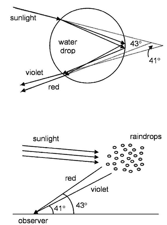
Many studies discuss the spectral colors produced in this way through a glass prism.[9] The underlying principle is the dispersion of light through glass—the index of refraction of glass increases as the wavelength decreases. Consequently, blue light refracts through a larger angle than red light at a glass-air (or water-air) interface (with intermediate wavelengths falling between these limits). Parallel front and rear interfaces, such as with a plain rectangular glass block, prevent the production of a spectrum. However, the nonparallel faces of a glass prism bring about and retain an angular displacement between the paths of differently colored light, producing individual colors beyond the prism. The refraction of white light by raindrops is responsible for rainbows.[6] They form when sunlight from behind an observer refracts on entering and exiting a raindrop (Fig. 1).
Fig. 1 The formation of a rainbow is caused by the refraction of sunlight by raindrops. The primary bow is produced by a single reflection within a drop (top image) combined with the dispersion of light associated with the variation of refractive index with wavelength. To observe a rainbow (bottom image), sunlight must come from behind the observer; the red portion forms a bow of 43° cone angle, the violet, a 41° cone angle.
Light forming a primary rainbow will have internally reflected once within each drop (assigning a distinct polarization to the spectral color), whereas light forming a secondary rainbow will have internally reflected twice.[10] The formation of supernumerary bows, existing on the inside of primary and secondary arcs and rather rarely observed, involves the additional mechanism of optical interference rather than solely refraction.[6]
Structural Color from Interference
Interference is by far the most common mechanism by which structural color is produced, both naturally and artificially. To describe it, we consider white light incident on a series of overlapping thin films of alternating high and low refractive index. Colored light may be strongly reflected by constructive interference between reflections from the individual interfaces forming this stack of films.[11] For this to occur, the resultant reflections from each interface must emerge from the stack with identical phases, i.e., the optical path differences between them must be an integral number of whole wavelengths. This condition is satisfied only by a specific color, or selection of colors, and is additionally influenced by the angle at which the light is incident and reflectivity is observed. The critical variables that determine which color is brightly reflected are the refractive indices of the films in the stack, their individual thicknesses, and the angle of incidence or observation. Any imposed variations on these variables alter the optical conditions that create the interference, thereby producing a reflection and transmission of a different color. The hue from such interference is clearly angle dependent, a property that is implicit in the definition of iridescent color produced by this mechanism. The most favorable narrow-band reflection condition is produced when the optical thickness of each layer in the stack corresponds to a quarter of the wavelength required for optimal reflectivity.[12] For a large number of such layers of high refractive index contrast, the reflectivity will be bright, saturated, and of a narrow spectral band. The brightness of this reflectivity will increase with the number of layers:[12] if the dimensions or indices of the system deviate from the quarter-wave condition (something that is common in natural systems), the reflected intensity may be proportionally reduced and the reflected wavelength band will adopt a more complex form.[13]
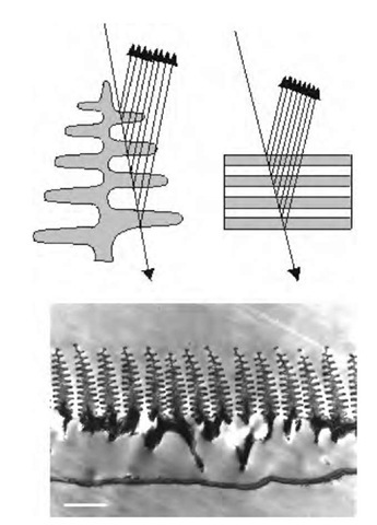
Among the innumerable examples of interference color in nature, those that are most vivid and spectrally pure tend to catch the eye most readily. An excellent example of such stunning structural color is the iridescence associated with several of the male Morpho butterflies that are indigenous to South and Central America.[14] The striking deep blue color of their dorsal wing surfaces is produced without the aid of any blue pigmentation, generated instead by optical interference within nanosized laminations in the wing scales[15] (Fig. 2).
There is a profound complexity associated with this Morpho multilayer system. Whereas conventional technological multilayer systems generally comprise continuous layering over what is often a substantial area, the Morpho laminations are distinctly discreet in character, comprising assemblies of up to 10 layers of high-index material called cuticle, separated by intervals of air. This high number of layers yields up to 85% blue light reflectivity despite the presence of some inherent optical absorption because of the presence of melanin pigmentation within the scale structure.[15] The laminations are built into long and near-parallel ridge structures that run the length of each iridescent scale and have a spatial periodicity of between 700 and 1200 nm. Analysis of the optics of such Morpho structure confirms that the observed layers of thickness approximately 80 nm, separated by ”layers” of approximately 100 nm of air, produce blue coloration at normal incidence through interference. The laminations appear to be embedded into the scale ridging to reflect the interference-produced blue light over a much broader angle than would be possible with flat or even curved continuous multilayers.[15] Variations in the design of this type of discrete multilayering in other butterfly species lead to more specific, angular-dependent reflection properties.[16]
Fig. 2 The top image is a schematic diagram that illustrates the multiple reflections associated with two types of multilayer structure; the left one is that of a Morpho butterfly iridescent scale while the right one represents more conventional flat and continuous layering. The bottom image shows a transmission electron micrograph of a cross section through a Morpho iridescent scale. (Scale bar: 700 nm).
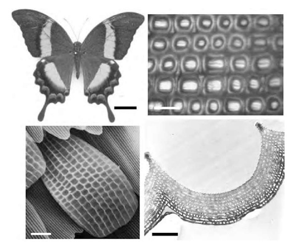
In the case of butterfly systems, one finds that a more complex structure (such as the sculpting of an otherwise continuous multilayer) may produce surprising effects. The butterfly Papilio palinurus exhibits structural color that is green to human vision. Close examination of the structure of its wing scales reveals a continuous multilayer that is shaped into juxtaposed concavities[17] (Fig. 3).
The multilayers follow the profile of these concavities in such a way that the physical dimensions of each layer remain approximately constant regardless of position across each concavity. This creates the effect of a curved multilayer with uniform layer dimensions, rather than a patterned multilayer of variable dimensions. This particular sculpting simultaneously produces two structural colors, namely, blue and yellow, which combine addi-tively to give the stimulus of a third color, the green that we observe.[17]
On close optical inspection (a color image of this detail is included in the on-line version of this article), a yellow color is produced at or near normal incidence via reflection from the flat center of each concavity, whereas a blue annulus is produced via a double reflection across each opposite side of the concavity. This is possible only because each angled side combines with the surface that is orthogonal to itself on the opposite side of each concavity. The blue annulus pattern is produced in this way: normally incident blue light reflected from one 45°-inclined surface is directed across the concavity to the opposite orthogonal surface from where it returns parallel to the incident direction. These pairs of inclined surfaces comprise nearly identical multilayering and are both inclined at approximately 45° to the direction of normally incident light on the scale surface. Accordingly, their spectral reflectivity characteristics are closely matched. Furthermore, through a double reflection from the opposite sides of each concavity, the polarization of only one of the colors may be rotated by 90°, a property that is thought to have evolved due to selection pressures associated with both cryptic and conspecific signaling.[18]
Fig. 3 Optical images showing the structural color (top left) and scale patterning (top right) of a P. palinurus butterfly, together with electron micrographs of one of its iridescent scales (bottom left) and the cross section through one of the concavities (bottom right) of such a scale. The green coloration of P. palinurus is produced through color mixing of juxtaposed yellow and blue regions (top right), both colors of which are produced from the same multilayer profile. (The on-line version of this figure is in color.) (Scale bars: top left, 1 cm; bottom right, 6 mm; bottom left, 10 mm; bottom right, 1 mm).
The examples of Morpho and P. palinurus are very advanced and complex multilayer systems. Similarly complex multilayer systems have been shown to exist in many different animal taxa, both terrestrial and aquat-ic.[19,20] For instance, the reflecting properties of some scarabaeid beetles are not based on periodicities in refractive index, but rather result from the helicoidal ordering of chitin microfibrils in planes that make up the insect integument.[21] Such cuticle is composed of laminar planes of chitin fibers, the axes of which are systematically rotated by a small amount with each consecutive plane. After a certain number of layers through the system corresponding to a specific distance perpendicular to the planes, the fiber axis is rotated 180° and is once again parallel to that of the first layer. In this way, two corresponding planes of cuticle occur over a distance that corresponds to half the helical pitch of the system. The consequent optical effect is that of constructive interference associated with consecutive planes that have unidirectional chitin axes and which are separated by half the helical pitch. Thus the system may be treated as a multilayer reflector, which reflects a band of wavelength of unpolarized polychromatic light in an analogous way to cholesteric liquid crystals. As a further consequence of the helical arrangement of chitin fibers, optical reflection from these taxa of beetles is circularly polarized. This is not due to any optical rotation arising at a molecular level owing to the l-amino acids of the cuticle protein and the d-amino sugar of the chitin. It instead arises at the supermolecular level and has been termed form optical rotation,[12] in contrast to the optical rotation exhibited by cholesteric liquid crystals. It should be noted, however, that similar helicoidal structure, found in many other iridescent insect and plant taxa,[22] is not responsible for their strong coloration and does not produce anomalous optical properties such as optical activity. For these species, inherent refractive index periodicity within the species’ exocuticle is responsible for the iridescence. For optically active beetles, the subwavelength pitch of the helical order is responsible for the observed form of optical activity and iridescent color.
Whereas the natural multilayer systems described so far are designed for rather narrow-band reflectivity, modifications to this design can produce broad-band color reflectivity, creating the appearance of gold or silver coloration. Three variations of multilayer design can generate this effect (Fig. 4).
The first of the three variations is known as a chirped system, in which the dimensions of the high- and low-refractive-index layers within the stack systematically increase or decrease through the system.[23] A variation on this, described as a chaotic reflector, produces a similar effect by randomly varying the thicknesses of high- and low-index material. In both of these systems, the range of layer dimensions developed by the specimen, combined with layer refractive index, determine the position and width of the reflected wavelength band and the quality of the silver or goldlike mirror on its surface.[24] The third type of layered system incorporates a composite of three overlapping but regular multilayer systems, each tuned to a different wavelength band.[25]
There are many applications for artificially manufactured multilayer films, ranging from antireflection coatings on optical instruments such as spectacles, binoculars, and telescopes to specific-band optical filters for transmission or reflection for wavelengths ranging from infrared to X-ray. Their design is based on production of constructive or destructive interference in alternating layers of high- and low-refractive-index dielectric materials deposited onto surfaces. Other multilayer devices that additionally facilitate polarized beam splitting accompany interference by Brewster angle reflection.[26]


