The EGF motif is a common structural domain found in many protein structures, especially those of proteins associated with blood clotting, fibrinolysis, neural development, and cell adhesion. The motif is characterized as a protein module of ~ 45 residues having six conserved cysteine residues (designated CI through CVI) that form three internal disulfide bonds with the connectivity CI-III, CII-IV and CV-VI. The motif was first found in epidermal growth factor (EGF), hence the name EGF motif or EGF-like domain. Although found as a single unit in EGF and transforming growth factor a, the EGF domain is often present as a repeating unit in much larger proteins. For example, the protein fibrillin has over 30 EGF domains. Furthermore, EGF motifs in proteins are frequently present in combination with repeating units of other modules such as the Kringle domain.
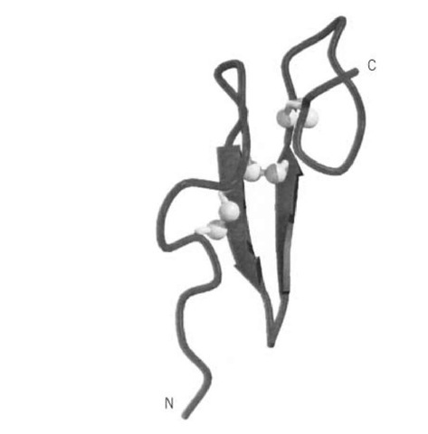
The sequence of the EGF motif is often recognizable from just the primary structure of a protein. The consensus sequence is X-X-X-X-Cys-X(2-7)-Cys-X(1-4)-(Gly/Ala)-X-Cys-X(1-13)-t-t-a-X-Cys-X-Cys-X-X-Gly-a-X(1-6)-Gly-X-X-Cys-X, where X is any amino acid (the number in brackets defining a variable number of X residues), a is an aromatic residue, and t is a nonhydrophobic residue (1). The tertiary structure of the EGF motif is also highly conserved and can be described as having a two-stranded antiparallel b-sheet scaffold upon which the three disulfide bonds are positioned (Fig. 1). The disulfide bonds link the N-terminal and C-terminal loop regions to the core. Binding of calcium ions to some classes of EGF domain further stabilizes the N-terminal region and is thought to cause the formation of long helical arrangements of multiple EGF motifs (2); this property may account for their proposed function of mediating protein-protein interactions.
Figure 1. Schematic representation of the backbone structure of an EGF domain (2). b-Strands are shown as arrows, and the three disulfide bonds are shown in light gray, with the sulfur atoms depicted as spheres. The N- and C-termini are labeled. This figure was generated using Molscript (3) and Raster3D (4, 5).

