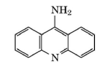
Acridines are fused linear tricyclic aromatic molecules of planar geometry. Aminoacridine (Fig. 1) was originally developed as a topical antibacterial agent and is one of the most widely used and studied acridines. It is a strong base (pKa 9.9) due to the resonant amino group and is largely ionized at physiological pH. Acridines are typical DNA-intercalating ligands, binding tightly but reversibly to double-stranded DNA by insertion of the aromatic chromophore between adjacent base pairs, with the long axis of the acridine parallel to the base-pair axis, ensuring maximum overlap in the binding site (1, 2). It was originally hypothesized by Brenner et al. (3) that acridine-induced mutations arise through the addition or deletion of a single base pair, an event described as frameshift mutagenesis.
Figure 1. Structure of the mutagen 9-aminoacridine.
9-Aminoacridine is a strong mutagen in the rII region of bacteriophage T4 (4), in the CI gene of lambda phage (5), at various loci in Escherichia coli (6), and at the hisC3076 locus in Salmonella typhimurium (7). This and related simple acridines cause frameshift mutagenesis, especially in monotonous runs of repeated sequences. In mammalian cells their activity is somewhat weak and variable. They appear not to be causing frameshifts (at least in the most commonly used mammalian assays), but are mutagenic through weak ability to break chromosomes (8).
Substitutions on the acridine ring profoundly affect the mutagenic characteristics of these compounds, which have been divided into seven subgroups according to their distinctive mutagenic characteristics (2). 9-Aminoacridine and related simple compounds form the first group, while proflavine and other 3,6-diaminoacridines form the second group. The 3,6-diamino groups increase DNA binding affinity, and these compounds resemble 9-aminoacridine in having frameshift mutagenic activity, but are also substrates for metabolic activation (9). They are also readily activated by visible light into other products that are mutagenic in the Salmonella mutagenicity test (Ames test) (10). Some of these compounds and also the third class of quaternized acridines have considerable affinity for mitochondrial DNA. The fourth group of acridines retain DNA intercalation activity, but also act as topoisomerase II (topo II) poisons (see DNA Topology).
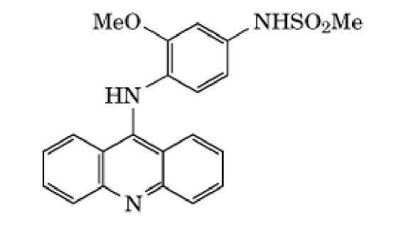
Type II topoisomerases produce double-strand breaks in DNA during replication and transcription, permitting strand passage through the transient break, which is then resealed by the enzyme. The potent antitumor activity of 9-anilinoacridines of the fourth group, such as amsacrine (Fig. 2), is thought to relate to their ability to stabilize the cleavable complex formed between topo II enzymes and DNA, thereby leading to an accumulation of double-strand breaks (11). These DNA breaks also result in these compounds showing strong clastogenic and recombinogenic properties. A clastogen is a physical, chemical or viral agent that produces chromosome breaks and other chromosomal mutagenesis. The fifth group of benzacridines are characterized by a higher susceptibility to metabolic activation, primarily through oxidative mechanisms (12). Benzacridines are primarily base-pair substitution mutagens, producing a spectrum of mutagenic events that is fundamentally different from that produced by other acridines and in which the acridine ring plays only a minor role. The sixth group of acridines, epitomized by those originally developed in the Institute of Cancer Research, Philadelphia, and widely known as ICR compounds (13), all carry an alkylating moiety, usually a nitrogen mustard. These compounds are not only potent frameshift mutagens in various organisms, but also cause base-pair substitution events in mammalian cells (2, 14). Although the nitroacridines can be distinguished as a seventh group, they may alternatively be considered as a subgroup of the previous class, because they can act as either simple acridines or as alkylators, depending upon the position of the nitro substitution and the nitroreductase capability of the organism concerned.
Figure 2. Structure of the 9-anilinoacridine amsacrine.


