To appreciate skull anatomy, take a moment and look at ‘ your own face in a mirror. The structures above the ; neck are designed for the acquisition and initial processing of nutrients, the exchange of respiratory gases, the acquisition of sensory information about light, sound, touch, odor, and taste, and the broadcast of information about your own thoughts and emotions. Sensory and motor information is processed and sent from here to coordinate body functions. Complex signals can be sent to others of our species via vocalizations and/or the contractions of facial muscles. The head is our window for contact, perception, and communication with our world, and the skull provides the framework, the organizing hub, for the head. Thus, the skull is interesting in itself. It is also fundamentally important in our picture of evolutionary biology. This article describes the skull morphology of the evo-lutionarily diverse group called marine mammals (see Reynolds et al, 1999).
I. Defining the Term “Skull”
The term “skull” is inexact. It has been used to describe the entire skeleton of the head. It has also been used to refer to only the cranium (plural, crania) or housing for the brain and sense organs, and the upper jaw (the part of “poor Yorick” that Hamlet held in his hand). This article uses the word skull to refer to the entire head skeleton, including the cranium and the derivatives of the first two visceral arches, i.e., the lower jaw (or mandible, which is composed of right and left dentaries) and the hyoid apparatus. The mandible and hyoid apparatus of marine mammals have received less attention in the literature, but they are particularly important in adaptations for feeding (see later).
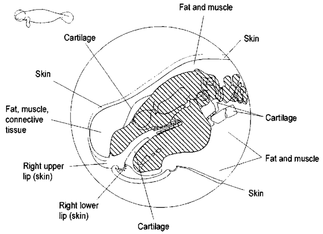
The skull acts as a foundation for the fat, muscle, skin, and vascular and sensory structures that form the head. Thus, the skull alone does not dictate the contours of the head. For example. the external profile of the manatee head differs from that of the underlying skull (Fig. 1), and the relationships between the bones and the soft tissues of the head vary among species (Fig. 2). Odontocete cetaceans have a melon, a fatty facial pad, the shape of which is only partly defined by the underlying bones (Mead 1975); the extreme difference in head and skull profiles may be found in the sperm whale (Phijseter macrocephalus). Contrarily, the dorsal surface of the right whale’s head follows closely that of the underlying skull, although the right whale also has huge lower lips that follow the contour of the upper jaw but are not predicted by the outline of the lower jaw.
The shape of the head may also influence the dynamics of locomotion and balance. The completely aquatic species (cetaceans and sirenians) have shorter necks and less need for “antigravitational” muscles that support the head than do terrestrial mammals (imagine the right whale moving its head around in the air the way a sea lion does).
II. Feeding and Swallowing
The specific characteristics of a skull (including dentition) often reflect the animal’s methods of feeding (Fig. 3). For example, the “typical” heterodont dentition (Kardong, 1998) of terrestrial mammals such as the dog is also found to various degrees in seals, sea lions, walrus (Odobenus rosmarus), sea otter (Enhydra lutris), and polar bear (Ursus maritimus).
Figure 1 Positions of skull features within the head vary among species. Here we illustrate hoty the midline contours of the manatee head differ from a left lateral illustration of the skull. The skeletal elements are cross-hatched to con trast them with soft tissues.
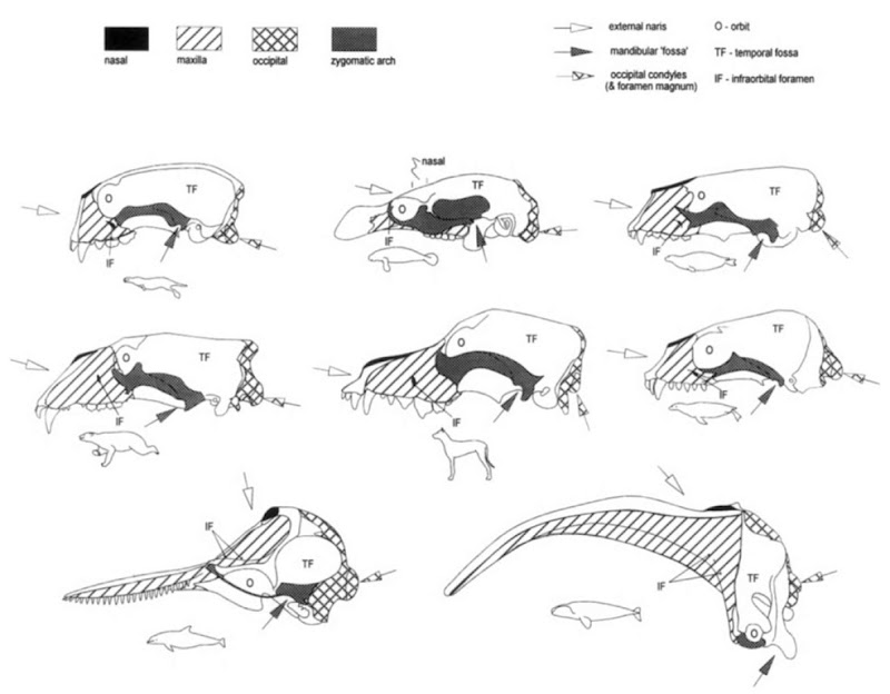
Figure 2 Skulls and first two cervical vertebrae (unless fused, as it is in cetaceans) of a selection of marine mammals for comparison with those of the dog. Each species is scaled so that the distances between the shoulder and the pelvis are similar; body cavities are therefore roughly similar in length, allowing one to compare head sizes with visceral volumes among species.
Figure 3 Feeding apparatus and typical food. Dominant tooth type is also given. Note that the embryonic teeth of the right whale have been replaced with horny plates of baleen in the upper jaw only.
Heterodonty is tooth shape differences in different parts of the mouth: incisors and canines rostrally and premolars and molars caudally. Although each of these tooth types may vary in shape, the definitions are specific and are related to tooth position in the bones of the upper jaw (Hildebrand, 1995). Incisors are found only in the incisive or premaxillary bone; canines are found in the maxilla, in or very near the suture with the pre-maxilla; premolars are deciduous cheek teeth (erupting from the maxillary bones) that are found rostral to the molars; and molars are nondeciduous cheek teeth. Typically, deciduous teeth are replaced vertically as they are pushed out by the developing permanent teeth deep to them. Each tooth shape may perform a distinct function, sort of a “Swiss Army mouth” (G. Early, personal communication). Incisors, if chisel like, are for slicing and chipping and, if pointed, for piercing. Long, pointed canines are good for capturing and piercing. Relatively blunt cheek teeth are good for crushing and grinding. Manatees, unlike all other marine mammals (including the dugong, Dugong dugon), have continuous, horizontal tooth replacement of their molars (Domning and Hayek, 1984).
In some species, all of the teeth have the same shape—this is the homodont condition. The homodont dentitions of odontocetes and manatees differ in shape and function (they are grasping teeth for dolphins and grinding teeth for manatees).
Baleen whales lose their embryonic teeth and acquire food by sieving plankton, small fish, and (in the case of gray whales, Eschrichtius robustus) benthic invertebrates using plates of horny (keratinized) baleen suspended from their upper jaws (Pivorunas 1979).
The lower jaw generally mirrors some of the features of the upper jaw, particularly dentition. Articulation of the mandible with the cranium (the temporomandibular joint or TMJ) also reflects feeding style. The lower jaws of the rorquals must support the large, pleated gular sac that fills with water and prey during lunge feeding (Lambertsen, 1983), whereas those of the skimmers, such as the right whales (Eubalaena spp.), support massive lower lips that guide an almost continuous stream of water past long baleen plates. Gray whales, which are bottom feeders, have relatively robust lower jaws. In addition to supporting their fish-grasping teeth, the lower jaws of odontocetes are fat filled and perform the acoustic function of receiving and guiding sound energy to the earbones (Norris and Harvey, 1974). The lower jaws of the herbivorous manatees (Trichechus spp.) support grinding cheek teeth. The mandible of the manatee also has large mental foramina to transmit the large nerves associated with the vibrissae (see later).
Feeding style may also be reflected in the shape of the rostrum, the zygomatic arch, and the temporal fossa. Relatively large temporal muscles and their fossae are typical of carnivorous mammals that tear or shear flesh without finely dividing that prey in die mouth and/or have teeth for killing and temporarily holding prey (Hildebrand, 1995). Carnivorous mammals have upper and lower tooth rows that do not slide horizontally but rather occlude with a chopping motion, using a hinge joint that is roughly in line with the tooth row (Fig. 3). In contrast, the TMJ in herbivores is typically above the tooth line (Fig. 3). Relatively large masseters and the robust zygomatic arches that support them are more typical of herbivorous mammals that use a crushing and rolling action to chew. This feeding style requires a TMJ that can slide and a large masseter to apply force along the extent of the cheek tooth row. TMJs of the mysticetes accommodate complex axial rotations of the individual dentary bones and absorb shock in lunge feeding of rorquals (Lam-bertsen, 1983), whereas the two dentaries of the odontocetes tend to fuse with age into a single compound bone.
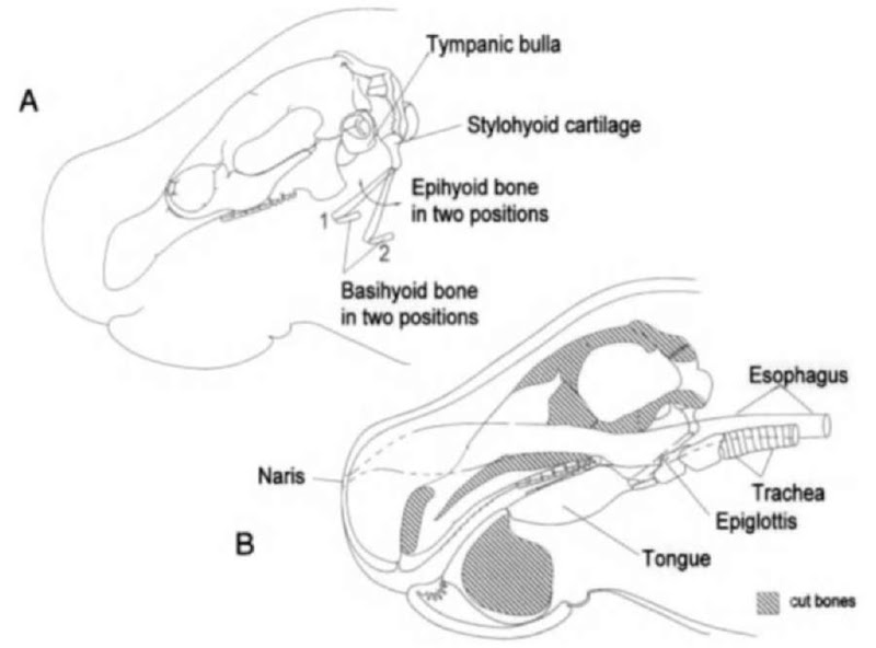
Feeding includes swallowing. How do the bones of the head accommodate swallowing? Hyoid bones provide the mechanical support of many of the muscles that act upon the tongue and the larynx. Swallowing requires the coordinated action of these muscles as food leaves the oral cavity and moves through the pharynx. The dorsal hyoid bones (epihyoids) are attached by the stylohyoid cartilage to the base of the skull at a position immediately caudal to the tympanic bones (Fig. 4A). They provide the primary hinge to depress and move the tongue cau-dad. The ventral hyoid bones (basihyoids) form a secondary hinge that allows sternohyoid (sternum to hyoid) and mylohyoid (mandible to hyoid) muscles to move the basihyoid bones forward and back (Fig. 4B). In suction feeders, such as the squid-eating beaked whales and pilot whales (Globicephala spp.), the hyoid apparatus and its associated muscles are massive.
The soft and hard palates that separate the respiratory system from the digestive system are almost unique to mammals and allow extended chewing time or suckling while breathing. The fleshy soft palate extends the bony margin of the nasal cavity (Fig. 5) and makes a very important contribution to the evolutionary forces that shaped mammal skulls. Prolonged chewing, while breathing, means there is additional time for food processing in the mouth. Teeth of mammals are more indicative of food type than is the case for most other vertebrates. The specificity of dentition and the hardness of teeth, which increase preservation in the fossil record, contribute significantly to our current understanding of the ecology and evolution of extinct species.
III. Bony Features vs Bones
One approach to studying the skull is to focus on bony features (Fig. 6). Bony features are morphological characters or landmarks of the skull that are formed by one or more bony elements (individual elements or bones may develop from one or more ossification centers). Bony features reflect evolutionary, developmental, and mechanical pressures in a grossly visible manner. In contrast to bony elements or skull bones, bony features are structures that incorporate one or more parts of individual bones. For example, the zygomatic arch, which supports the masseter muscle and helps close the jaws, may be composed of one, two, or three bones. The rostrum or muzzle may be elongate and may include the nasal bones. Thus, to characterize individual skulls without having to identify individual bones, biologists can use the morphology of bony features such as zygomatic arch shape and composition; rostrum length; orbit size, shape, and position; and jaw articulation.
Figure 4 (A) Lateral view of the manatee cranium with the hyoid apparatus in two positions to illustrate its range of motions. Muscles between the basihyoid and the sternum move the hyoid apparatus down and back. Muscles between the tongue and basihyoid move the hyoid apparatus up and forward. (B) Left midsaggital section of the manatee head. Movements of the hyoid apparatus influence the trajectories of air and food through the head and into the trachea and esophagus, respectively.
Figure 5 Comparisons of the morphological adaptations of the mammalian head that allow respiration while food is in the mouth. Separation of oral and nasal cavities accommodates prolonged chewing and allows teeth to be modified accordingly. The dog and manatee (upper tivo on the left) are represented schematically in the upper right. In the dolphin (lower left and right), further modification is shown to accommodate the migration of the respiratory opening to the top of the head.
Take, for example, the small zygomatic arch of the right whale and the relatively delicate one of the dolphin. In these species the masseter muscles are relatively small and the temporal muscles are relatively massive. Compare these zygomatic arches with the massive ones in the manatee and polar bear.
In general, large, forward-facing orbits are characteristic of predators that relv on vision as their primary sensory modality’, whereas laterally facing orbits are more typical of nonpredatorv species. Also note how the orbits of most species in Fig. 6 are open caudally, in contrast to those of the fully aquatic mammals. In species in which the orbit is open, there is a postor-bital ligament caudal to the eye that extends between the pos-torbital process of the frontal bone and zygomatic arch; these postorbital processes, most evident in the polar bear, serve as sites for muscle attachment.
Figure 6 Selected bony features are emphasized. The rostrum, composed of the premaxilla, maxilla, and occasionally the nasal bone, forms the “face” of each species. The zygomatic arch, which supports the masseter muscle, may be composed of a single bone, thejugal, or parts of as many as three bones in some species. Arrows indicate directions of airflow at the external naris, vertebral column articulation at the occipital condyle, and lower jaw artiadation at the mandibular fossa. Processes extending from the frontal may be present to help form the dorsocaudal aspect of the orbit, referred to as postorbital processes of the frontal: absent in the seal, small in the otter, and relatively large in the dolphin.
The positions (on the top of the skull) of the external nares may reflect respiratory adaptations to diving, feeding, and locomotion. Note the positions of the external nares in fully aquatic species. The internal nares or passageways for air through the cranium are bordered by the nasal bones in all species.
The occipital condyles position the head on the neck and influence the flexibility at the terminal end of the neck (Figs. 2 and 6). Some of the species with short necks have two or more fusions of cervical vertebral (Flower, 1885; Slijper, 1979; Rommel, 1990), placing the base of the skull very near the shoulder joint and the thoracic cavity. Species with long necks may have a wide range of neutral head positions and may also have a greater range of movement than fully aquatic species (King, 1983).
IV. Ground Plan of Skull Bones
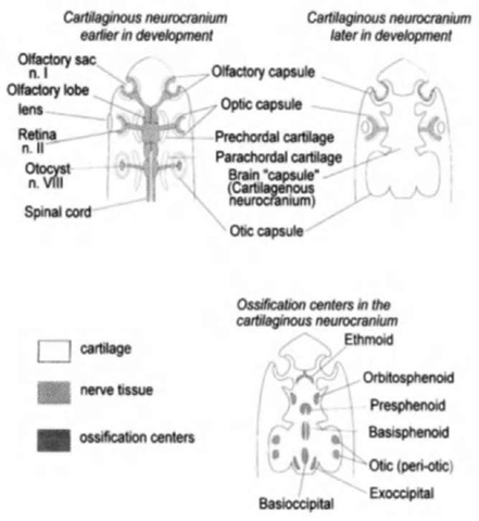
What other factors shape the skull? In all vertebrates, the skull bones develop from ossification centers in a basic pattern that partially or completely encloses the brain plus the sensory organs of olfaction, vision, hearing, and balance (Fig. 7). Bones that are prefonned in cartilage are referred to as endochondral or replacement bones; those deposited directly as bone within tough connective tissue membranes are referred to as dermal or membrane bones. The distinction between endochondral and dermal bones is valuable in establishing homologies with skull bones of the lower vertebrates.
Figure 7 Schematic illustration of ventral views of the developing vertebrate skidl (modified after Kent and Miller, 1997). The basic plan of encapsulation of the senses and brain is illustrated in the top two drawings. The lower drawing illustrates the ossification centers that will eventually become the replacement bones of the cranium.
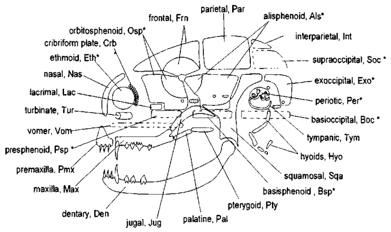
How does one determine which elements of the skull, i.e., which of the 40-some bony elements, are homologous between species? A systematic approach that allows one to compare homologous elements (Fig. 8) is a particularly useful way to avoid the “mental indigestion” (Romer and Parsons, 1977) of having to memorize all the individual bones.
In some species, individual bones fuse (ankylose) to form compound bones;1 these include the occipital, temporal, and sphenoid “bones.’ Of particular interest is the temporal “bone,” which is a compound bone made up of many separate bony elements and/or ossification centers (see Kent and Miller, 1997). In many mammals, the bulk of the temporal is a single unit with no visible sutures between the bony elements once skeletal maturity is reached. Thus, it is common to refer to the temporal as a bone, but this is not the case in several of the marine mammals, particularly cetaceans and sirenians.
The exploded diagram of the cranium of the Florida manatee illustrates the overlapping and/or abutting margins that make up the skull sutures (Fig. 9). Note that the ear bone complex—made up of the periotic and tympanic bones plus the middle ear ossicles (only the malleus is visible in this figure)— is illustrated as a single unit. Another compound bone is the occipital, which in some species is composed of the basioccip-ital, exoccipitals, supraoccipital, and inter(post) parietal (Jollie, 1973; Kellogg, 1928).
Figure 10 is a left lateral view of the individual cranial bones for our representative marine mammals. Contrast this illustration with Fig. 6 to reinforce the distinction between bones and bony features; see Fig. 2 to compare skull size to total body size. The abbreviations are the same as in the schematic generic cranium (Fig. 8) and the exploded manatee cranium (Fig. 9). Pick any one bone and compare it with the homologous structures in all of the crania.
V. Sutures
The regions between adjacent bones are referred to as sutures. In the exploded illustration of the manatee skull (Fig. 9), the hatched regions represent sutures or regions of overlap; skull bones can meet in several ways and attach by more than one type of material (e.g., cartilage and connective tissue).
In Fig. 11, the sutures of the dog skull are compared with those of the manatee skull. Note that different types of sutures may be found between the same bones in different species. The type of suture generally reflects the mechanical forces necessary to accommodate whatever biological constraints are !We have used three different terms incorporating the word “bone.” Bones or bony elements are discrete ossifications that can be traced phylogenetically. Compound bones are structures that appear to be single units in adults because the sutures between the component bones have been resorbed or are not apparent. Bony features are gross features, such as the zygomatic arch, that are made up of discrete and distinct bones or parts of bones (e.g., the zygomatic process of the squamosal bone).
Figure 8 Left lateral schematic of the mammalian skull illustrating relative bone positions. Most skull bones are bilaterally paired—the unpaired skull bones are positioned along the midline and are dotted in this illustration. This schematic approach has been used for more than 100 years and provides a framework in which to compare a wide variety of mammalian skulls (modified after Flower, 1885; Kent and Miller, 1997; Evans, 1993). Recall that the nose, eyes, and ears are encapsidated early in development; these three sensory areas are represented by circular regions in the schematic. Endochondral bones (first laid down as cartilage) are marked with an asterisk.
Figure 9 Exploded cranium of the Florida manatee. The overlapping and/or abutting margins of bones that make up the sutures of the cranium are shown.
Figure 10 Left lateral illustrations of individual cranial bones of selected marine mammals and the dog. Abbreviations are the same as in Fig. 8. Use Fig. 8 to help visualize how each species has modified the basic plan of mammalian skull morphology.
Figure 11 Suture types in the dog and manatee. Definitions are from a variety of sources, and names in parentheses are from Schaller (1992). Suture types are defined by their shape. FLA, plane or butt joint (harmonious, sutura plana); an approximately straight suture with nearly squared-off margins. SQA, squamous or scarf joint (sutura squamosa); a suture with tapered overlapping margins. FOL, foliate joint (sutura foliata); a regular suture in which adjoining bones interleave. SER, serrate joint (sutura serrata); an irregular suture in which adjoining bones interlock. SYN, synchondrosis; a joint that has persistent cartilage between bones (synchondroses cranii).
Figure 12 Sagittally sectioned crania of the manatee. The upper illustration shows the number and complexity of individual bones. The lower drawing is simplified to illustrate four sutures that give some indication of relative age in the manatee. (A) Between the presphenoid bone and the basisphenoid bone. (B) Between the basisphenoid bone and basioccipital bone. Sutures labeled A and B are the first to close and may reflect growth of the braincase. (C) Between the basioccipital bone and the two exoccip-ital bones (bilaterally paired sutures). (D) Between the two exoccipital bones and the supraoccipital bone. Sutures labeled C and D ankylose later in life and probably reflect changes in the mechanics of head and neck morphology as the animal matures.
He established principles that allow one to use changes in the skeleton that occur during an animal’s life to estimate the age of a specimen by comparing it to others. The sutures of a skull can be used for relative aging in some species, e.g., the sutures that allow brain growth ankylose after the brain stops growing. In the manatee, five skull sutures (two associated with the occipital are bilaterally paired) are used to estimate relative age (Fig. 12). The first two (A and B) appear to reflect the changes in brain size and ankylose within the first few years. The three associated with the occipital bone appear to be more related to musculoskeletal changes that occur during juvenile and adult life.
Development of the skull bones proceeds at a pace different from the development of the soft tissues of the head. Bone is constantly being remodeled and thus it can indicate aspects of the evolutionary forces that work to separate species. This plasticity is reflected in the way individual skull bones form around vessels and nerves. The resulting openings, or foramina (singular, foramen), are often phylogeneti-cally conserved and so can be used to establish homologies of the same bones in very different species. An individual nerve or vessel may be completely surrounded by a bone or bones of the skull, resulting in a specific foramen. Because this process occurs early in the development of the individual and appears to have the same essentials in all vertebrates, we use these cranial nerve foramina to help identify the cranial bones (Fig. 13).
VI. Telescoping
Telescoping is a process often cited describing the skulls of cetaceans. The term, coined by Miller (1923), refers to the elongation of the rostral elements and the dorsorostral movement of caudal elements (Kellogg, 1928; Miller, 1923; Rommel, 1990). The relative movement of skull bones in cetaceans creates sutures with considerable overlap of adjacent bones. If the skull were sectioned, one could observe as many as four different bones overlapping each other; this overlap resembles old-fashioned collapsible telescopes. In dolphins, the external nares have been displaced to the dorsal apex of the skull so the nasal bones are located just caudal to the external nares but dorsal to the braincase. The premaxillary and maxillary bones have been extended at the rostral tip and also pulled up and back over the frontal bones to maintain their relative positions with the nasal bones. The narial passages are essentially vertical in cetaceans, which eliminates the nasal bones as roofing bones of the nasal passages. The nasal bones are, instead, relatively small vestiges that lie in depressions of the frontal bones; the roof of the cetacean mouth is not the floor of the nasal passages as it is in most other mammals. Telescoping is different in odontocete and mysticete cetaceans (Fig. 14). Changes in the mysticetes are dominated by a ven-trocaudal extension of the maxillary bones, whereas in the odontocetes, the premaxillary and maxillary bones are shifted more dorsocaudally (Kellogg, 1928). Interestingly, whereas the odontocete facial muscles have moved up and back over the eye, the temporal muscles of the mysticetes have moved up and forward over the eye. Thus, the temporal fossae of mysticetes are very different from those of odontocetes. Contrast the morphological differences associated with telescoping in cetaceans (Fig. 14) with the homologous structures illustrated in Fig. 6.
Figure 13 Openings, or foramina, of the skull. Foramina can be used to establish homologies of the same bones in different species; each foramen (or foramina) associated with 1 or more of the 12 cranial nerves (labeled I through XII) has a name that is used (fairly) consistently by vertebrate morphologists. Thus the cribriform plate, found at the anterior margin of the brain-case, is associated with the olfactonj nerves (this will be referred to parenthetically as I—olfactory n.) in all of the species that have a sense of stnell [even odontocetes, which do not have olfactory nerves as adults, have these perforations (Rommel, 1990)]. The second cranial nerve, associated with the optic nerve, passes through the optic foramen and usually perforates the orbitosphenoid bone (II—optic n.). The orbital fissure is usually at the orbitosphenoid bone-alisphenoid bone suture (also anterior lacerate foramen; III—oculomotor n., IV— trochlear n., Vq—ophthalmic branch of trigeminal n., VI—ab-ducens n.). The foramen rotundum (Vmx—maxillary branch of the trigeminal n.) and the foramen ovale (Vmn—mandibular branch of the trigeminal n.) perforate the alisphenoid bone. The stylomastoid foramen is located at the tympanic bone-basioc-cipital bone suture (VII—facial n.). (Nerve VIII—vestibulocochlear n. is not shown; it perforates the periotic bone through its internal auditory meatus.) The jugular foramen (also posterior lacerate foramen) is at the exoccipital bone-basioccipital bone suture (IX—glossopharyngeal n., X—vagus n., XI—accessory n.). The hypoglossal foramen usually perforates the exoccipital (XII—hypoglossal ». ). An additional cranial nerve (O— terminal n.) was discovered after the numbering system was developed. This nerve is found rostral to the olfactory nerve of the species illustrated; it has been described only in odontocetes and is not illustrated here.
Additionally, the remodeling associated with telescoping is reflected in the number and positions of the cranial nerve foramina in the maxilla. Consider the nerves that are associated with the muscles of the face: the (sensory) trigeminal nerve (V) and the (motor) facial nerve (VII). The right and left facial nerves control such muscle activity as facial expression in the dog, feeding in the manatee, and focusing of sonar pulses in the dolphin. The trigeminal nerves signal the brain to coordinate the muscular activities in the same region. The relatively large sizes of these two nerves in the dolphin and manatee reflect the importance of these neuromuscular actions. The nerve diameters are reflected by the size of the foramina (the infraorbital foramina) through which the trigeminal nerve pierces the maxilla (Fig. 15). As the bones and muscles of the face are reshaped to accommodate the evolution of a melon and its need for complex mechanical manipulation, the sensory and motor nerves are moved up and over the orbit. The homologous (and thus same-named) opening, which is infraorbital in most species, is now actually above (or supraorbital) the orbit in cetaceans!
VII. Additional Influence of Central Nervous System Morphology on the Skull
The shape and dimensions of the central nervous system strongly influence skull morphology. Consider the shape of the brain (Fig. 16) and how it might influence the shape of the braincase (region enclosing the brain). Note the arrangement and sizes of the cranial nerves and envision how the skull foramina would reflect these and their accompanying vessels. In some species, additional vascular adaptations for diving have influenced the development of foramina to excessive proportions (e.g., the foramen magnum is a relatively close fit to the spinal cord; but enlarged arteries and veins surrounding the central nervous system in cetaceans, phocids, and manatees have necessitated enlargement of the openings).
Figure 14 Dorsal (A) and lateral (B) views illustrating telescoping in odontocetes (left, e.g., Tursiops) and mysticetes (right, e.g., Eubalaena). Telescoping refers to the elongation of the rostral elements [both fore and aft in the case of the premaxillary and maxillary bones (Pmx and Max), the vomer, and mesorostral cartilage], the dorsorostral movement of the caudal elements [particularly the supraoccipital bone (Soc)], and the overlapping of the margins of several bones. This overlap or sliding over each other of these elements resembles old-fashioned telescopes. One result of telescoping is the displacement of the external nares (and the associated yiasal bones) toward the dorsal apex of the skull—up and over the rostral margin of the brain! Telescoping is actually quite different in odontocete and mysticete cetaceans; in most odontocetes the rostrum is dorsally concave, whereas in mysticetes the rostrum is ventrally concave. The temporal fossae of the mysticetes have moved up and fonvard over the eye; the temporal fossae in odontocetes are in a more typical mammalian position. Relatively more bone mass is moved up and over the orbit in odontocetes, whereas relatively more bone nwss is moved down and under the orbit in mysticetes. In the lower schematic (C), arrotvs indicate the directions of relative movement as each shdl is remodeled to accommodate the brain and the respiratory, feeding, and acoustic apparatus of the two types of cetaceans. Note that the odontocete brain makes up a larger percentage of the cranial volume than the brain of the mysticete; in C the brains are sealed to fit in the lateral views of the crania in B above them.
In conclusion, we can state that a glance at a skull tells a lot about an organism: how it senses its environment, how it feeds, how big its brain is, and so on. Instead of becoming bogged down in trying to memorize names, study a skull with an open mind (pun intended) about adaptations and function.





![Openings, or foramina, of the skull. Foramina can be used to establish homologies of the same bones in different species; each foramen (or foramina) associated with 1 or more of the 12 cranial nerves (labeled I through XII) has a name that is used (fairly) consistently by vertebrate morphologists. Thus the cribriform plate, found at the anterior margin of the brain-case, is associated with the olfactonj nerves (this will be referred to parenthetically as I—olfactory n.) in all of the species that have a sense of stnell [even odontocetes, which do not have olfactory nerves as adults, have these perforations (Rommel, 1990)]. The second cranial nerve, associated with the optic nerve, passes through the optic foramen and usually perforates the orbitosphenoid bone (II—optic n.). The orbital fissure is usually at the orbitosphenoid bone-alisphenoid bone suture (also anterior lacerate foramen; III—oculomotor n., IV— trochlear n., Vq—ophthalmic branch of trigeminal n., VI—ab-ducens n.). The foramen rotundum (Vmx—maxillary branch of the trigeminal n.) and the foramen ovale (Vmn—mandibular branch of the trigeminal n.) perforate the alisphenoid bone. The stylomastoid foramen is located at the tympanic bone-basioc-cipital bone suture (VII—facial n.). (Nerve VIII—vestibulocochlear n. is not shown; it perforates the periotic bone through its internal auditory meatus.) The jugular foramen (also posterior lacerate foramen) is at the exoccipital bone-basioccipital bone suture (IX—glossopharyngeal n., X—vagus n., XI—accessory n.). The hypoglossal foramen usually perforates the exoccipital (XII—hypoglossal ». ). An additional cranial nerve (O— terminal n.) was discovered after the numbering system was developed. This nerve is found rostral to the olfactory nerve of the species illustrated; it has been described only in odontocetes and is not illustrated here. Openings, or foramina, of the skull. Foramina can be used to establish homologies of the same bones in different species; each foramen (or foramina) associated with 1 or more of the 12 cranial nerves (labeled I through XII) has a name that is used (fairly) consistently by vertebrate morphologists. Thus the cribriform plate, found at the anterior margin of the brain-case, is associated with the olfactonj nerves (this will be referred to parenthetically as I—olfactory n.) in all of the species that have a sense of stnell [even odontocetes, which do not have olfactory nerves as adults, have these perforations (Rommel, 1990)]. The second cranial nerve, associated with the optic nerve, passes through the optic foramen and usually perforates the orbitosphenoid bone (II—optic n.). The orbital fissure is usually at the orbitosphenoid bone-alisphenoid bone suture (also anterior lacerate foramen; III—oculomotor n., IV— trochlear n., Vq—ophthalmic branch of trigeminal n., VI—ab-ducens n.). The foramen rotundum (Vmx—maxillary branch of the trigeminal n.) and the foramen ovale (Vmn—mandibular branch of the trigeminal n.) perforate the alisphenoid bone. The stylomastoid foramen is located at the tympanic bone-basioc-cipital bone suture (VII—facial n.). (Nerve VIII—vestibulocochlear n. is not shown; it perforates the periotic bone through its internal auditory meatus.) The jugular foramen (also posterior lacerate foramen) is at the exoccipital bone-basioccipital bone suture (IX—glossopharyngeal n., X—vagus n., XI—accessory n.). The hypoglossal foramen usually perforates the exoccipital (XII—hypoglossal ». ). An additional cranial nerve (O— terminal n.) was discovered after the numbering system was developed. This nerve is found rostral to the olfactory nerve of the species illustrated; it has been described only in odontocetes and is not illustrated here.](http://lh5.ggpht.com/_NNjxeW9ewEc/TNGplKZjSpI/AAAAAAAAPqc/ggwKRfuH-IM/tmp1DF105_thumb_thumb.png?imgmax=800)
![Dorsal (A) and lateral (B) views illustrating telescoping in odontocetes (left, e.g., Tursiops) and mysticetes (right, e.g., Eubalaena). Telescoping refers to the elongation of the rostral elements [both fore and aft in the case of the premaxillary and maxillary bones (Pmx and Max), the vomer, and mesorostral cartilage], the dorsorostral movement of the caudal elements [particularly the supraoccipital bone (Soc)], and the overlapping of the margins of several bones. This overlap or sliding over each other of these elements resembles old-fashioned telescopes. One result of telescoping is the displacement of the external nares (and the associated yiasal bones) toward the dorsal apex of the skull—up and over the rostral margin of the brain! Telescoping is actually quite different in odontocete and mysticete cetaceans; in most odontocetes the rostrum is dorsally concave, whereas in mysticetes the rostrum is ventrally concave. The temporal fossae of the mysticetes have moved up and fonvard over the eye; the temporal fossae in odontocetes are in a more typical mammalian position. Relatively more bone mass is moved up and over the orbit in odontocetes, whereas relatively more bone nwss is moved down and under the orbit in mysticetes. In the lower schematic (C), arrotvs indicate the directions of relative movement as each shdl is remodeled to accommodate the brain and the respiratory, feeding, and acoustic apparatus of the two types of cetaceans. Note that the odontocete brain makes up a larger percentage of the cranial volume than the brain of the mysticete; in C the brains are sealed to fit in the lateral views of the crania in B above them. Dorsal (A) and lateral (B) views illustrating telescoping in odontocetes (left, e.g., Tursiops) and mysticetes (right, e.g., Eubalaena). Telescoping refers to the elongation of the rostral elements [both fore and aft in the case of the premaxillary and maxillary bones (Pmx and Max), the vomer, and mesorostral cartilage], the dorsorostral movement of the caudal elements [particularly the supraoccipital bone (Soc)], and the overlapping of the margins of several bones. This overlap or sliding over each other of these elements resembles old-fashioned telescopes. One result of telescoping is the displacement of the external nares (and the associated yiasal bones) toward the dorsal apex of the skull—up and over the rostral margin of the brain! Telescoping is actually quite different in odontocete and mysticete cetaceans; in most odontocetes the rostrum is dorsally concave, whereas in mysticetes the rostrum is ventrally concave. The temporal fossae of the mysticetes have moved up and fonvard over the eye; the temporal fossae in odontocetes are in a more typical mammalian position. Relatively more bone mass is moved up and over the orbit in odontocetes, whereas relatively more bone nwss is moved down and under the orbit in mysticetes. In the lower schematic (C), arrotvs indicate the directions of relative movement as each shdl is remodeled to accommodate the brain and the respiratory, feeding, and acoustic apparatus of the two types of cetaceans. Note that the odontocete brain makes up a larger percentage of the cranial volume than the brain of the mysticete; in C the brains are sealed to fit in the lateral views of the crania in B above them.](http://lh3.ggpht.com/_NNjxeW9ewEc/TNGpvbUQfiI/AAAAAAAAPqk/kzLxd8nCV_w/tmp1DF106_thumb_thumb1.png?imgmax=800)
