The postcranial skeleton includes all the bones and cartilages caudal to the head skeleton. With a few exceptions, ” our description is limited to the bones of this region. The postcranial skeleton is subdivided into axial components (the vertebral column, ribs, and steniebrae, which are “on” the midline) and appendicular components (the forelimbs. hindlimbs. and pelvic girdle, which are “off’ the midline). The skeleton supports and protects soft tissues, controls modes of locomotion, and determines overall body size and shape. The marrow of some bones may generate the precursors to certain blood cells, Skeletal elements may store lipids (particularly in the Cetacea) and calcium (particularly in the Sirenia) and thus influence buoyancy. Because bones are remodeled continuously in response to biochemical and biomechanical demands over the life span of the marine mammal, they offer information that can help interpret life history events after death.
Skeletons of seven different species of marine mammals are discussed: the Florida manatee (Trichechus manatus latirostris), the harbor seal (Phoca vitnlina), the California sea lion (Zalo-phus californianus), the North Atlantic right whale (Eubalaena glacialis), the common bottlenose dolphin (Tursiops truncatus), the polar bear (Ursus maritimus), and the sea otter (Enlujdra lutris). The domestic dog (Canis familiaris) skeleton is used to provide a familiar reference for comparison with marine mammal skeletons. These species were chosen, in part, because much is known about thein and they provide a wide range of biomechanical morphologies. The manatee, dolphin, and right whale represent the many species that have lost their hindlimbs and are permanently aquatic; the other species may spend at least some of their life cycles on land. Note that the seven species are representative of generalized types—even within species, sexual dimorphism may produce different features.
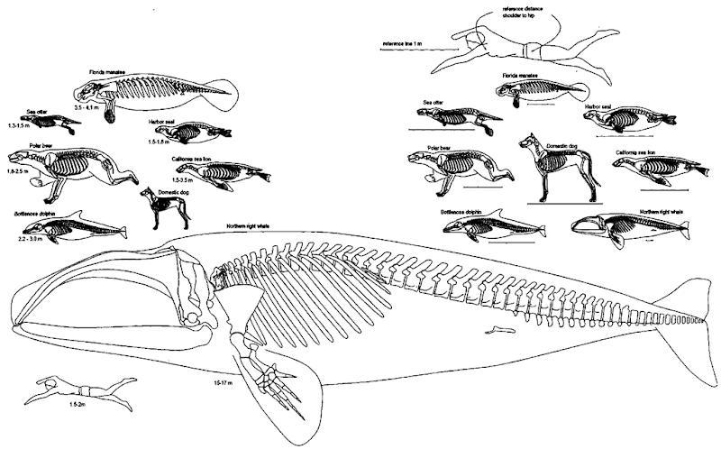
Visualize the articulated (assembled) skeleton by first considering individuals in an absolute sense (left and lower parts of Fig. 1) and then in a relative sense (upper right of Fig. 1). Consider what decisions morphologists must make when comparing the “size” of any one feature among individuals that difier substantially in size. Compare the two methods of scaling by using the human swimmer as an example. Because human sensory systems are comparative and relational comparisons (such as proportions and percentages) are intuitive, we use the relative scheme scale comparisons among species for most of this article.
We will refer to the manatee when describing individual morphological structures but refer to the seven selected marine mammal species when discussing marine mammals in general [for details on the same structures in the dolphin, see Rommel (1990); for the true seals, see Pierard (1971) and Pier-ard and Bisaillon (1978); for the dog, see Evans (1993); for mammals in general, see Flower (1885)].
I. Axial Skeleton
A. Vertebral Structures
A few structural terms are necessary to better grasp the functional morphology of the vertebral column. Each vertebra (plural, vertebrae) has several “standard” parts (Fig. 2A). The body or centrum (plural, centra) of each vertebra is a subcylindrical structure that forms die primary mechanical support of the vertebral (spinal) column (Fig. 2B). Several kinds of projecting parts or processes may extend from the centrum and provide attachment sites, protection for soft tissues, or both. The largest and most common lateral processes are transverse processes, which tend to be long and robust in the lumbar vertebrae (and in the first few caudal vertebrae of permanently aquatic species). In some regions of the vertebral column (e.g., cervical), there may be two transverse processes extending from each side of a single vertebra.
Dorsal to the centrum is an arch of bone (the neural arch), which forms the sides oi a neural canal. The neural arch protects the spinal cord. Interestingly, in some marine mammal species, the neural arch is considerably enlarged to accommodate relatively large masses of vascular tissue and/or fat that may be juxtaposed to the dura mater of the spinal cord. Because of these enlargements, the neural canal may not reflect the dimensions of the spinal cord as it does in most terrestrial species (Giffen, 1992). Additionally, each neural arch may extend dorsally as a neural spine (spinous process). The neural spines function as levers to increase the mechanical advantage of the epaxial (epiaxial) muscles to dorsiflex the tail.
Relative motion between adjacent vertebrae is controlled, in part, by the size and shape of the intervertebral disks (Fig. 2C). Intervertebral disks resist compression when skeletal muscles force vertebrae together. Intervertebral disks are composite structures with a fibrous outer ring, the annulus jibro-sus, and a semiliquid inner mass, the nucleus pulposis. The inner, jelly-like nucleus forms a dynamic joint that can adjust its shape to support complex forces of bending. The outer elastic fibers of the disk are arranged in a resilient ring, or annulus. The same basic design of surrounding a deformable core with elastic fibers is used in baseballs and golf balls.
Figure 1 Selected marine mammal skeletons (the Florida manatee, the harbor seal, the California sea lion, the North Atlantic right whale, the bottlenose dolphin, the polar bear, and the sea otter) compared with the skeleton of the domestic dog. Drawings on the left and bottom are scaled so that 1 m on any one species equals 1 m on all the others in this group. The range of adxdt total body lengths (snout tip to tail tip; note that human height is measured differently) is given in meters beneath each drawing. This group is sized to fit the right whale onto the page. Skeletons in the group on the upper right are scaled so that the distance between the shoulder and the hip joints is equal in all seven species. Thus, the body cavities of this group are approximately the same length. A reference line representing 1 m is given below each of these drawings; note that it is different for each skeleton in this scaling scheme. Both groups are sized so that the dolphins on the right and left are equal in length.
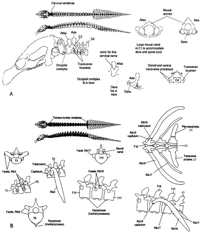
Figure 2 (A) The manatee and its vertebrae. In this and. subsequent illustrations of specific features, we illustrate a particular region of the vertebral column with dorsal and lateral views of that region. (B) Parts of a vertebra. The body, or centrum, of each vertebra is the primary mechanical support of the vertebral column. Vertebral centra help prevent the body of the animal from collapsing when large body muscles contract. Dorsal to the centrum is an arch of bone, the neural arch; within the arch is the neural canal. The neural arch protects the spinal cord and it may extend dorsally as a neural spine or spinous process. (C) The flexible region between adjacent vertebral bodies functions as a tough joint. Intervertebral disks (made of jibrocartilage) have two parts: the inner nucleus pulposus and the outer annulus fibrosus. (D) Zygapophyses are the articular facets that constrain motions between adjacent vertebrae. The cranial pair on each vertebra are prezygapophyses and the caudal pair are postzygapophyscs. The region of articulation between prezygapophyses and postzygapophyses of adjacent vertebrae is typically a synovial joint if the facets overlap. In some regions of the vertebral column the facets do not overlap and may be connected by collagen fibers.
Flexibility of the vertebral column depends, in part, on the thickness of the disks. Disk thickness varies within and among species, and intervertebral disks represent a substantial proportion (10 to 30%) of the length of the vertebral column. Relaxed (neutral) curvature of the vertebral column is vertebral, not intervertebral. That is, the shape of the spinal curve is determined by the shapes of individual vertebrae. Intervertebral disks provide flexibility, whereas curvature is provided by (non-parallel) vertebral body faces (Fig. 3).
In addition to the intervertebral disks, adjacent vertebrae may have other surfaces of articulation (facets). Zygapophyses (singular, zygapophysis) may be located on the neural arch, neural spine, and/or the transverse processes. Zygapophyses are bilaterally paired articular facets, found on the cranial and caudal aspects of each vertebra (Fig. 2D). The cranial pair are termed prezygapophyses, and the caudal pair are postzy-gapophyses. The region of articulation between prezygapophyses and postzygapophyses is typically a synovial joint if the facets overlap.
Zygapophyses in the neck and cranial thorax tend to be oriented horizontally to allow axial rotation (twisting) and side-to-side motions. Zygapophyses in the caudal thorax and tail tend to be oriented vertically to facilitate up-and-down motions. In some regions of the vertebral column (particularly the tails of most species and under the dorsal fin of dolphins), adjacent vertebrae may have reduced or absent zygapophyses, possibly reflecting the increased flexibility or influence of nonvertebral structures on the mobility of these regions. Test these zy-gapophyseal constraints on yourself by bending and twisting your body—note how your thorax (rib-bearing region) flexes (most of the axial rotation and some lateral flexion occurs here) when compared with your lumbar (low back) region (where most of the allowable dorsoventral flexion occurs).
B. Vertebral Column
Traditionally, the typical mammalian vertebral column is separated into five regions, each of which is defined by what is or is not attached to the vertebrae. These regions are cervical, thoracic (dorsal in the older literature), lumbar, sacral, and caudal (Fig. 3). The vertebral formula is an alpha-numerical abbreviation for the numbers of vertebrae in each region; note that some regions have similar numbers of vertebrae in most species, whereas other regions have very different numbers of vertebrae in different species. For example, C6:T17:Ll:S0:Ca25 describes the 6 cervical, 17 thoracic, 1 lumbar, 0 sacral, and 25 caudal vertebrae in the Florida manatee. Individual vertebrae in each region are also referred to by their position using one or two (regiospecific) letters and a number. For example, the third cervical vertebra is designated as C3, the first thoracic as Tl, the first lumbar as LI, the second sacral as S2, and the tenth caudal as CalO. In some species the morphological distinctions among vertebrae from each region are unambiguous, whereas in others, the distinctions among vertebrae in adjacent regions are less evident (Fig. 3).
The total number of vertebrae, excluding the caudal vertebrae, is surprisingly close to 30 in most mammals (Flower and Lydekker, 1891). With this in mind, examine the manatee vertebral column and contrast the numbers of different vertebrae it has with the number of vertebrae in the other selected species (Fig. 3).
C. Cervical Region
Cervical vertebrae (Fig. 4A) are located cranial to the rib-bearing vertebrae of the thorax. Some cervical vertebrae have movable lateral processes known as cervical ribs, none of which make contact with the sternum. Most mammals have seven cervical vertebrae, but all sirenians and the two-toed sloth (Choloeptis) have six and the three-toed sloth (Bradijpus) has nine. Serial fusion (ankylosis) of two or more cervical vertebrae is common in cetaceans, but all cetaceans have seven cervical vertebrae. Contrary to what has been reported in some published works, cervical vertebrae in some cetaceans (e.g., the narwhal Monodon monoceros, beluga Delphinapterus, and river dolphins) are unfused and provide considerable neck mobility.
The first two cervical vertebrae have unique names: the atlas (CI) and the axis (C2). Unlike all other vertebrae, the atlas and axis are not separated by an intervertebral disk but rather by a synovial joint, similar to the skull-atlas joint, where the occipital condyles of the skull articulate with the atlas (Fig. 4A). The shape of this joint dictates the range of motion between the skull and the first vertebra. Typical head motions on the atlas are up and down and side to side (Fig. 4A). The axis has an elongated centrum, the odontoid process or dens, that extends into and sometimes beyond the foramen of the atlas. The shape of the dens restricts motions between the atlas and the axis to twists or rotations parallel to the long axis of the body. Test these constraints on yourself by twisting your head and neck. Head nodding, as in expressing “yes,” is constrained by the atlas; head shaking, as in expressing “no,” is constrained by the axis.
Cervical vertebrae provide support for the head and can aid in feeding and locomotion. As a group, they can, in some cases, flex to provide a wide range of postures. Typically, permanently aquatic marine mammals have short necks, even if they have seven cervical vertebrae. In contrast, pinnipeds have relatively long necks, and the external appearance of a short neck in true seals is misleading. Close comparison of the skeletons of the true seal and sea lion reveals that they have quite similar neck lengths, although the distribution of body mass is different. True seals often hold their heads close to the thorax, which causes the neck to form a deep “S” curve in the neck. This provides true seals with a “slingshot potential” for grasping prey (or careless handlers).
Cervical vertebrae may have distinct transverse foramina that perforate the bases of the transverse processes and run parallel to the long axis of the body. Transverse foramina protect the arteries that supply the head and neck from kinking when the neck is flexed. Transverse foramina are particularly distinct in cervical vertebrae of carnivorous marine mammals (and, of course, the dog). A pair of notches (or foramina) in the neural arch of the atlas may be present (Fig. 4A); this opening is for the first spinal nerve that exits die neural canal between the back of the skull and the bulk of the neural arch of CI. Rarely, one finds cervical ribs, which are movable extensions of the ventral transverse processes. Cervical ribs do not extend to the sternum.
Figure 3 Vertebral columns of the seven selected marine mammal species: the vertebral column of the dog is similar enough to the vertebral columns of the bear and other carnivorous species to exclude it from this illustration. There are typically five separate regions: cervical, thoracic, lumbar: sacral, and caudal. In this illustration, the first thoracic vertebrae for all the species illustrated are aligned. Each sktdl and vertebral column is scaled to similar shoulder—”hip” distances. Sacral vertebrae are, by definition, associated with an attached pelvis; therefore, penna-nently aquatic species with (unattached) pelvic vestiges have no sacral vertebrae. The vertebral formula, an alpha-numerical abbreviation for the numbers of vertebrae in each region, is provided below each vertebral column. The numbers given are for the specimens illustrated; some of these numbers vary between individuals of each species.
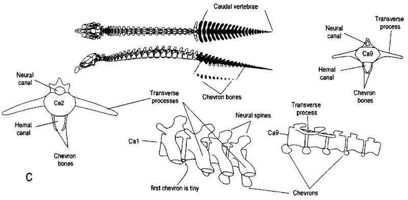
Figure 4 (A) Cervical vertebrae. At the tap of this illustration, these vertebrae are filled in on the dorsal and lateral views of the manatee skeleton. The first and second cervical vertebrae are uniquely named the atlas and the axis, respectively. Cervical vertebrae may have transverse foramina, which perforate the bases of the transverse processes. Typical skull motions on the atlas are tip and down and side to side. The axis has an elongated centrum, the dens, that extends into the large neural canal of the atlas. The shape of the dens restricts motions between the first two vertebrae to “twists” or rotations parallel to the long axis of the body. (B) Thoraco-lumbar vertebrae. Thoracic vertebrae support the ribs, which anchor the diaphragm muscles. Some ribs are double headed and articulate with their respective, vertebrae via capitula and tubercula. (continues)
Figure 4 (Continued) Each capitulum articulates with one or more costal facets on the vertebral centrum at or near the intervertebral disk. Each tuberculum articulates ivith a facet on the transverse process of its respective vertebra. Lumbar vertebrae are trunk vertebrae tvithout ribs: they have pronounced transverse processes called pleurapophyses. In manatees, there may be a rib on one side and a transverse process on the other side of the same lumbar vertebra. (C) Caudal vertebrae a re found in the tail. At the top of this illustration, these vertebrae are filled in on the dorsal and lateral views of the manatee skeleton. In permanently aquatic marine mammals that have no direct attachment between the pelvic vestiges and the vertebral column, the transition between lumbar and caudal vertebrae is defined by the presence of chevron bones. Chevron bones are ventral intervertebral ossifications. By definition each is associated with the vertebra cranial to it.
D. Thoracic Region
The thoracic region (commonly referred to as the thorax) is defined by the presence of more or less movable (i.e., not fused to the respective vertebrae) ribs (Figs. 3, 4B, and 5). Thoracic and lumbar vertebrae support the trunk—the region occupied by the thorax (with the lungs and heart) and the abdomen (with the remaining viscera). By definition, the first thoracic vertebra has a pair of ribs that, unlike cervical ribs, extend to the sternum. Most thoracic vertebrae have ribs that possess two segments. The proximal, dorsal segment is attached to the vertebral column and is termed a vertebral rib (Fig. 5). A distal, ventral segment is attached to the sternum and is termed a sternal rib (Romer and Parsons, 1986). A joint between the vertebral rib and the sternal rib allows flexibility of the thorax. The distinction between vertebral and sternal ribs is more common in birds and some reptiles, but it is less common in mammals because most mammals have cartilaginous sternal ribs termed costal cartilages. This distinction is made because, unlike most other mammals, odontocete cetaceans have bony rather than cartilaginous sternal ribs (bony sternal ribs are also found in the armadillo, Priodontes).
Thoracic vertebrae are constrained by their rib attachments and, thus, tend to be much less flexible than lumbar and caudal vertebrae. Typically, the thorax is relatively more flexible ax-ially and laterally than it is dorsoventrally because zygapophy-ses tend to be oriented horizontally.
As suggested earlier, the mechanics of motion of the thoracic vertebrae are different from those of either cervical vertebrae or postthoracic vertebrae. Thoracic vertebrae have dual functions of longitudinal support and support of the diaphragm muscles. This dual function is reflected in the arrangement, size, and shape of the zygapophyses. The last thoracic vertebrae (here, we use first and last to designate cranialmost and caudalmost ribs to avoid complicated sentences confusing caudal as a direction and caudal as a region of the vertebral column) have ribs associated with them, but these last ribs may not be attached to their respective vertebrae (floating ribs), and there is little or no indication on their respective vertebrae to indicate their presence. The last ribs of the cetaceans are closely associated with the superficial aspects of the robust hypaxial muscles and may have little to do with support of the diaphragm, whereas those of manatees are imbedded in the hypaxial muscles and may provide support for the diaphragm.
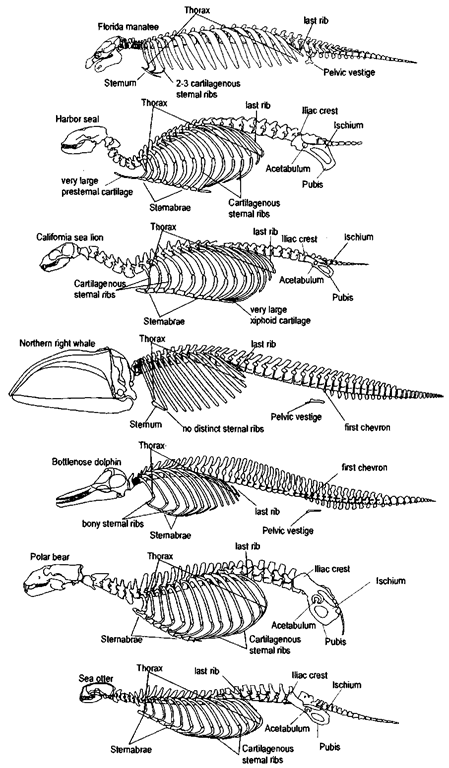
Figure 5 Axial skeletons plus pelvises of the seven selected marine mammal species; because the axial skeleton and pelvis of the dog are similar to those of the bear and of other carnivorous species, the dog’s skeleton was excluded. In this illustration, the first thoracic vertebrae for all the species illustrated are aligned. Vertebral ribs extend from the vertebral column to join the sternal ribs, which extend from the sternum. The sternum develops from a series of sternebrae. In some species (i.e., humans, right whales, manatees), the steniebrae fuse into a single bony unit. Each skidl and vertebral column is scaled to similar shoulder-hip distances. Permanently aquatic species (manatee, dolphin, and right whale) have pelvic vestiges. Because the pelvic bones of these three species are not in the same location in relation to the first chevron bones, the first caudal vertebrae (defined by the first chevrons) of these three skeletons are not aligned. Distinct sexual dimorphism has been described for the pelvic vestiges of manatees and dugongs.
The attachment sites of ribs on the vertebrae are called rib facets or costal facets (ribs = costae) if the region of attachment has a distinct articulation (Fig. 4B); rib facets are located on the vertebral centrum, on the transverse processes, or on both. Some rib attachments are made via long connective tissue fibers; the ribs do not actually touch the respective vertebra, and there is no obvious structure on the (cleaned) vertebrae to suggest rib attachment. For this reason, dogmatic adherence to narrow definitions of numbers of thoracic or lumbar vertebrae or both should be avoided. Cetaceans and pinnipeds have very mobile rib cages. Surprisingly, diving mammals have considerable “tilt” to their ribs. In these species, the double-headed ribs (see later) have joints that are aligned to allow for substantial swing of the attached vertebral ribs in the plane of rotation. This extreme mobility of the rib cage can help accommodate the lung volume changes that accompany changes in depth so that ribs will not break. It may also have evolved to accommodate rib cage dynamics caused when abdominal muscle contractions prevent caudal displacement of the diaphragm during inspiration; in this case, ribs could be moved forward to increase lung volume.
Some thoracic vertebrae have ventral projections called hypapophyses—not to be confused with chevron bones, which are intervertebral and are structurally not part of the vertebrae (Figs. 3 and 4C). In the manatee, the diaphragm is firmly attached along the midline of the central tendon to hypapophyses. Hypapophyses also occur in some cetaceans (i.e., Kogia, the pigmy and dwarf sperm whales) in the caudal thorax and cranial lumbar regions; it is assumed that these hypapophyses increase the mechanical advantage of the hypaxial muscles.
The neural spines of thoracic vertebrae are often longer than those in any other region of the body (Figs. 3 and 5). Long neural spines provide mechanical advantage to neck muscles that support a head cantilevered in front of the body. Therefore, species with large heads tend to have long neural spines; however, the buoyancy of water negates the need for such long neural spines in permanently aquatic species.
Embryonically, ribs and transverse processes are homologous structures, but the presence of a movable joint ultimately distinguishes a rib from a transverse process in an adult. The unfinished joint may be indicative of developmental age. In some species (i.e., the manatee), there may be a movable “ri-blet” (pleurapophysis, pleura = side or rib) on one side and an attached “transverse process” on the other side of the same (typically the first lumbar) vertebra. Pleurapophyses are more common in some of the lower vertebrates (Romer and Parsons, 1986; Kardong, 1998).
A rib may attach to its respective vertebra (Fig. 4B) at one or more locations (e.g., centrum or transverse process, or both). Typically the cranialmost ribs have two distinct regions of articulation with their juxtaposed vertebrae. The cranialmost articular part of a double-headed rib is the head or capitulum (plural, capitula) and it articulates (via a synovial joint) with the body (or bodies) of one or more vertebrae. The tuberculum (plural, tubercula) is the second point of attachment of a double-headed rib; it articulates (sometimes with a synovial joint, other times by connective tissue fibers) to a transverse process of the respective vertebra. Since the capitula of some double-headed ribs touch two vertebrae, the vertebra with the transverse process attachment is used to identify the respective vertebra.
The caudahnost ribs have single attachments and are referred to as single headed. In most mammals, the single-headed ribs have lost their tubercula and are attached to their respective vertebrae by the capitulum at the centrum. In contrast, the single-headed ribs of cetaceans lose their capitula and are attached to their respective vertebrae by their tubercula at the transverse processes (as occurs in some reptiles). Thus, single-headed ribs of cetaceans attach to vertebrae farther from the midline than single-headed ribs of other mammals. Additionally, the transverse processes of the thoracic vertebrae in cetaceans can be longer than those of other mammals. The last ribs often “float” free at one or both ends, i.e., they are attached to neither their associated vertebra nor the sternum. These ribs tend to be significantly smaller than the ones cranial to them and are often lost in preparation of the skeleton.
Sternum The sternum is formed from bilaterally paired serial elements, called sternebrae (the sternal equivalent of vertebrae; Fig. 5). The paired elements fuse on the midline, occasionally imperfectly, leaving foramina in the sternum. The first and last sternebrae are called the manubrium and xiphis-ternum, respectively. In some species (e.g., some odontocetes, all mysticetes, and all sirenians), sternebrae fuse into a single unit. Sternal ribs extend from the sternum to join the vertebral ribs. True seals have an elongate presternal cartilage extending cranially from the manubrium. Some species, particularly the California sea lion and its close relatives, have enlarged xiphisternal cartilaginous extensions.
E. Lumbar Region
Lumbar vertebrae are trunk vertebrae that typically do not bear movable ribs (Fig. 3). Recall that ribs and transverse processes develop from the same embryonic stmctures. Occasionally a distinct pleurapophysis may be found in this region. As noted previously, pleurapophysis are commonly found in manatees: there will be a “normal” transverse process on one side and a movable riblet on the other side of the same vertebra. These lumbar ribs are clearly distinct from thoracic ribs (Fig. 4B).
As already noted, the number of lumbar vertebrae is usually closely linked to the number of thoracic vertebrae; an increase in number in one section typically means a reduction in the other. For example, there are 19 thoraco-lumbar vertebrae in all species of Artiodactyla, whereas there are 20 or 21 in Carnivora; compare these numbers with those of the selected marine mammals (Figs. 3 and 4B). Interestingly, the scaling of the skeletons to similar shoulder-hip joint distances helps us avoid some of the “distractions” that are inherent in the variation among species in size, number, and shape of the other regions of the body.
Typically, lumbar vertebrae are more flexible dorsoventrally than they are laterally and they may have no axial flexibility. Some of the mobility of lumbar vertebrae may be constrained by ribs that, although not attached to these vertebrae, are angled backward and extend beyond the tips of some lumbar vertebrae (Figs. 4B and 5).
F. Sacral Region
There are at least two commonly accepted definitions for sacral vertebrae: (a) serial fusion of postlumbar vertebrae, only some of which may attach to the pelvis (the human sacrum), and (b) only those vertebrae that attach to the ileum, whether or not they are fused serially (Fig. 3). Both definitions have merit. In species in which there is contact between the vertebral column and the pelvis, it is relatively easy to define sacral vertebrae. However, it is not easy in cetaceans and sirenians (dugongs have a ligamentous attachment between the vertebral column and the pelvic vestiges) because there is no direct attachment between the pelvic vestige and the vertebral column and there is no vertebral fusion. Thus, permanently aquatic species have (by either definition) no sacral vertebrae. Within a species, the number of serially ankylosed vertebrae may vary, particularly with age. The number of sacral vertebrae is commonly two to five, but there are exceptions.
G. Caudal Region
Caudal (cauda = tail) vertebrae are found in the tail (Fig. 4C). The number of caudal vertebrae varies widely. Long tails usually require numerous caudal vertebrae (up to a maximum of 46 in one African pangolin, Manis gigantea). Florida manatees have between 22 and 27 caudal vertebrae, and bottlenose dolphins and right whales both have 26; for cetaceans, the minimum is 13 in Caperea marginata (pygmy right whale) and the maximum is 49 in Phocoenoides dalli (Dalls porpoise). Note that caudal vertebrae of cetaceans extend to the tip of the tail (fluke notch), whereas manatee vertebrae stop 3-9% of the total body length (as much as 17 cm in a large specimen) from the fluke tip. Caudal vertebrae range from being robust, important locomotory structures in permanently aquatic species to being relatively small, unimportant pieces in pinnipeds and the polar bear.
Chevron Bones The chevron bones or chevrons (Fig. 4C) are ventral intervertebral ossifications found only in the caudal region. They are relatively large in permanently aquatic species but can be found as small ossifications in many other species, including the dog. By definition, each is associated with the vertebra cranial to it [note that there is some controversy over which is the first caudal, see Rommel (1990)]. Pairs of chevron bones form arches, creating a ventral channel called the hemal canal. Within the hemal canal, the blood vessels (caudal arteries and veins) that supply the tail are protected from occlusion when the tail is flexed. In some cetaceans the ventral aspects of each chevron bone pair fuse and may then function to increase the mechanical advantage of the hypaxial muscles to flex the tail ventrally.
II. Appendicular Skeleton
A. Pectoral Limb Complex
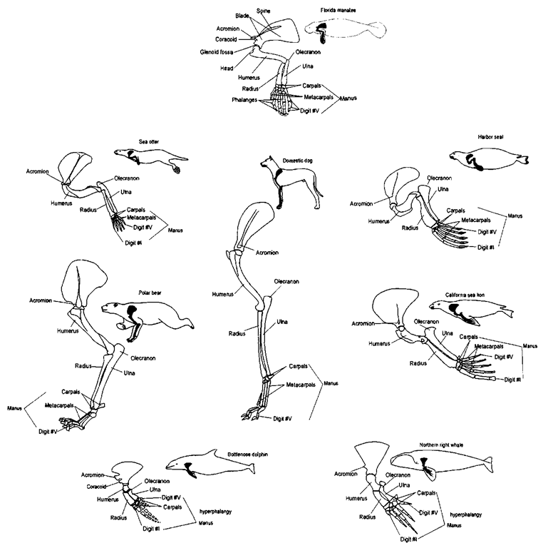
The forelimb (Fig. 6) is referred to as a flipper in perma-nendy aquatic species (in true seals and sea lions the hindlimb is also referred to as a flipper). The forelimb includes the scapula, humerus, radius and ulna, and manus.
The scapula is attached to the axial skeleton only by muscles; there is no functional clavicle in marine mammals (Klima et al, 1980; Strickler, 1978). The scapula consists of an essentially flat (slightly concave medially) blade with an elongate scapular spine extending laterally from it. The scapular spine is roughly in the center of the scapular blade in most mammals. However, in cetaceans, the scapular spine is close to the cranial margin of the scapular blade, and both the acromion and the coracoid extend beyond the leading edge of the blade. The distal tip of the spine is the acromion.
The humerus has a ball-and-socket articulation in the glenoid fossa of the scapula; this is a very flexible joint. The humerus articulates distally with the radius and ulna; this is also a flexible joint in most mammals, but it is constrained in cetaceans. The olecranon is a proximal extension of the ulna that increases the mechanical advantage of the triceps muscles that extend the forelimb. In species like the polar bear and the sea lion, the olecranon is robust; however, in the Cetacea it is relatively small. The radius and ulna of manatees fuse at both ends as the animal ages. This fusion prevents axial twists that pronate and supinate the manus. The radius and ulna of Cetacea are also constrained but are not typically fused. The iorelimbs of the sea otter are very mobile, allowing for the manipulation food.
The distal radius and ulna articulate with the proximal aspect of the manus. The manus includes the carpals, metacarpals, and phalanges. There are five “columns” of phalanges, each of which is called a digit. The digits are numbered, using Roman numerals, starting from the cranial aspect (the thumb; associated with the radius). Note that the longest digit may be different in different species (Fig. 6).
In many of the marine mammals, the “long” bones of the pectoral limb (humerus, radius, and ulna) are relatively short, and the phalanges are elongated. Cetaceans are unique among mammals in that they have more phalanges than any of the other mammals (Fig. 6); this condition is known as hyperpha-langy (Howell, 1930). The number varies between individuals of each species: pilot whales (Globicephala spp.) have the most (14), the bottlenose dolphin has a maximum of 9, and the right whale has a maximum of 7.
Notice the “plane” in which each manus is oriented (Fig. 6). The sea otter’s manus is much like that of humans and has similar dexterity. The polar bear manus is much like that of the dog and other terrestrial quadrupeds. Permanently aquatic species hold the manus in a plane roughly parallel to the midsagittal plane of the body or parallel to the nearest body surface.
B. Pelvic Limb Complex
The typical mammalian pelvis is made of bilaterally paired bones: ilium, ischium, and pubis (Fig. 7). Each of the halves of the pelvis attaches (via the ilium) to one or more sacral vertebrae. The crest of the ilium, which is a prominent landmark, flares forward and outward beyond the region of attachment between the sacrum and the ilium. The two halves of the pelvis join at the pubic symphysis on the ventral midline. In permanently aquatic marine mammals, there is but a vestige of a pelvis to which portions of the rectus muscles of the abdomen may attach. Additionally, the male reproductive organs may be supported by these vestiges. Occasionally, in some of the large whales, a hindlimb articulates with the pelvic vestige.
Figure 6 The forelimb (pectoral appendage) includes the humerus, radius and ulna, and mantis. The forelimb attaches to the pectoral girdle, which is made of bilaterally paired scapulae. The scapula is a flat (slightly concave medially) blade with an elongate scapular spine extending laterally from it. The distal tip of the spine is the acromion process. The humenis has a ball-and-socket articulation with the glenoid fossa of the scapula. The humenis articulates with the radius and ulna at the elbow. The radius and ulna articulate with the proximal aspect of the manus at the wrist. The manus includes the carpals, metacarpals, and phalanges. There are five “columns” of phalanges; each “column” is called a digit. The digits are numbered, using Roman numerals, starting from the cranial aspect (associated with the radius, in humans—the thumb).
Figure 7 The pelvic appendage (hindlimb) and pelvic girdle. The typical mammalian pelvis is made of bilaterally paired coxae attached to one or more sacral vertebrae. Each coxa is made of three elements: the ilium, ischium, and pubis. Each half of the pelvis attaches to one or more sacral vertebrae. The crest of the ilium is a prominent landmark; it flares forward and outward beyond the region of attachment between the sacrum and the ilium. The tivo halves of the pelvis join at the ventral midline pubic symphysis. In permanently aquatic marine mammals there is but a vestige of a pelvis. The hindlimb, if present, is attached to the pelvic girdle via a ball (femoral head)-and-socket (acetabulu m) joint at the hip. The proximal limb bone is the femur. Distally, the femur articulates with the tibia and the fibula at the knee joint. The tibia and fibula distally articulate with the pes at the ankle. Note that the pes is oriented in differen t “planes” in some of the selected species. The pes is made of the tarsals proximally, the metatarsals medially, and the phalanges distally.
The hindlimb, if present, is attached to the vertebral column via a ball-and-socket joint at the hip. The proximal limb bone is the femur, whose head articulates with the socket of the pelvis, the acetabulum. Distally the femur articulates with the tibia and the fibula (at the knee joint). The tibia and fibula articulate distally with the pes or foot (Fig. 7). The pes is composed of the tarsals proximally, the metatarsals medially, and the phalanges distally. Note that the digits of the sea lion terminate a significant distance from the tips of the flipper. Of the chosen group of marine mammals, the limbs of the polar bear are closest in gross appearance to those of the terrestrial mammals. In contrast, many of the marine mammals have relatively short “long” bones (femur, tibia, and fibula) and relatively long phalanges. The femurs of some phocids (e.g., the harbor seal) are so short that the knee cannot contact the ground.
Note that the pes is oriented in different “planes” in some of the selected species (Fig. 7). The pes of the polar bear is oriented in a fashion similar to that of the dog. The sea otter, seal, and sea lion all orient the pes parallel to the sagittal plane when swimming. The sea lion can rotate its hind flippers to “walk” on land, and the sea otter can manipulate its hind flippers to hold food. Compare the positions of the first and last digits in the sea otter with those of the seal and sea lion and note how the sea otters fibula crosses the tibia to achieve this orientation.
Sexual Dimorphisms In many mammals the adult males are larger than the adult females, and among marine mammals, this dimorphism is extremely pronounced in certain pinnipeds and the sperm whales. In contrast, the adult females of the baleen whales and some other species are larger than the adult males. In permanently aquatic marine mammals, there may be sexual dimorphisms in the pelvic vestiges (Fagone et al, 2000). The penises of mammals are supported by tough fibrous structures, the crura, which attach to the pubic bones of the pelvis. The muscles that engorge the penis with blood are also attached to the pelvis, and presumably the mechanical forces associated with these muscles influence pelvic vestige size and shape, particularly in manatees. Males in some groups of mammals, particularly the carnivores, have a bone within the penis (the baculum) that helps support the penis.
III. Sutures and Epiphyses
Many bony features change as the individual develops and matures; these features can be used as milestones in life history studies, and in some cases, they can be fairly accurate estimators of age. Some postcranial elements grow from separate ossifications centers at the margins of the bones (Fig. 8A). Epiphyses (epi-, upon, and pkysis-, grow or generate) are regions at the ends of bones (most often at or near a joint) associated with longitudinal growth in mammals. Epiphyses allow the portion of the bone near a joint to grow at a different rate than the rest of the bone. Growth occurs at a cartilaginous plate between the epiphysis and the diaphysis (dia-, between; body or shaft). At the completion of growth, the epiphysis fuses with the rest of the bone, and the cartilage plate disappears. Eventually the suture between the two parts disappears. Skeletal maturity is defined (for most mammals) when the vertebral epiphyses are all fused to their respective centra. Because most marine mammals are large, their epiphyses are more evident than those of smaller terrestrial mammals.
The best-studied epiphyses are those of the long bones. There are, however, other epiphyses that are less well known. For example, the proximal finger bones may have epiphyses (Fig. 8B). The pattern of epiphysial fusion in the flipper can be used to determine the relative age of an individual when compared to the fusion pattern in other animals of a known age. Occasionally, epiphyses are found on the proximal ends of ribs, on the scapula at or near the coracoid, or at the tips of neural spines and transverse processes.
Among the epiphyses most important to mammalian osteologists are those located at each end of the vertebral centrum (Fig. 8D). Most mammals have indistinct bony plates at each end of their growing vertebrae. These plates are vertebral epiphyses. Manatees (and humans) are among the few species of mammals that have distinct or no vertebral epiphyses. In manatees, the vertebral epiphyses are delicate irregular films of bony tissue found within the cartilaginous surfaces at the ends of the centrum. After these vertebral sutures are completely fused, the skeleton cannot grow in length and the individual is considered skeletally mature. This may or may not coincide with other forms of maturity, such as reproductive maturity.
Nonepiphyseal sutures can also be used to estimate the age of parts of the postcranial skeleton (Fig. 8C). For example, the two halves of the neural arch ankylose at the dorsal midline to form the base of the neural spine. These sutures may persist well into adulthood in cetaceans.
There is much more to learn about the postcranial skeleton. See Pabst et al (1999) for additional functional morphology on marine mammals. Young (1975) for information on mammals in general, and Alexander (1994) for vertebrates. For principles on size and scaling see Calder (1966) and Schmidt-Nielsen (1984).
