The marine environment presents mammals with unique physiological challenges. Most obviously, the ocean’s high salt content and the inavailability of sources of fresh drinking water make it highly desiccating. The kidney is the sole osmoregulatory organ in mammals, so it must excrete excess minerals in urine while at the same time conserving water to prevent dehydration. Moreover, it must maintain osmotic homeostasis of the body fluids even while diving, when depressed cardiovascular function may compromise many physiological processes. To meet the special needs of mammals living in the sea. the kidneys differ in some significant and interesting ways from those of most terrestrial species.
I. General Structure of the Mammalian Kidney
In all mammals, the fundamental functional units are the millions of nephrons that transform plasma filtered from the blood into urine, which contains metabolic toxins and excess minerals that are then excreted from the body. Macroscopically, the kidney is organized into an outer tissue layer, the cortex, that surrounds an inner region, the medulla. In the simplest kidneys, such as those of rodents, the medulla forms a single cone-shaped papilla (unipapillary kidney). In other species, the kidney can have multiple papillae, forming a “multipapillary” kidney. This type of kidney is also referred to as “multireniculate,” with the reniculi corresponding to the medullary papillae and their associated cortical tissue (Sperber, 1944).
Multireniculate kidneys can be of two types (Oliver, 1968). In a “compound” multireniculate kidney, the cortex is continuous and encloses all of the papillae. The urine produced by each of the papillae is drained through the collecting ducts (the terminal segment of the nephron) into a funnel-like structure, the pelvis, that surrounds all of the papillae. This type of kidney is found in only a few species of mammals, including humans and beavers.
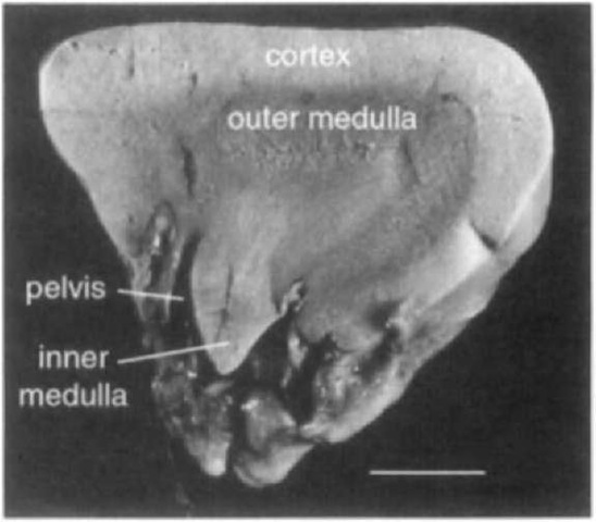
The “discrete” multireniculate kidnev is actually a single organ composed of many miniature kidneys, the reniculi, enclosed in a sheet of connective tissue and peritoneum (the renal capsule) to form a contiguous renal mass (Fig. 1). Each of the reniculi typically resembles a simple unipapillaiy (or bi-papillary) kidney, with its own blood supply and renal pelvis (Figs. 2 and 3). The inicrocirculatory organization and arrangement of the nephrons in each reniculus are essentially the same as those in a simple unipapillaiy kidnev. Urine produced by each of the reniculi drains from the pelvis into ureteral tubules that connect to the main ureter, which carries the urine to the bladder. The reniculi can number in the hundreds or even thousands in each of the paired kidneys. Discrete multireniculate kidneys are found in almost all marine mammals, including cetaceans, pinnipeds, otters, and polar bears (Ursus mar-itimus), but in only a few noniiiarine species.
II. Structure of the Kidneys of Marine Mammals
A. Cetaceans and Pinnipeds
The division of the multireniculate kidney into discrete, independent units is developed to the greatest degree in cetaceans and pinnipeds, which are considered here together because of their general similarities in renal morphology. There is no published information about the renal structure of the walrus (Odobenus rosmnnis), although the regular harvesting of this animal historically and even in the present would seem to provide abundant opportunity for study.
Figure 1 The discrete multireniculate kidney of the Cape clawless otter (Aonvx capensis) with its covering of perirenal fascia. The outlines of the reniculi are evident on the surface of the kidney. Scale bar: 10 mm.
Figure 2 A unipapillary reniculus from the kidney of the Cape clawless otter Like the typical mammalian kidney, there is an outer covering of cortical tissue, and the medulla is divided into outer and inner regions. The inner medulla forms a conical papilla that contains the longest loops of Henle. Urine drains from the nephrons into the pelvis, and from there to the bladder via the ureter. Scale bar: 2.5 mm.
In cetaceans and pinnipeds, the renicular lobulation of the kidney is clearly evident as grooves on its surface. In some species, the reniculi are rather loosely packed within the renal capsule and are separated by considerable connective tissue (perirenal fascia), whereas in others the packing is tight and the reniculi are more hexagonal than round, minimizing the space between them. Especially in cetaceans, they can be arranged in grape-like clusters on branches of the ureter (Abdelbaki et al., 1984).
Although most reniculi in cetaceans and pinnipeds are usually unipapillaiy, reniculi with two or three medullary pyramids are not uncommon, and reniculi with as manv as seven pyramids have been noted in the North Atlantic right whale, Eubalaena glacialis (Maluf, 1989). In these multipapillary reni-culi, which themselves resemble a compound multireniculate kidney, the medullary tissue forming each papilla appears to be functionally discrete, whereas the cortex can either have divisions corresponding to each papilla or can be continuous, forming a capsule that encloses all the papillae within the reniculus.
Figure 3 A bipapillary reniculus from the kidney of the Cape clawless otter. The cortex is continuous, surrounding both medullae. The two medullary papillae are functionally separate, but the urine drains into a common pelvis. Scale bar: 3 mm.
In the typical bean-shaped, unipapillary kidney of terrestrial mammals, the renal artery enters the kidney, and the renal vein and ureter exit, from the hilus, which is located at an indentation 011 the concave medial surface of the kidney. In cetaceans and pinnipeds, the comparable vessels (i.e., renicular artery and vein) are similarly arranged in the individual reniculus. In some pinnipeds, the entire multireniculate kidney is likewise bean shaped, with a hilus-like medial indentation where the renal artery enters and the renal vein and ureter exit. In other pinnipeds and most of the cetaceans, the renal mass is elongate, and the renal artery and vein enter toward the anterior end and the ureter exits more caudally.
A unique feature of the kidneys of cetaceans and pinnipeds is the presence of a conspicuous sporta periinedullaris, a fibro-muscular sheet that separates the cortex and the medulla. First described in cetaceans, it was for a time thought to be unique to this group. Subsequently, however, it has been found to be well developed in both cetaceans and pinnipeds. Because it is present but poorly developed and inconspicuous in nonmarine mammals, it appears to be a feature of the kidney that is related to a marine existence.
The sporta perimedullaris lies at the corticomedullary junction and has perforations that allow penetration of tubular and vascular elements from the cortex into the medulla. In longitudinal cross section of the reniculus, the sporta perimedullaris usually appears as short ribbons of connective tissue and smooth muscle fibers. The function of the sporta perimedullaris remains unknown, but the presence of smooth muscle fibers suggests that, like the renal pelvis, it may be spontaneously contractile, perhaps assisting in expulsion of the urine from the kidney.
B. Otters
There are published accounts of renal structure for three species of otters (Sperber, 1944; Beuchat, 1996, 1999): the sea otter (Enhijdra lutris), which is exclusively marine; the river otter (Lutra lutra), which occurs only in fresh water; and Africa’s Cape clawless otter (Aanyx capensis), which like the manatee inhabits both freshwater and marine environments. Despite their very different osmoregulatory requirements, all three species have discrete multireniculate kidneys that most resemble those of pinnipeds. Lobulation of the kidney is readily apparent externally, and both unipapillary and bipapillary reniculi have been noted in the Cape clawless otter (Figs. 1-3).
C. Polar Bears
Nothing is known about the renal anatomy of the polar bear, the only ursid that can be considered truly at home in the sea. However, discrete multireniculate kidneys have been described in all other bear species examined (i.e., grizzley, U. arctos; sun bear, U. malatjanus; and sloth bear, Melursus ursinus; Sperber, 1944). Studies of kidney function in polar bears are lacking.
D. Sirenians
The most exclusively marine of the recent sirenians is the extinct Steller’s sea cow (Hydrodamalis gigas). The only descriptions of its kidney are from publications by Steller from the 1750s in which he notes that the kidney resembled “those of seal and sea otter,” with numerous reniculi (Sperber, 1944; Maluf, 1989). From this the kidney would seem to be of the discrete multireniculate type. There were no studies of renal function in Steller’s sea cow before its extinction.
The dugong (Dugong dugon) is also highly marine. Its kidney, however, is unlike that of any other marine mammals, bearing a greater resemblance to the crest-type kidney of the dog. It is elongated but lacks superficial grooves and internal division into reniculi. There is a continuous cortex and medial hilus. The medulla is divided into cranial and caudal regions, each forming a long crest with girdle-like dorsal and ventral extensions that are suggestive of transverse lobulation.
Manatees are found in a range of salinities from seawater to freshwater. The Amazonian manatee (Trichechus inunguis) is restricted to freshwater. The West Indian manatee (Trichechus manatus) and the West African manatee (T. senegalensis) are the only mammals besides the Cape clawless otter that naturally occur in both freshwater and marine environments. Sperber (1944) described the kidneys of T. manatus and T. senegalensis as appearing “to represent a transitional stage between kidneys with undivided cortex and divided medulla, and the renculi kidneys.” In these species, the kidney seems to most closely resemble a compound multireniculate design, with 6-11 medullary papillae. Although a continuous cortex surrounds all of the medullae, there are folds of cortex (plicae corticales) and septa that separate adjacent medullary regions, producing the external appearance of lobulation in the adult (Maluf, 1989). The papillae are not conical, forming instead at their tips a concave surface that protrudes into a large, muscular renal pelvis. There is no inner medulla. The kidney is bean shaped, with the renal artery and vein and the ureter located together at the indentation of the hilus.
III. Size of the Kidney
The mass of the kidney increases with body size in mammals (Beuchat, 1996). In marine mammals that have discrete multireniculate kidneys, the number of reniculi in each kidney increases as well. In the harbor porpoise (Phocoena phocoena), for example, the kidneys of an adult (body length = 1.6 m) weigh 150-200 g and contain roughly 300 reniculi that range in weight from 0.15 to 0.6 g each. In an 11-m right whale, one of the kidneys can weigh 32 kg and contain 5400 reniculi, each with an average weight of 2.6 g.
In general, the kidneys of cetaceans and pinnipeds are larger than those of similarly sized terrestrial mammals (Beuchat, 1996). This may be a consequence of the division of the kidney into reniculi, with the connective tissue surrounding each reniculus accounting for at least part of the difference in mass.
IV. Urinary-Concentrating Ability
All marine mammals examined to date can produce urine that is at least as concentrated as seawater (1000 mosM), and most can do substantially better than this. Measurements from cetaceans (six species) range from 1081 to 1737 mosM and those from pinnipeds (five species) and the sea otter are even higher, from 2000 to 2750 mosM (Beuchat, 1996). The highest value recorded from a manatee is 1100 mosM.
Although these osmolalities are far less than those achieved by some desert rodents, in which the urine can exceed 8000 mosM, they are surprisingly high considering the structure of the kidney in most marine mammals. The countercurrent multiplier theory predicts that the concentrating ability should increase with increasing length of the loop of Henle. In species with discrete multireniculate kidneys, the nephrons have veiy short loops of Henle because the individual reniculi are much smaller than an appropriately sized nonmultireniculate kidney would be for that species (Beuchat, 1996). How marine mammals produce such concentrated urine with such short loops of Henle remains a paradox.


