Introduction
Low back pain is thought of as having no structural correlates in radiographic findings. But an associated deconditioning syndrome is assigned by back pain complaints accompanied by functional deficits, especially peak force and performance deficits of deep trunk muscles. We were aiming at investigating if there might be comparable relations between spinal mal-alignment and complaints in chronic low back pain patients. And if spine shape aberrations were in fact associated with low back pain, could they be used to determine exercise programs for an active low back pain therapy, as is generally known for diagnostic screening procedures and low back pain therapy monitoring based on muscle function deficits? Seeking for exercise induced adaptations, we intended to find statistical correlations indicating some kind of specificity for those individualized exercise programs which are based on initial findings in spinal alignment and trunk muscle function.
Our scientific approach involved two aspects that were important for both practical applications and scientific analysis methods in the field of low back pain treatment and research. First of all, our spine shape assessment was non-invasive, and therefore suitable for screening and monitoring without any risks for patients and volunteers. And secondly, indirect spine shape assessment by means of video raster stereography allowed an easy access to multivariate statistical analysis approaches. Therefore, variable interdependencies could be taken into account which might have covered significant effects in earlier investigations.
Background
From an economic point of view, low back pain (LBP) is one of the most emerging and cost-pushing health disorders in the western world, and for the majority of cases neither direct organic signs nor structural correlates can be identified (Waddell et al., 1980). According to McGill (2007, p. 5), more than 80% of all patients with back complaints suffer from nonspecific low back pain. He suggests that, besides other factors, insufficient diagnosis procedures may contribute to the current uncertainty regarding the true incidence of specific low back pain issues.
Several influencing factors are discussed to be essential in the etiology of low back pain, such as psycho-social components (Waddell et al., 1980), and organic mechanisms in terms of spinal instability due to ligament function and deficits in neuromuscular coordination and compensation: neutral zone spinal instability hypothesis (Panjabi, 1992).
With respect to these biomechanical and social-medical findings, and being aware of muscular dysfunction in LBP patients compared to pain free volunteers (Cady et al., 1979; Denner, 1997; McNeil et al., 1980), reconditioning of muscle function and neuromuscular coordination patterns is supposed to be a successful intervention mode in the therapy of low back pain (Denner, 1997; McGill, 2007; Panjabi, 1992; Waddell et al., 1980), especially when segmental stabilization is taken into account (Ljunggren et al., 1997; O’Sullivan, Twomey & Allison, 1997; Richardson, Hodges & Hides, 2004).
Beside deficits in muscle function of LBP patients, there are anthropometric risk factors for the development and progredience of LBP which deal with spinal shape asymmetries in the frontal plane (Balague, Troussier & Salminen, 1997) and the alignment of the lumbosacral transition in the sagittal plane (Adams, Mannion & Nolan, 1997; Lewit, 1991, p. 60). Video raster stereographic back shape reconstruction offers a valid and reliable and – in contrast to radiographic screening procedures – a non-invasive, non-aggressive high-resolution system for spine shape assessment in screening and monitoring (Drerup & Hierholzer, 1994).
Recent video raster stereographic investigations of the spinal form of male and female LBP patients and pain free volunteers revealed spine shape parameters indicating LBP by means of multivariate factor analyses: trunk imbalance and trunk inclination (Schroder, Stiller & Mattes, 2010). While a more extended trunk inclination should be considered to be due to the higher age of the patients (Gelb et al., 1995; Kobayashi et al, 2004; Takeda et al., 2009), trunk imbalance remained as a marker for low back pain. Additionally, there was some evidence for a flatter lumbar lordosis in male patients, revealed by means of discriminant analyses (Schroder, Strubing & Mattes, 2010). With female patients, too, pelvis torsion and pelvis tilt were found to be indicating low back pain (Schroder, Stiller & Mattes, 2011). It is highly probable that video raster stereography offers some possibilities in the process of differential diagnosis of sacroiliac disorders (Foley & Buschbacher, 2006).
Furthermore, there was some evidence for non-parametric signs in the spinal alignment of back pain patients with vertebral blockades (Schroder, Farber & Mattes, 2009) or a lumbar facet joint syndrome (Schroder, Strubing & Mattes, 2010). These findings and some specific kind of profile of spinal shape parameters should be helpful for diagnosis procedures in the field of orthopaedic practioneers. This work is in process.
The findings mentioned above might provide an opportunity to create therapeutic exercise programs based on spinal form deviation signs, comparable to individualized exercise programs based on muscle function deficits (Denner, 1997). So far, specific correlations between adaptations of muscle function and clinical out-come parameters could hardly be established (Mannion et al., 2001b; 2001c). Nevertheless, first results of a pilot study seemed to show specific adaptations following individualized exercise programs, e.g. trunk imbalance decreased mainly in patients who showed extraordinary values in the frontal plane before a short-term training period of ten weeks. This specific decrease correlated with pain reduction and was accompanied by increases in peak forces of trunk muscle strength (Schroder et al., 2009).
In general, spinal form adaptations are difficult to prove by means of statistical calculations (Kuo, Tully & Galea, 2009), because they depend on the degree of mal-alignment, and adaptations are varying considerably among individuals (Weifi, Dieckmann & Gerner, 2003; Weifi & Klein, 2006). Age and gender also seem to be influencing factors for the degree of spinal form adaptations in some parameters (Schroder & Mattes, 2010). Correlations between clinical out-come and muscle function increases are augmented, when spinal form adaptations are taken into account in multiple regression models.
Methods
Study design
First of all, a cross-sectional study was conducted to identify spine shape parameters associated with low back pain. Secondly, a pre-post-effect analysis was carried out, seeking for exercise induced adaptations in the process of reconditioning.
Subjects
At least 405 subjects could be examined, 213 patients suffering from low back pain (LBP) and 192 volunteers – most of them freshmen at the University of Hamburg – serving as controls (CON). The controls were included if there was no diagnosis dealing with back pain complaints, no serious back pain history for two years, and no back pain at all in the last six months.
Participants were divided into female and male subsamples. Due to the large sample size, the observed – relatively small – differences in anthropometric parameters between patients and controls were almost significant, except for the body weight of the males (tab. 1).
|
age [y] |
height [m] |
weight [kg] |
BMI [kg/m2] |
|
|
LBP females |
50,5 |
1,68 |
67,9 |
24,2 |
|
SD (n=129) |
14,2 |
0,06 |
6,0 |
1,6 |
|
LBP males |
47,6 |
1,83 |
82,4 |
24,6 |
|
SD (n=84) |
15,3 |
0,06 |
6,0 |
1,4 |
|
CON females |
26,5 |
1,70 |
65,7 |
22,8 |
|
SD (n=79) |
4 7*** |
0,06* |
6,5* |
1 4*** |
|
CON males |
27,6 |
1,85 |
82,2 |
24,0 |
|
SD (n=113) |
4 4*** |
0,05** |
5,5 |
1 2*** |
Table 1. Anthropometric data of low back pain patients (LBP) and pain free controls (CON) (mean ± standard deviation; LBP vs. CON: * p<0,05; ** p<0,01; *** p<0,001 Student’s t-test)
Female patients were significantly older (t = -17,636; p < 0,000), had a slightly smaller body height (t = 2,475; p = 0,014), a slightly larger body weight (t = -2,517; p = 0,013) than the female controls and also showed a slightly higher body mass index (t = -6,353; p < 0,000).
Male patients were significantly older (t = -11,668; p < 0,000), had a slightly smaller body height (t = 2,395; p = 0,018), a nearly identical mean body weight (t = -0,330; p = 0,742), and showed a slightly higher body mass index (t = -3,298; p < 0,001), too (tab. 1).
Patients were included after clinical and radiographic examinations by an orthopaedic physician (Buchholz & Partner, Hamburg, Germany), who qualified the pain syndrome as chronic unspecific back pain (LBP), when no correlation to structural signs could be established and when patients suffered from low back pain for a time period of six months minimum. In fact, back pain history varied from six months to more than nine years (average: 8 months) and most of the patients had gone through several treatment trials before. Specific signs, such as vertebral fractures, spinal surgery, severe scoliosis or acute sciatic symptoms were exclusion criteria, as well as a back pain state of more than 5 points in the CR10 pain scale reacing from zero to ten points (Borg, 1998) at examination time.
107 of those patients mentioned above went through an exercise therapy program and were re-examined in a post-test. Treatment effects could be analysed for 61 female patients (57%), and for 46 males (43%). Females were 48,7 ± 14,1 years of age, body height was 1,70 ± 0,07 m, body weight was 67,8 ± 10,7 kg, and their body mass index (BMI) was 23,6 ± 3,3 kg/m2. Males were of the same age (49,6 ± 14,3 years), but naturally higher (1,80 ± 0,07 m) and heavier (81,4 ± 12,9 kg), while the body mass index was comparable (24,9 ± 2,9 kg/m2) to the females, and not indicating obesity.
Spine shape assessment
Spine shape parameters were calculated by means of video raster stereography (Formetric®-System1), a high resolution back shape reconstruction device (reconstruction error 0,2 to 0,5 mm; resolution 10 pts./cm2) (Drerup & Hierholzer, 1994). Reproducibility of back shape reconstruction was proved. Reliability coefficients (ICC: Intra Class Correlation) were ranging between 0,99 and 0,91 for the sagittal plane, and between 0,82 and 0,69 for the frontal plane. For the coronal plane, reliability was 0,81 (Mohukum et al., 2009; Schroder & Mattes, 2009; Schroder, Reer & Mattes, 2009) (tab. 2).
Specific back surface landmarks - like the vertebra prominens (VP), the beginning of the rima ani representing the sacrum point (SP), and the right and left lumbar dimple (DR, resp. DL) representing the position of spinae iliaca posterior superior (SIPS) of the pelvis – were recognized automatically to build up a Cartesian coordinate system. This coordinate system served as calibration reference frame for a three-dimensional surface reconstruction using triangulation equations that ensured a valid correlation between back shape reconstructions and radiographic assessments of the anatomy of spine and pelvis characters 2 (Drerup & Hierholzer, 1985; 1987a; 1987b) (fig. 1).
|
Spine shape parameter |
Short/ ICC |
Explication |
|
Trunk imbalance [mm] |
Tr-Imb ICC=0,82 |
Plumb deviation from vertebra prominens to midpoint between dimples in the frontal plane (fig. 2) |
|
Trunk inclination [mm] |
Tr-Inc ICC=0,91 |
Plumb deviation from vertebra prominens to pelvis position/ midpoint between dimples in the sagittal plane (fig. 2) |
|
Pelvis tilt [mm] |
P-Tilt ICC=0,81 |
Deviation of the axis of lumbar dimples to the floor line in the frontal plane (fig. 2) |
|
Pelvis torsion [°] |
P-Tors ICC=0,69 |
Relative torsion between left and right side pelvis bones (os ilium) in the frontal-transversal plane |
|
Vertebral side deviation [mm] |
Side-rms ICC=0,71 |
Average deviation of vertebral bodies in the frontal plane (rms from vertebra prominens to midpoint between dimples) |
|
Vertebral rotation (rms) [°] |
Rot-rms ICC=0,81 |
Average rotation of vertebral bodies in the transversal plane (rms from vertebra prominens to midpoint between dimples) |
|
Kyphosis angle (ICT-ITL) [°] |
KA-max ICC=0,91 |
Maximum thoracic angle calculated from ICT and ITL triangles (fig. 2) |
|
Lordosis angle (ITL-ILS) LA-max [°] ICC=0,99 |
Maximum lumbar angle calculated from ITL and ILS triangles (fig. 2) |
|
Table 2. Spine shape parameters, short-cuts with Intra Class Correlation coefficient (ICC), and a description of anatomy and corresponding geometry
Fig. 1. Video raster stereography with camera and projector system (left), projection lines on the back surface with vertebra prominens (VP) and lumbar dimples (DL+DR) high-lighted -here with optical markers only for demonstration (middle), and video raster stereography back surface reconstruction with landmarks recognized automatically (red dots) and plane curvatures representing convex (red areas) or concave (blue areas) back shape profiles (right).
For a better understanding of geometry and corresponding anatomical landmarks, spine shape parameters were illustrated in an animation, especially for the sagittal plane (fig. 2).
Fig. 2. Spine shape in the sagittal plane: kyphosis angle (KA-max) and lordosis angle (LA-max) with inflectional points of the curvature from cervical to thoracic spine (ICT), from thoracic to lumbar spine (ITL) and from lumbar to sacral spine (ILS) and three dimensional animation of back surface with lumbar dimples (yellow dots – with arrows representing the direction of the mathematical normal on each dimple’s plane) and spinous processes like vertebra prominens (VP) and sacrum point (SP) marking the beginning of the rima ani (green dots).
Trunk muscle peak force assessment
Torques of the superficial trunk muscles were assessed by means of isometric peak forces (sensor sample rate 100 Hz, sensibility 0,85 mV/V, signal smoothing by a sliding average over 0,3 sec) in a test chair that allowed data acquisition in all three dimensions (extension-flexion, lateral flexion, axial rotation) (Myoline®)3, while patients or volunteers had to be fixed only once for all test contractions in a universal standard position. Reproducibility was verified, and reliability coefficients were ranging between 0,85 and 0,94 for trunk muscle testing in all three dimensions (Schroder, Reer & Mattes, 2009).
Pain documentation
Pain was described by means of the CR10 pain scale questionnaire, an instrument for self-rated pain and exertion, evaluated by Gunnar Borg (1998). The CR10 pain scale (0=nothing at all, 0,5=extremely weak, 1=very weak, 2=weak, 3=moderate, 5=strong, 7=very strong and 10=extremely strong) combined categorical and rational aspects of the phenomenon pain -for a valid assessment with respect to the non-linear relation between pain state and semantic expressions for its description4. Reliability had been verified earlier, and coefficients ranged between 0,78 to 0,99 (Borg, 1998, pp. 41-43).
Treatment
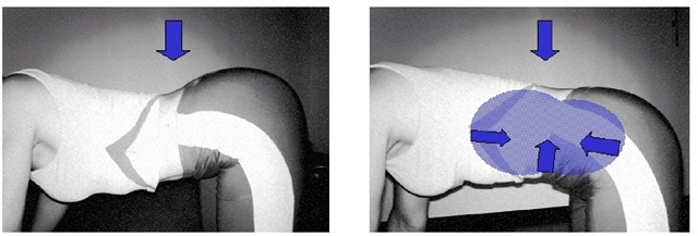
About 50% of all low back pain patients (n=107) went through an individualized exercise program for a time period of 10 to 12 weeks from pre- to post-testing. There were 18 training sessions altogether, normally two sessions per week. Every session took 60 minutes and followed a fixed schedule of seven phases: a systematic ergometer warm-up (5 min), functional strengthening (2 to 4 exercises) and stretching (4 to 6 exercises), as well as physiotherapist pulley and weight training (4 to 6 exercises) using standard training devices. But the exercise program was dominated by Segmental Stabilization Training (SST), which was learned and re-learned in every session (2 to 3 min) in a basic exercise (fig. 3), and which was applied in several static (2 to 4 exercises) and dynamic (2 to 3 exercises) tasks with an emphasis on the special SST-coordination5 pattern.
Fig. 3. Coordination pattern of Segmental Stabilization Training (SST)
All exercises were performed for one to three sets, with an intensity that allowed 10 to 15 repetitions or 20 to 30 seconds of static resistance, respectively. Number of sets and reps (volume and intensity and the choice of exercise itself (content) were determined by individual findings in the pre-test and anamnesis information right before starting the intervention. Training took place in the field of out-patient rehabilitation in groups of three to five patients and was conducted and controlled by at least one physiotherapist (Schroder & Farber, 2010).
Statistics
Data were described as mean ± standard deviation (SD), mean ± CI (95% confidence interval) for figure 4, and mean ± SEM (68% confidence interval meaning the Standard Error of the Mean) for figure 5. Normal distribution was proved using the Kolmogorov-Smirnov-test.
For the cross-sectional study, a factor analysis (SPSS 12: principle components extraction, Kaiser-normalisation with varimax rotation) was conducted to explore a spine shape structure model of almost independent factors, determined by video raster stereography spine shape parameters, seeking for differences between low back pain patients and pain free controls. In a second multivariate approach, discriminant analyses (SPSS 12) were calculated for males and females to reveal spine shape parameters being able to separate low back pain patients from pain free volunteers. At least, these extracted parameters were analysed for significant differences between patients and controls by means of univariate procedures (Student’s t-test), and a spine shape profile was illustrated for males and females with or without low back pain.
For the analysis of treatment effects in the sample of patients who went through an exercise program, three-way ANOVAs (SPSS 12: within-subjects factor: pre vs. post exercise program, between-subjects factor for gender: female vs. male and between-subjects factor for age: under 60 years vs. over 60 years) were calculated. Bivariate Pearson correlations and linear multiple regression models based on pre-post-differences were calculated to analyse interdependencies of variables monitored in the process of reconditioning.
Significance was accepted for p-values of p<0,05 *. Differences showing p-values of p<0,01 ** or p<0,001 *** were deemed very significant.


