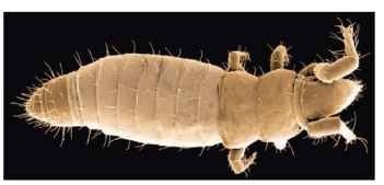
The Protura are a group of minute, almost unpigmented soil arthropods (Fig. 1). With a body length of merely 0.2-2.6mm in adult size, they inhabit moist crevices within soil, predominantly forest humus, but also leaf litter, moss, and occasionally

FIGURE 1 Scanning electron microscopy of a proturan (family Acerentomidae) found in subtropical rainforest litter from Lamington National Park, Queensland, Australia.
rotting wood, where they probably feed on fungal mycelia. Apart from the eventual hardened, yellowish pigmented body parts, living specimens are nearly translucent and peristaltic movements of the gut can easily be observed. Living specimens also perform characteristic movements with the abdomen in that the small postabdomen is recurrently invaginated into the preabdomen, probably for the purpose of ventilation. These regular ventilatory movements of the postabdomen become especially rapid when the animals are stressed and attempt to escape from a threat.
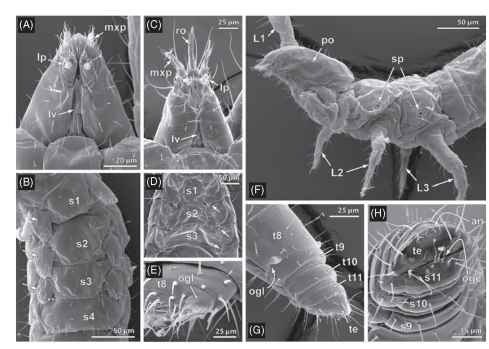
Proturans are distinguished from all other hexapods by an extraordinary set of unique features. They resemble insects in their tag-mosis, but each tagma shows distinctive modifications (Fig. 2A-H). The proturan head has the insect-typical set of mouthparts (mandibles, first maxillae, and a labium formed by medially fused second maxillae) but with neither eyes nor antennae (Fig. 2F). The thorax is composed of three segments, each bearing one pair of walking legs, but the anteriomost pair is usually carried forwards above the head and equipped with numerous sensilla as if to replace the antennae functionally. The apparent substitution of the antennae by the forelegs correlates with a backward shift of the head onto the prothoracic segment, which therefore is hardly recognizable in at least dorsal view. Proturan locomotion is accordingly mainly “tetrapod” and usually slow although the forelegs, which are still equipped with unpaired pretarsal claws, can be used in locomotion, as when climbing through fungal mycelia or moss and escaping predatory threats. The abdomen is composed of eight large segments (preabdomen) followed by four conspicuously smaller segments (postabdomen). The 12-segmented abdomen accordingly seems to be one segment longer than in all other hexapods (Fig. 2G and 2H). The segmental nature of the ultimate abdominal segment, however, has not yet been confirmed. The alleged twelfth abdominal segment is sometimes considered to represent an unusually large nonsegmental “telson”, which in all other hexapods merely forms minute sclerites (“laminae anales”) around the anus, if at all. This alternative view is supported by the postembry-onic development of the abdomen, during which three penultimate segments are formed in front of the “telson” (or ultimate segment). This anamorphic development of the Protura is unique among hexa-pods. The same holds true for vestiges of leg-derived appendages on the first three abdominal segments (Fig. 2B and 2D), which primarily are still segmented and distally taper off into an eversible vesicle. The sternites and leg-bases of the appendicular vestiges on the first three abdominal segments remain clearly separated, whereas in the ” cerci-bearing hexapods” (Cercophora, i.e., Diplura and Insecta) they consistently fuse and form so-called coxosternites.

FIGURE 2 Morphological characteristics of the Protura exemplified by Eosentomon sp. (Eosentomidae: A-B, F-H) and Acerentomon sp. (Acerentomidae: C-E). (A) Ventral view of a typical eosen-tomid head, showing entognathy, linea ventralis (lv), and reduced palps of the first (mxp) and second (lp) maxillae. (B) Ventral view on an eosentomid preab-domen, showing segmented vestiges of appendages (arrows) on the first three abdominal segments. (C) Ventral view of an acerentomid head, showing the labrum prolonged to a rostrum. (D) Ventral view of an acerentomid preabdomen, showing two-segmented appendicular vestiges on the first abdominal segment and short, unsegmented appendicular vestiges on second and third abdominal segment. (E) Dorso-lateral view on eighth abdominal segment, showing the opening of the abdominal defensive glands (ogl) covered by a pectinate lid. (F) Lateral view on an eosentomid head and thorax (left pro-thoracic leg L1 removed), showing position of the cephalic pseudoculus (po) and spiracles on the laterotergites of meso- and metathorax. (G) Dorso-lateral view on an eosentomid postabdomen, showing the smaller ultimate segments (t9-t11) followed by a segment-like “telson” (te). (H) Frontal view on the postabdomen, showing the transverse opening of the genital chamber (ogc) and the terminal, transverse slit forming the anus (an). Abbreviations: an, anus; L1-3, locomotory legs on thoracic segments 1-3; lv, linea ventralis, lp, labial palp; mxp, maxillary palp; ogc, orifice of genital chamber; ogl, orifice of abdominal defensive gland; po, pseudoculus; ro, rostrum; s1-11, sternites of abdominal segments 1-11; sp, spiracle; t8-11, tergites of abdominal segments 8-11; te, “telson.”

FIGURE 3 Consensus tree inferred from 52 morphological characters depicting the relationships among the ten currently recognized families of the Protura. The three main subgroups according to current classification are highlighted in gray. Numbers in brackets refer to the number of species described thus far (compiled by Andrzej Szeptycki, Institute of Systematics and Evolution of Animals, Polish Academy of Science; see http://www.isez.pan.krakow.pl). Asterisks indicate families that may not represent monophyletic groups.
The apparent primitiveness of the abdomen relative to the states in remaining hexapods forms the name-giving character of the Protura (“primitive-tails”). Their relatedness to insects, however, has repeatedly been questioned and still is doubted. Upon their discovery and first description by Filippo Silvestri in 1907, pro-turans were considered as intermediate forms between myriapods and insects and were therefore classified by Anton Berlese in 1909 as ” Myrientomata.” Traditional phylogenetic hypotheses recognize the Protura as sister group of the Collembola (Ellipura-hypothesis) based on the shared absence of caudal appendages (name-giving character), a corresponding entognathous state of the mouthparts, and the presence of a cephalic longitudinal groove called a linea ventralis (Fig. 2A and 2C), along which secretions of the salivary glands and/or labial kidney pass backwards toward the cervical region. In the light of a crustacaean origin of hexapods, the Ellipura-hypothesis gains additional support from the presence of paired lateral sense organs on the head capsule (postantennal organ in Collembola, pseudoculus in Protura; Fig. 2F) that are innervated by the pro-tocerebrum. Molecular data instead currently favor a sister group relationship between Protura and Diplura (Nonoculata-hypothesis). Apart from the absence of eyes (name-giving character), this clade is usually stated to have no unambiguous morphological support. The following correspondences of the Protura and Diplura are nevertheless remarkable: the first immature instar (hatchling) is an immobile, nonfeeding prelarva (pupoid stage); mandibles are primarily denticulate and devoid of a molar plate; the thoracic sterna primarily form Y-shaped apodemes; Malpighian (excretory) organs, when present, are differentiated as short papillae; and the gonopores show a corresponding segmental (or intersegmental, respectively) position in both sexes. Current embryological and some ultrastructural data, in contrast, still raise doubts on whether proturans are most closely related to hexapods at all.
Proturans are distributed worldwide and seem to be lacking at the polar regions only; a faunistic overview is presented in Szeptycki (2007). About 750 proturan species have been described thus far and assigned to ten (or seven, respectively) families (Fig. 3 ). By far the most species-rich and abundant genus is Eosentomon (Eosentomidae), followed by Acerentulus (Berberentulidae) and Acerentomon (Acerentomidae).
COMPARATIVE ANATOMY AND CLASSIFICATION
The monophyly of the Protura is supported by plenty of unambiguous apomorphies. Apart from the absence of antennae, most noteworthy are (1) the relatively small size of the head (microcephaly) that anteriorly tapers off into a narrow, immobile labrum; (2) the enigmatic head endoskeleton (fulcro-tentorium); (3) the position of the tracheal spiracles on thoracic nota; (4) the presence of abdominal defensive glands; (5) the formation of a genital chamber housing external genitalia; and (6) the structure of the spermatozoa.
The microcephalic state of the head seems to be correlated with a sucking mode of food uptake. Although details on feedings habits are still unknown, the pipette-shaped head can be assumed to optimize the uptake of scratched-off particles or fluid food. The head endoskeleton is largely cuticular and resembles the X-shaped cor-potentorium of pterygote insects. The prelarval stage demonstrates that it is formed by paired cuticular rods that arise next to the mouth opening; in subsequent stages, these rods are coalesced and form an X-shaped “”ulcro-tentorium.” The homology of its components to paired anterior and posterior tentorial apodemes in remaining hexa-pods is likely but still disputed.
Meso- and metathorax are the only body segments that primarily have one pair of spiracles each, but they are not located within the pleuron as commonly found in the cerci-bearing hexapods (Diplura and Insecta), but are located mid-laterally on the tergites (Fig. 2F). Subgroups of the Protura are characterized by the loss of the tra-cheal system.
Paired abdominal defensive glands of variable size are dorsally located in the preabdomen and open to the exterior via a more or less conspicuous orifice at the hind-margin of the eighth abdominal tergite (Fig. 2E and 2G). These glands seem to compensate the relatively slow “)etrapod” movements by the production of repellents that efficiently act as a deterrent to at least predacious mites.
In both sexes, external genitalia, the so-called squama geni-talis (putative copulatory organ in males, ovipositor in females), are formed within a genital chamber that opens to the exterior through a transverse slit between the eleventh abdominal sternum and the “telson” (Fig. 2H). Comparative muscular studies support the view that the proturan external genitalia are formed by the ninth abdominal segment, but their appendicular nature and homology with insect genitalia still remain unclear.
Flagellate spermatozoa show a strikingly variable axonemal pattern in proturans, but a common, distinctive feature of the axoneme is the absence of central microtubules (9 + 0 to 14 + 0-patterns). Many species show spermatozoa devoid of a flagellum, which is generally considered to be due to convergent loss.
Present uncertainties on proturan interrelationships, however, hardly permit final conclusions on character evolution within the Protura. Cladistic analyses of proturan phylogeny are still in the very beginning. Three major subgroups are currently distinguished (Fig. 3), among which the Sinentomata are particularly interesting for combining characteristics of both Eosentomata and Acerentomata.
The Eosentomata (or Eosentomoidea) uniquely share lens-like pseudoculi with a central foramen and are further characterized by aflagellate spermatozoa and elongate maxillary glands that extend into the thorax, by defence gland orifices with inconspicuous lids (Fig. 2G), and by details of their external genitalia. Most species of the Eosentomata maintain denticulate mandibles (except Styletoentomon with stylet-shaped mouthparts), a tracheal system with paired spiracles on the meso- and metathorax (except the Chinese Antelientomon, sole genus of the Antelientomidae), complete Y-shaped apodemes on the thoracic sterna, and segmented leg-derived appendages with terminal eversible vesicles on all of the first three abdominal segments.
Among the remaining proturans, denticulate mandibles and a tra-cheal system are only present in the East-Asian Sinentomon species, whereas three pairs of segmented abdominal appendages are maintained in the East-Asian Fujientomon species only (as well as in some Hesperentomon species). Sinentomon species are unique among the Protura in having no cephalic linea ventralis. Based on features of the aflagellate spermatozoa and the presence of striated pseudoculi, among others, the Sinentomidae and Fujientomidae were united in the Sinentomata, but this classification is not supported in current cladistic analyses. The structure of the pretarsal claws of the walking legs and a transverse, variably striated cuticular band on both tergum and sternum of the eighth abdominal segment indicate that the Sinentomidae and Fujientomidae form a presently unresolved unit with the Acerentomata. Molecular analyses, in contrast, retrieve the Sinentomata as a basal grade leading to a unit composed of Eosentomata and Acerentomata.
Members of the Acerentomata (or Acerentomoidea) generally have pointed or stylet-shaped mouthparts and share a special type of pseudoculus with a posterior foramen, incomplete or missing sternal apodemes on the thoracic segments, unsegmented appendages devoid of eversible vesicles on at least the third (and in some subgroups also the second) abdominal segment (Fig. 2D), and conspicuous, more or less pectinate lids (“combs”) roofing the orifice of the abdominal defensive glands (Fig. 2E) . Compared to Eosentomata and Sinentomata, acerentomoid proturans seem to be plesiomorphic in maintaining flagellate spermatozoa in most of the subgroups. The interrelationships among the acerentomoid subgroups still largely remain unclear, since some of the traditionally recognized families seem to have no unambiguous morphological support.
BIOLOGY
Information on proturan biology is still scarce, since attempts to rear proturans under laboratory conditions succeeded just a few years ago (see Machida, 2006). The insemination mode remains unclear, but a direct sperm transfer is likely as males produce no spermato-phores. The egg period (embryogenesis) of Baculentulus densus (Berberentulidae) was estimated to take about 2 months, but might be shorter in other species. Hatchlings (prelarvae) are in a resting stage and show depauperate mouthparts and walking legs as well as only nine abdominal segments (or eight segments plus “telson”). The subsequent postembryonic stages comprise three mobile and feeding immature instars that consecutively add abdominal segments in front of the ultimate segment (or “telson”): larva I keeps 9 abdominal segments,larva II shows a 10-segmented abdomen, and larva III (maturus junior) the full set of 12 abdominal segments as in the adult. Full maturity is generally achieved after the molt of the maturus junior except for male acerentomids that include a short stage termed preimago with incomplete genitalia. It is not known whether adult proturans still molt. The postembryonic development takes about 3-5 months, and some proturans accordingly show more than one generation per year. Parthenogenetic species have been recorded as well.
Proturans reared under laboratory conditions recently confirmed that fungal mycelia, especially mycorrhiza, form their main food source. In forest humus, proturans may be amazingly abundant and show population densities up to 90,000 individuals per square meter. Some species are equally abundant at depths of about 25 cm below the humus level.
COLLECTING AND SPECIMEN PREPARATION
The most efficient collecting technique for proturans is their extraction from soil samples by Berlese-Tullgren (or Kempson) apparatus. Because of their small size, specimens preserved in 75% ethanol need to be studied by a compound light microscope for proper species identification. The most important taxonomic characters refer to the chaetotaxy and forelegs, but the shape of the mouthparts, modifications of the maxillary gland canal (Filamento di Sostegno), the pseudoculi and abdominal appendages, as well as the female external genitalia may be likewise relevant to distinguish the species. Determination keys and diagnostic descriptions of all pro-turan genera known thus far were compiled by Nosek (1978). North-American genera can readily be identified with the keys provided by Copeland and Imadate (1990). An online catalogue of all known valid species, including synonyms, is regularly updated by Andrzej Szeptycki (Polish Academy of Sciences).
