Specialized Terms
apitherapy Medicinal use of the honey bee or its products.
debridement Removal of dead or contaminated tissue from a wound.
entomopathogenic Causing disease or death in insects.
maggot therapy Therapeutic myiasis; treatment of wounds by introducing live fly larvae into the wound.
myiasis Infestation of fly larvae on or in a vertebrate host.
necrotic; necrotic tissue Dead; as in, dead tissue.
Throughout history, humans have used insects and their products therapeutically. Ingested, injected, or topically applied, insects have been used to treat an assortment of respiratory, gastrointestinal, cardiac, neuromuscular, and infectious diseases. To this day, therapeutic insects are prescribed worldwide. Medicinal maggots and honey bee venom therapy are two commonly prescribed insect-based treatments.
INTRODUCTION AND HISTORICAL OVERVIEW
Many insects are ingested for their medicinal value as well as their nutritional value. Roasted, boiled, or powdered cockroaches (Blattodea: Blattidae) have been ingested by people of many cultures to treat respiratory diseases. Various beetles (Coleoptera) are noted to be useful in the treatment of intestinal diseases. The blister beetle, Lytta vesicatoria (Spanish fly), is the source of cantharidin, a vesicant that was ingested in Europe as an aphrodisiac. Stinkbugs (Heteroptera: Pentatomidae) in China and termites (Isoptera) in India were used for the same function.
Up until a few decades ago, patients with neurosyphilis often were cured with an inoculation of malaria (Plasmodium spp.). The syphilis pathogen (Treponema pallidum) was killed by the recurrent fevers and possibly also other unidentified interactions with the malaria parasites. The malaria was then eradicated with quinine or another species-specific antimalarial agent. Initially, malaria inoculation was achieved by transferring the blood of a malaria-infected neurosyphilitic to a nonparasitized neurosyphilitic. This practice was soon replaced by mosquito-transmitted inoculations. Eventually, syringes replaced mosquitoes as the preferred method of inoculation.
” Caterpillar fungus ” (dong chong xia cao) is a Chinese moth larva (Hepialidae: Hepialus oblifurcus) infected with an entomopatho-genic (insect-killing) fungus, Cordyceps sinensis (Clavicipatales:
Ascomycotina). Ingestion of the caterpillar fungus reportedly strengthens and rejuvenates the body. At a cost of approximately $1000 per kilogram, this tonic achieved international notoriety in 1993 when fungus-drinking Chinese athletes set new world track records. Caterpillar fungus is often prepared as a broth; both the broth and the caterpillar are eaten.
Arthropods have played a role in wound care for centuries. The use of large ant or beetle mandibles for holding together wound edges has been documented in many countries. Honey and spider-webs both have been used to dress wounds and prevent infection. Maggot therapy—the topical application of specific, disinfected blowfly larvae (especially Phaenicia, Lucilia, and Phormia) to treat infected wounds—is currently practiced in over 35 countries.
MAGGOT THERAPY
The practice of maggot therapy is based on observations that wounds naturally infested with maggots (wound myiasis) often are free of infection and debris. For centuries, European military surgeons described how the maggot-laden wounds of soldiers not promptly removed from the battlefield often appeared clean, once the larvae were wiped away. Soldiers’ maggot-infested wounds were observed to heal better than wounds that had not been infested. After his own observations of wound myiasis on the battlefields of World War I, William Baer intentionally placed blowfly larvae into the chronic wounds of his patients at Johns Hopkins and Children’s Hospital in Baltimore. Baer first presented his results in 1929; by 1935, thousands of physicians and surgeons had embraced this practice. Many hospitals maintained their own therapeutic fly colonies; other practitioners obtained maggots from pharmaceutical companies.
Maggot therapy all but disappeared during the 1940s. The reasons for this probably include the development of antibiotics and the refinement of surgical techniques that came about during World War

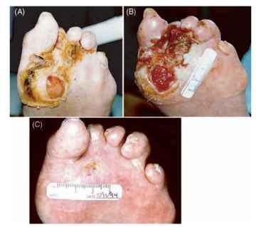
FIGURE 1 The left foot of a 73-year-old man who was treated for 3 years by orthopedic and podiatric surgery for his chronic foot ulcers: (A) before, (B) during, and (C) 1 year after maggot debridement therapy.
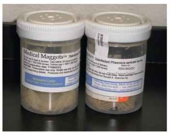
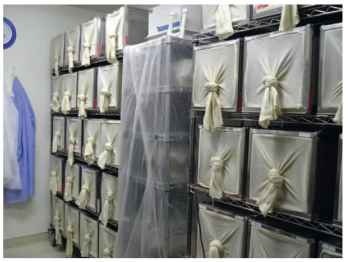
II. Over the next several decades, therapeutic myiasis was performed only rarely, and only as a last resort, in patients who failed to respond to aggressive surgical and antibiotic treatments. But the 1980s brought about the realization that surgery and antibiotics could not cure all wounds. Many microbes were by now resistant to the once omnipotent antimicrobials. In the 1990s, maggot therapy was reintroduced as a treatment for nonhealing wounds, after a series of controlled clinical studies demonstrated better responses to maggot therapy than to conventional medical and surgical treatments (Fig. 1). Today, medical grade larvae are produced by 24 laboratories worldwide, and sent to thousands of centers in over 40 countries for treating wounds (Figs. 2 and 3).

FIGURE 2 In the United States, medicinal maggots are sold under the brand name Medical Maggots™.

FIGURE 3 This facility (insectary) for breeding medicinal maggots produces nearly one million eggs per week. The eggs are disinfected in a separate section of the laboratory, at which point they become an FDA-regulated medical device and are handled according to Good Manufacturing Procedure (GMP) standards of the medical device industry.
Maggots effectively treat open wounds by removing dead and infected tissue (debridement), killing bacteria (disinfection), and stimulating the wound to heal. To understand the procedure of maggot therapy, it is necessary to review the natural history of the fly. Many species of blowflies (Calliphoridae) naturally “blow” or lay their eggs on carrion, feces, or the dead (necrotic) tissue of a living host. Upon hatching, the larvae secrete and excrete their digestive enzymes, liquefying the dead organic tissue so that they can ingest it. The larvae will be satiated within 3-7 days (depending on the temperature and abundance of food) and the maggots leave the host to pupate underground or in some other protected area. One to three weeks later, adult flies eclose (emerge). Therapeutic maggots—blowfly larvae that have been made “germ-free” (disinfected)—are placed on wounds at a density of 5-10 cm~2. Larvae are retained on the wound for about 48 h in cage-like dressings. After one or more such cycles of treatment, the wound is often free of necrotic tissue and able to accept a skin graft or heal spontaneously. Treatments can be administered in the hospital, clinic, nursing home, or at home.
Maggot therapy has been used to treat pressure ulcers, venous stasis ulcers, diabetic foot ulcers, burns, traumatic wounds, and non-healing postsurgical wounds. Compared with conventional wound therapy, medicinal maggots are credited with more rapid debride-ment and wound healing. Maggot therapy has reportedly saved numerous limbs from amputation and other surgical procedures. Other advantages of maggot therapy include its simplicity, safety, and relatively low cost. Efforts are underway to isolate the active constituents in the maggots’ secretions, but thus far no isolate has been found to equal the efficacy of the live larvae.
BEE VEENOM THERAPY
Honey bee (Apis mellifera) venom, propolis, royal jelly, beeswax, and honey all are used therapeutically. Medicinal use of any of these products can be considered to be “apitherapy,” but many authorities use the term specifically to denote the clinical use of honey bee venom itself (known more precisely as bee venom therapy). Bee venom therapy has been used successfully to treat rheumatological disorders (rheumatoid and psoriatic arthritis, gout, fibromyalgia), neurological diseases (multiple sclerosis, chronic pain syndromes), immunological diseases (scleroderma, systemic lupus erythematosis), and other chronic illnesses.
Bee venom contains a multitude of polypeptides, enzymes (phos-pholipase A2, hyaluronidase), and biologically active amines (his-tamine, dopamine, and noradrenaline). The mechanisms by which venom exerts its beneficial actions are unknown, but might include an anti-inflammatory effect resulting from alterations seen in pituitary and adrenal gland function, local effects on the nerves and blood vessels, and stimulation of acupuncture-like pathways. A combination of these and other mechanisms may explain the diversity of benefits attributed to venom therapy.
Some therapists administer the treatments in the form of an increasing number of bee stings; other practitioners inject a partially purified extract of the bee venom, in gradually increasing doses. Serious toxic reactions to the venom are uncommon because patients are educated about the signs and symptoms of venom reactions; they are also supervised closely following treatment and are given ready access to medical care after leaving the therapist’s office or apiary.
